LEHNINGER PRINCIPLES OF BIOCHEMISTRY - VOL. 2. BIOENERGETICS AND METABOLISM - 2014
PART II. BIOENERGETICS AND METABOLISM
19. OXIDATIVE PHOSPHORYLATION AND PHOTOPHOSPHORYLATION
19.4. The Role of Mitochondria in Thermogenesis, Steroid Synthesis, and Apoptosis
The primary function of Cell/35.html">Mitochondria is ATP synthesis. However, mitochondria also participate in processes that, in certain Tissues and under specific circumstances, become critical. In adipose tissue, mitochondria generate heat to protect vital Organs from hypothermia; in the Adrenal Glands and Gonads, mitochondria are involved in the synthesis of Steroid Hormones; and in virtually all tissues, they participate in apoptosis (programmed cell death).
Uncoupling of Respiration and Phosphorylation in Brown Adipose Tissue Generates Heat
We mentioned earlier that when the supply of ATP to a cell is adequate, The rate of respiration decreases. There is one interesting exception to this general rule. Most newborn mammals, including human infants, possess a specialized adipose tissue called brown adipose tissue (p. 588). Its function is to prevent ATP synthesis during fat oxidation and instead produce heat, thereby maintaining the newborn's body Temperature at the proper level. This specialized Adipose tissue is indeed brown because it is packed with mitochondria containing high levels of Cytochromes, whose heme groups absorb strongly in the visible spectrum.
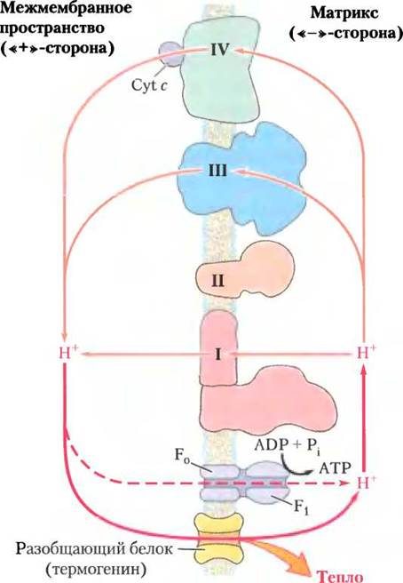
Brown adipose tissue mitochondria are structurally similar in many ways to those of other mammalian Cells, but their inner membrane contains a uncoupling protein, thermogenin (the product of the UCP1 Gene). Because of thermogenin, H+ ions pumped out of the mitochondria by electron transport return to the matrix bypassing the F0F1 enzyme complex (Fig. 19-34). As a result, the Free energy of Electron transport is used not for ATP synthesis, but for heat production, which helps maintain the required body temperature (see Fig. 23-17). In some hibernating animals, the uncoupling of respiration and phosphorylation in brown adipose tissue mitochondria generates the heat needed to maintain body temperature during hibernation (see Box 17-1). We will return to The Role of thermogenin when discussing The regulation of body weight in Chapter 23.
Class="center">Figure 19-34. Heat production by uncoupled respiration and phosphorylation in mitochondria. The uncoupling protein (thermogenin), localized in The inner mitochondrial membrane, provides specific pores for H+ ions. H+ ions pumped out of the mitochondria during electron transport return to the mitochondrial matrix through these pores, bypassing ATP synthase. Consequently, the energy of the proton gradient is dissipated as heat rather than being used for ATP synthesis.
Mitochondrial P-450 Enzyme Systems Catalyze Steroid Hydroxylation
Mitochondria are the site of biosynthetic reactions leading to The formation of steroid hormones, including Sex Hormones, glucocorticoids, mineralocorticoids, and vitamin D. These substances are synthesized from Cholesterol or related precursors through a series of hydroxylation Reactions Catalyzed by Enzymes of the cytochrome P-450 family, each of which contains a heme group (the ability of heme to absorb light at a wavelength of 450 nm gave this enzyme family its name). In these hydroxylation reactions, one atom of molecular oxygen is incorporated into the substrate, while the second is reduced to H2O.
R-H + O2 + NADPH —> R-OH + Н2O + NADP+
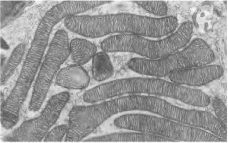
The P-450 family comprises several dozen enzymes, all of which are localized on the inner mitochondrial membrane with their catalytic sites facing the matrix. Steroidogenic cells (cells that synthesize Steroids) contain mitochondria specialized for steroid synthesis; such mitochondria are typically larger than those in other tissues and have a more developed and convoluted inner membrane surface (Fig. 19-35).
Figure 19-35. Mitochondria of the adrenal cortex specialized for steroid synthesis. As seen in this electron micrograph of a thin section of adrenal tissue, numerous mitochondria with abundant cristae are concentrated here, providing a large inner membrane surface area where P-450 enzyme systems are localized.
The electron transport pathway in the mitochondrial P-450 system is relatively complex; it involves a flavoprotein and an iron-sulfur protein that transfer electrons from NADPH to the P-450 heme (Fig. 19-36). The Structure of the P-450 enzyme family ensures their substrate Specificity and allows the heme group to react directly with O2.
Figure 19-36. Electron transfer pathway involving the cytochrome P-450 system in adrenal mitochondria. Two electrons are transferred from NADPH to an FAD-containing flavoprotein, adrenodoxin reductase, which passes them one at a time to adrenodoxin, a small, Water-soluble 2Fe-2S protein. Adrenodoxin delivers single electrons to the cytochrome P-450 hydroxylase, which interacts directly with O2 and the substrate (R-H) to yield the products: H2O and R-OH.

Another large family of P-450 enzymes is found in The Endoplasmic reticulum of hepatocytes. These enzymes catalyze reactions similar to those mediated by mitochondrial P-450 enzymes, but their substrate range is extremely broad, encompassing various hydrophobic compounds, many of which are xenobiotics (substances not found in nature, produced exclusively through Industrial processes). The P-450 enzymes of the endoplasmic reticulum exhibit broad and overlapping substrate specificities. Hydroxylation of hydrophobic compounds increases their water solubility, facilitating their excretion from the body in the urine via the Kidneys. Substrates of the P-450 oxygenase family include many common drugs. Reactions catalyzed by P-450 enzymes shorten the half-life of drugs in the bloodstream and diminish their therapeutic efficacy. Due to genetic variations, the Complement of endoplasmic reticulum cytochromes P-450 differs slightly among individuals, and some enzymes in this family also display variable induction levels, for instance, in response to alcohol consumption. Therefore, considering a patient's genetic profile and medical history is crucial when prescribing medications and determining dosages; however, such personalized approaches are rarely implemented in clinical practice due to economic constraints, though they may become feasible in the future. ■
Mitochondria Play a Major Role in Initiating Apoptosis
Apoptosis, or programmed cell death, involves the regulated demise of individual cells when required by the Organism (such as during normal embryonic development), while preserving cellular components such as Amino Acids, NUCLEOTIDES, and others. Apoptosis can be triggered by external signals acting on Plasma Membrane Receptors, as well as by internal events such as DNA damage, viral infection, oxidative stress caused by ROS, or other stressors like heat Shock.
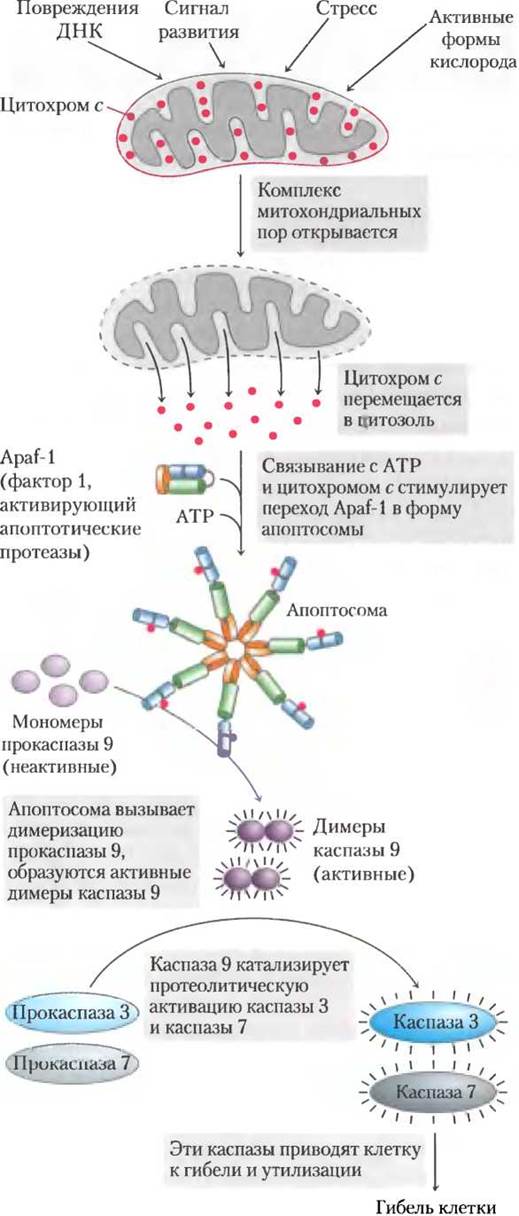
Mitochondria play a vital role in triggering apoptosis. When stress signals the destruction of a cell, one of the earliest changes is an increase in the permeability of the outer mitochondrial membrane, allowing cytochrome c to escape from the intermembrane space into the Cytosol (Fig. 19-37). This increase in membrane permeability is driven by the opening of so-called giant pores formed by protein subunits in the outer mitochondrial membrane. The opening and closing of these pores are modulated by Proteins that promote or inhibit apoptosis. Upon entering the cytosol, cytochrome c interacts with monomers of the protein Apaf-1 (apoptotic protease activating factor-1), leading to the formation of an apoptosome composed of seven Apaf-1 molecules and seven cytochrome c molecules. Within the apoptosome, procaspase-9 is activated to form caspase-9, a member of the family of specialized proteases (caspases) involved in apoptosis. The Active Site of all proteins in this family contains a Cysteine residue, and they cleave proteins specifically after aspartate residues on the carboxyl-terminal side of the Amino Acid Sequence; hence their name — caspases (derived from cysteine-dependent aspartate-specific proteases). Activated caspase-9 initiates a proteolytic cascade in which one caspase activates another, which in turn activates a third, and so on (see Fig. 12-51). The involvement of cytochrome c in apoptosis is a classic example of moonlighting, where an established enzyme takes on an additional function.
Figure 19-37. The role of cytochrome c in apoptosis. Cytochrome c is a small, water-soluble mitochondrial protein located in the intermembrane space, where its primary function is to shuttle electrons between complexes III and IV of the Respiratory Chain. However, it performs an entirely distinct secondary function within The Cell by triggering apoptosis through the Activation of a family of proteinases called caspases.
Summary of section 19.4 The Role of Mitochondria in Thermogenesis, Steroid Synthesis, and Apoptosis
■ In newborns, Electron transport along the Respiratory Chain and ATP synthesis are uncoupled in brown adipose tissue mitochondria; consequently, the free energy released during Fatty acid oxidation is dissipated as metabolic heat.
■ During the synthesis of steroid hormones in steroidogenic tissues (adrenal glands, gonads, Liver, and kidneys), hydroxylation reactions take place in specialized mitochondria.
■ During apoptosis, protein degradation is mediated by the proteolytic enzyme caspase-9, which is activated by cytochrome c released from the intermembrane space into the cytosol upon receiving an apoptotic signal.
Last update: 06/08/2026
Editorial and Educational Adaptation: This material has been compiled based on the primary/original source text. The project team performed an editorial review, corrected technical inaccuracies, structured sections, and adapted the content for an educational format.
What was processed:
- elimination of formatting defects (OCR errors, structural breaks, corrupted characters);
- editorial organization of content;
- standardization of terminology in accordance with academic sources;
- verification of factual statements against the original source text.
All mentions of the author, publication year, and origin of the primary text have been preserved in accordance with the source.