BIOCHEMISTRY: A TEXTBOOK FOR UNIVERSITIES - E. S. Severin - 2004
SECTION 1. STRUCTURE, PROPERTIES, AND FUNCTIONS OF PROTEINS
IV. Protein Functioning
Each individual protein, possessing a unique Primary Structure and conformation, also exhibits a unique function that distinguishes it from all other Proteins. The set of individual proteins performs a multitude of diverse and complex Functions within The Cell.
A prerequisite for protein functioning is the binding of another substance, referred to as a "Ligand." Ligands can be either low-molecular-weight substances or macromolecules. The interaction between a protein and a ligand is highly specific, which is determined by The structure of the protein region known as the protein-ligand binding site or the Active Site.
A. The Active Site of Proteins and the Selectivity of Ligand Binding
The active site of a protein is a specific region of the protein molecule, typically located in a depression (a "pocket"), formed by amino acid residues assembled within a specific spatial area during The formation of the tertiary structure, and capable of binding to a ligand in a complementary manner. In the linear sequence of the polypeptide chain, the residues that form the active site can be located at a considerable distance from one another.
The high Specificity of protein-ligand binding is ensured by the complementarity between the STRUCTURE OF THE protein's active site and the structure of the ligand (Fig. 1-25).
Class="center">Fig. 1-25. Protein-ligand interaction. A and B — non-complementary interaction and disruption of bonds between the protein and the ligand; C — Complementary interaction between the protein and the ligand.

Complementarity refers to the spatial and chemical correspondence of interacting molecules. A ligand must be capable of entering and spatially matching the conformation of the active site. This match may be partial, but due to the conformational lability of the protein, the active site is capable of minor adjustments and "fits" the ligand. Furthermore, bonds must form between the Functional groups of the ligand and The amino acid residues that constitute the active site, holding the ligand in place. The bonds between the ligand and the protein's active site can be either non-covalent (ionic, hydrogen, hydrophobic) or covalent.
1. CHARACTERISTICS OF THE Active Site
The active site of a protein is a region relatively isolated from the surrounding protein environment, formed by amino acid residues. Within this region, each residue, through its individual size and functional groups, shapes the "relief" of the active site.
The combination of such Amino Acids into a single functional complex alters the reactivity of their radicals, much like the sound of a musical instrument changes within an ensemble. Therefore, the amino acid residues that make up the active site are often referred to as an amino acid "ensemble."
The unique Properties of the active site depend not only on The chemical properties of the amino acids that form it, but also on their precise spatial orientation relative to one another. Therefore, even minor disruptions in the overall protein conformation resulting from point Mutations in its primary structure or environmental changes can alter the chemical and Functional Properties of the radicals forming the active site, impair protein-ligand binding, and disrupt its function. During Denaturation, the active site of proteins is destroyed, leading to the loss of their biological activity.
Frequently, the active site is formed in such a way that Water access to the functional groups of its radicals is restricted, thereby creating conditions for the binding of the ligand to the amino acid radicals.
In some cases, the ligand attaches to only a single atom possessing specific reactivity, such as the binding of O2 to the iron in Myoglobin or Hemoglobin. However, The properties of this atom to selectively interact with O2 are determined by the properties of the radicals surrounding the iron atom within the heme group. Heme is also found in other proteins, such as Cytochromes. Nevertheless, the function of the iron atom in cytochromes is different; it serves as a mediator for Electron transfer from one substance to another, with the iron alternating between ferrous (divivalent) and ferric (trivalent) states.
The protein-ligand binding site is often located between domains. For example, the proteolytic enzyme Trypsin, which is involved in the Hydrolysis of peptide bonds in dietary proteins within the intestine, has 2 domains separated by a cleft. The inner surface of the cleft is formed by amino acid radicals from these
domains that are located far apart in the polypeptide chain (Ser177, His40, Asp85).
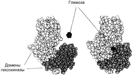
Different domains within a protein can move relative to one another upon ligand interaction, which facilitates the subsequent functioning of the protein. As an example, we can examine the action of hexokinase, an enzyme that catalyzes The transfer of a phosphoryl group from ATP to a glucose molecule (during its phosphorylation). The active site of hexokinase is located in a cleft between two domains (Fig. 1-26). Upon binding of hexokinase to glucose, the surrounding domains move closer together, trapping the substrate and thereby facilitating its subsequent phosphorylation.
Fig. 1-26. Binding of hexokinase to glucose.
The fundamental property of proteins that underlies their functions is the selective attachment of specific ligands to defined Regions of the protein molecule.
2. Diversity of Ligands
✵ Ligands can be inorganic substances (often Metal Ions) and organic substances, low-molecular-weight and high-molecular-weight compounds;
✵ there are ligands that undergo chemical modification upon binding to the protein's active site (substrate alterations within the enzyme's active site);
✵ some ligands bind to the protein only during its functioning (e.g., O2 transported by hemoglobin), whereas other ligands remain permanently bound to the protein, playing an auxiliary role in its activity (e.g., iron as a structural component of hemoglobin).
In cases where the amino acid residues forming the active site cannot ensure proper protein function, non-protein molecules may attach to specific regions of the active site. For instance, the active site of many Enzymes contains a metal ion (cofactor) or an organic non-protein molecule (coenzyme). The non-protein moiety firmly bound to the protein's active site and essential for its function is termed a "prosthetic group." Myoglobin, hemoglobin, and cytochromes feature an iron-containing heme as their prosthetic group (heme-containing proteins are discussed in detail in section 4, while Cofactors and Coenzymes are covered in section 2).
The assembly of protomers into an oligomeric protein is a prime example of high-molecular-weight ligand interaction. Each protomer, bound to the others, acts as a ligand for them, just as they do for it.
Sometimes, the binding of a ligand alters the protein conformation, thereby creating a binding site for other ligands. For example, upon binding four Ca2+ ions at specific sites, the protein calmodulin acquires The ability to interact with certain enzymes, modulating their activity.
3. Affinity of the Active Site for a Ligand
The Rate of protein-ligand interaction is determined by the concentrations of the protein and ligand in solution, as well as the degree of complementarity between the protein and the ligand.
Dissociation constant — a quantitative measure of the active site's affinity for a ligand.
Since protein-ligand binding is a reversible process, it can be described by the following equation:
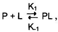
where P is the protein, L is the ligand, PL is the protein-ligand complex, K1 is the association rate constant for the protein-ligand binding, and K-1 is the dissociation rate constant for the PL complex.
When the rates of complex formation and dissociation are equal, the system is said to be in a state of equilibrium:
[Р] [L] К1 = [РL] К-1.
Hence:
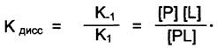
The ratio of the dissociation rate of the [РL] complex to its formation rate is termed the dissociation constant (Kd) of the [РL] complex. The smaller the Kd, the greater the number of ligand molecules bound to the protein, the higher the complementarity between P and L, and the stronger the affinity of the ligand for the protein. In other words, an inverse proportionality exists between Kd and ligand-protein affinity.
Occasionally, the reciprocal of Kd, known as the binding constant (Kb) or association constant, is used to describe protein-ligand binding.
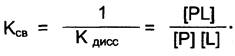
A direct proportionality exists between Kb and the affinity of the ligand for the protein.
Dependence of Protein Saturation on Ligand Concentration at Constant Protein Concentration
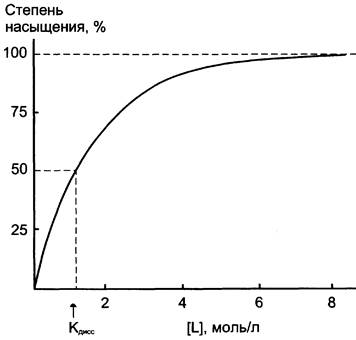
At a constant protein concentration, increasing the ligand concentration leads to a rise in the [PL] complex concentration. This relationship follows a hyperbolic curve (Fig. 1-27). The curve approaches a plateau when, at a certain ligand concentration, all protein molecules are bound to the ligand (protein saturation is achieved). The degree of protein saturation with a ligand can be expressed by the following equation: degree of saturation = [PL]/[P0] x 100 (where P0 is the protein concentration prior to ligand addition). At half-saturation of the protein with the ligand, the concentrations of [PL] and [P] are equal, and from the dissociation constant equation given above, it follows that Kd = [L], meaning Kd is numerically equal to the ligand concentration at which 50% of the protein is complexed with the ligand. Therefore, the saturation curve allows one to determine Kd and evaluate the affinity of the given protein for the ligand.
Fig. 1-27. Protein-ligand saturation curve.
Relationship between [PL] Complex Formation and Protein Concentration in the Presence of Excess Ligand
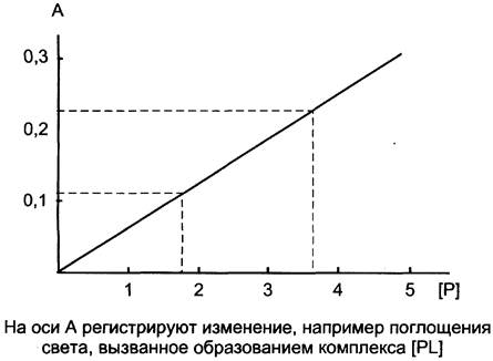
As stated above, at increasing ligand concentrations, protein saturation is limited by the protein's own concentration. In the presence of excess ligand, all protein molecules exist as the [PL] complex. However, increasing the protein concentration causes The amount of [PL] to rise in direct proportion to the protein concentration. The concentration of the [PL] complex can be monitored, for instance, by measuring Light absorption. Given that its amount is proportional to the protein concentration, the resulting graph can be used to determine the protein concentration in solution (Fig. 1-28).
Fig. 1-28. Graph of the dependence of changes in light absorption, reflecting the concentration of the [PL] complex, on the protein concentration P.
B. Substances Affecting Protein function
Although the Interaction of a ligand with the active site of a protein is highly specific, it is always possible to find another substance that will interact with the protein in a similar manner. A ligand that interacts with a protein and disrupts its function is called a "protein inhibitor." If this substance is structurally similar to the ligand, it is referred to as a structural analog of the ligand; it also interacts with the active site of the protein. An analog that replaces the natural ligand in the active site of a protein and diminishes its function is called a "competitive protein inhibitor."
1. Pharmaceuticals as modulators of Protein Functions
Analogs of natural protein ligands are used in medicine as therapeutic agents. Such drugs have found widespread application in regulating the transmission of excitation across synapses.
Signal transmission from nerve to nerve or from a nerve to an effector organ occurs via synapses using chemical molecules called Neurotransmitters. A neurotransmitter released by nerve endings during the passage of an impulse must interact with high specificity with receptor proteins on the postsynaptic membrane. However, by modifying the Chemical Structure of the neurotransmitter, substances can be obtained that also bind to the receptor while altering the physiological effect—either decreasing or enhancing it. In pharmacology, such substances are termed "antagonists" and "agonists," respectively.
Receptor protein inhibitors in cholinergic synapses
As an example, consider drugs that disrupt Nerve Impulse transmission through cholinergic synapses, where acetylcholine serves as the neurotransmitter. Cholinergic receptor proteins are structurally heterogeneous and capable of binding ligands other than acetylcholine. They are divided into 2 major groups:
✵ M-cholinergic receptors, named for their ability to selectively interact with muscarinist (a fly agaric toxin);
✵ N-cholinergic receptors, which selectively bind nicotine.
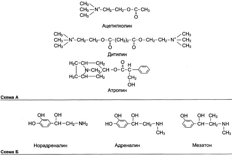
N-cholinergic receptors are present in neuromuscular junctions, where their interaction with acetylcholine causes Muscle contraction. To induce muscle relaxation during endoscopic examinations and various surgical Procedures, structural analogs of acetylcholine that act as inhibitors of these receptors are used. An example of such a substance is dithilin (suxamethonium), which belongs to a group of medicinal substances known as muscle relaxants (agents that cause muscle relaxation). These properties were originally discovered in curare poison, which is why these drugs are also referred to as curare-like agents (see Scheme A on p. 44).
The best-known specific inhibitor of M-cholinergic receptors is atropine. Atropine is an alkaloid found in certain plants, such as deadly nightshade (belladonna), henbane, and thornapple (datura). It binds to M-cholinergic receptors located on the membrane of effector Cells in the region of parasympathetic nerve endings. Atropine prevents their interaction with acetylcholine (acting as an antagonist of the natural ligand), thereby eliminating the effects of parasympathetic nerve stimulation.
Since acetylcholine binding to M-cholinergic receptors causes the contraction of many smooth Muscles, atropine (as a pharmaceutical preparation) relieves muscle spasms (acting as a spasmolytic). In addition, it reduces acetylcholine-stimulated secretion of glands (bronchial, digestive, sweat).
M-cholinergic receptors are present in various regions of the Central Nervous system. An overdose of atropine can cause motor and speech excitation.
Pharmaceutical substances as stimulators of protein functions
However, certain structural analogs of receptor protein ligands are not inhibitors; instead, they elicit physiological effects similar to or stronger than those of natural ligands. Their stronger and more prolonged effect is often due to the fact that modified ligands are inactivated and degraded more slowly in the body. For example, mesaton (phenylephrine) is structurally similar to the neurotransmitters of the sympathetic nervous system (norepinephrine and epinephrine). Mesaton increases vascular tone and Blood pressure, which is why it is used in hypotension and collapse. It is less susceptible to inactivation by enzymes, thus producing a more prolonged and potent effect than its natural analogs (see Scheme B).
2. Poisons as specific ligands for certain proteins
Some poisons, upon entering The Human Body, bind tightly to specific proteins, inhibit them, and thereby cause disruptions in biological functions.
For example, α-neurotoxins from cobra and krait venoms specifically interact with cholinergic receptors of postsynaptic membranes, blocking their function and exerting a curare-like effect. α-Neurotoxins are small proteins with a Molecular Weight of about 7000 D (65–70 amino acid residues). Their tertiary structure is stabilized by 4 or 5 specific Disulfide Bonds (depending on the toxin species). The affinity of neurotoxins for cholinergic receptors is very high (Kdiss = 10-11). Evidently, numerous bonds are formed between the toxin and the receptor, resulting in their virtually irreversible binding.
It must be kept in mind that the boundary between drugs and poisons is often blurred, and The Effect of their action depends on the dose of the administered substance. Thus, drugs prescribed in doses exceeding therapeutic levels can act as poisons—i.e., cause severe Metabolic Disorders and impairments of bodily functions—whereas poisons in microdoses are frequently used as therapeutic agents. For instance, atropine, which is widely used to relieve smooth muscle spasms, causes central nervous system excitation at high doses, and Sleep progressing into a coma at even higher doses. The well-known antihypertensive drug clonidine causes collapse when overdosed.
Last update: 06/08/2026
Editorial and Educational Adaptation: This material has been compiled based on the primary/original source text. The project team performed an editorial review, corrected technical inaccuracies, structured sections, and adapted the content for an educational format.
What was processed:
- elimination of formatting defects (OCR errors, structural breaks, corrupted characters);
- editorial organization of content;
- standardization of terminology in accordance with academic sources;
- verification of factual statements against the original source text.
All mentions of the author, publication year, and origin of the primary text have been preserved in accordance with the source.