ZOOLOGY OF INVERTEBRATES IN THREE BOOKS - BOOK 2 - G.I. Shcherbak - 1996
PHYLUM ANNELIDA
CLASS OLIGOCHAETA
Oligochaetes inhabit freshwaters, soil, and more rarely (about 200 species) marine environments. The majority of oligochaetes are burrowing forms; some live on the bottom surface among decaying organic matter, while a small number of species parasitize the gills of crustaceans. About 5,000 species are known. In the freshwaters of Ukraine, about 200 species of oligochaetes have been recorded, and 33 species are part of the fauna of the Black and Azov Seas. Soil oligochaetes are less studied here, with approximately 35 species of earthworms known. Most oligochaetes range in size from 0.5 mm to 40 cm, though some species of tropical earthworms (Megascolides australis) can reach 3 m.
In their general body plan, oligochaetes are closely related to Polychaeta. These are exclusively polymeric Annelids that have lost parapodia and most appendages of the prostomium. They exhibit homonomous metamerism, although mature individuals possess a ring-like expansion on certain body segments—the clitellum, the epidermis of which secretes the cocoon material. They are hermaphroditic, with reproductive Organs restricted to specific body segments. Development is direct, without metamorphosis.
Class="center">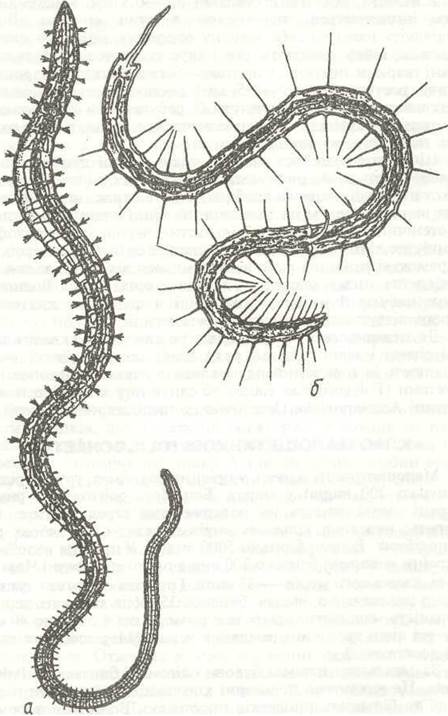
Fig. 19. Oligochaetes:
a - sludge worm Tubifex tubifex (family Tubificidae); b - Stylaria lacustris (family Naididae)
The body of oligochaetes is more or less elongated and nearly cylindrical (Fig. 19). The prostomium typically lacks appendages and eyes, and only in some marine species (family Naididae) are paired eyes present. Occasionally, the prostomium is extended into a proboscis. The peristomium is absent; the Mouth is located on the first trunk segment (Fig. 20). The number of segments ranges from 5–6 to 500–600. Parapodia are reduced, leaving behind paired setae in their place. Each segment, except the first, generally bears four bundles of setae: two dorsal and two ventral. Sometimes the dorsal bundles are reduced or, more rarely, may disappear altogether.
Typically, each bundle contains a pair of setae, whereas in aquatic forms there are from 2 to 10–15 setae of diverse shapes (needle-like, hook-shaped, pectinate, etc.). In some tropical forms (genus Pheretimania), the number of setae can reach up to 150, forming rings around each segment. Setae participate in the locomotion of the worms: burrowing forms brace them against the walls of their burrows, while aquatic species use them for swimming. Special setae are located on reproductive segments near the male genital pores; these widen the openings of the partner's spermathecae during copulation. In addition, near the spermathecal openings, there are specialized sharp, thick, knife-like setae with a longitudinal groove that forcefully press into the partner's Skin, presumably directing sperm into the spermathecae.
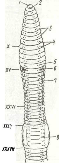
Fig. 20. Anterior region of the body of the earthworm Lumbricus terrestris from the ventral side:
1 - prostomium; 2 - mouth; 3 - ventral setae; 4 - spermathecal pores; 5 - female genital pore; 6 - male genital pore; 7 - seminal groove; 8 - clitellum; 9 - X - XXXVII - segments
The cutaneous-muscular sac (Fig. 21) has a typical annelid Structure consisting of a thin elastic cuticle, a skin epithelium (epidermis), and two Muscle layers: a thin circular layer and a thicker longitudinal layer. Connective Tissue Cells lie loosely between the muscle fibers. In the regions where setae emerge, invaginations of the cuticle and epidermis form seta-bearing follicles. The skin of oligochaetes is rich in glandular cells situated among the epithelial cells. The mucus they secrete lubricates the body and, in soil-dwelling oligochaetes, keeps it moist—a vital condition for cutaneous respiration. Numerous mucous and proteinaceous unicellular glands are located in the epithelium of the clitellum; they secrete the cocoon material.
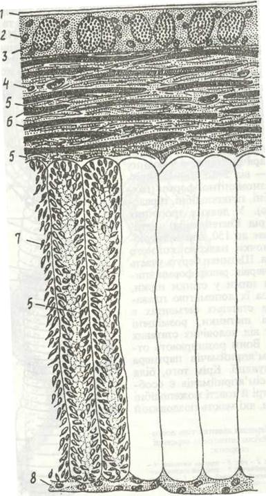
Fig. 21. STRUCTURE OF THE cutaneous-muscular sac of Lumbricus terrestris:
1 - cuticle; 2 - epidermis; 3 - mucous gland; 4 - Nucleus of a CONNECTIVE TISSUE Cell; 5 - connective tissue; 6 - circular muscle fibers; 7 - longitudinal muscle fibers; 8 - peritoneal epithelium
Beneath the cutaneous-muscular sac lies a layer of peritoneal epithelium that tightly adheres to it and lines the secondary body cavity, or coelom (Fig. 22).
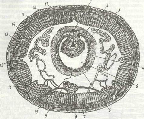
Fig. 22. Diagram of a cross-section through the middle part of the body of Lumbricus terrestris:
1 - dorsal Blood vessel; 2 - typhlosolar vessels; 3 - typhlosole; 4 - intestinal and vascular plexus; 5 - midgut; 6 - spermatogenic cells; 7 - ventral nerve cord; 8 - subneural vessel; 9 - ventral blood vessel; 10 - mesentery; 11 - setigerous follicle; 12 - metanephridium; 13 - coelomic epithelium; 14 - longitudinal musculature; 15 - circular musculature; 16 - epidermis; 17 - cuticle
In the middle and posterior segments of many soil-dwelling species (such as Lumbricus), unpaired dorsal pores located near the intersegmental furrows connect the coelom with the external environment. Coelomic fluid oozes through these pores, which is believed to lubricate the body and facilitate the animal's movement through the soil. Excretory corpuscles are also expelled from the body cavity through the Pores in the posterior region of the body.
Oligochaetes move through the contraction of the Muscles of the cutaneous-muscular sac, with the coelomic fluid acting as a hydrostatic Skeleton. Oligochaetes can crawl and swim; burrowing forms move continuously, cutting passages through the soil. Burrowing occurs differently than in forms with an unpartitioned body cavity (for example, priapulids). Septa between the segments prevent fluid from surging through the entire body; instead, it flows only within 2–5 segments via openings in the dissepiments.
The Digestive System begins with the oral opening leading into a muscular Pharynx, into which numerous Salivary Glands empty. The pharynx transitions into a narrow Esophagus, which may expand into a crop at its posterior end, followed by a muscular gizzard (Fig. 23). These regions belong to the ectodermal foregut. In species that pass soil through their gut, there are three pairs of calciferous glands that open successively into one another and via a pair of ducts into the esophagus. They are packed with calcium carbonate crystals; their main function is to remove carbonates from the blood and bind excess carbon dioxide in the blood by forming soluble bicarbonates. Additionally, lime enters the esophagus, where it neutralizes humic acids contained in the soil that are harmful to the digestive organs. The walls of the midgut are rich in ciliated and secretory cells, where food is digested and absorbed. In soil-dwelling oligochaetes, the DORSAL SIDE OF the midgut features a longitudinal groove-like protrusion into the intestinal lumen—the typhlosole—which significantly increases the surface area of the gut (Figs. 22, 23). It terminates near the posterior end of the body, where the midgut transitions into a short hindgut that opens via the anus on the pygidium. The outer walls of the intestine are covered with chloragogen tissue, the cells of which perform an excretory function by accumulating dissimilation products. Most oligochaetes feed on PLANT AND ANIMAL remains, passing large quantities of soil through their guts. Aquatic inhabitants feed on microscopic Algae, Protozoans, and Bacteria.
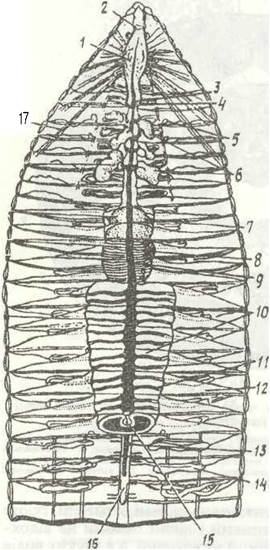
Fig. 23. Internal anatomy of Lumbricus terrestris (dissection from the dorsal side):
1 - pharynx; 2 - supraoesophageal ganglion; 3 - oesophagus; 4 - hearts; 5 - seminal receptacles; 6 - Seminal Vesicles; 7 - crop; 8 - gizzard; 9 - dorsal blood vessel; 20 - midgut; 11 - lateral vessels; 12 - dissepiment; 13 - ventral vessel; 14 - metanephridium; 15 - typhlosole; 16 - ventral nerve cord; 17 - radial pharyngeal muscles
The excretory system is typically represented by a single pair of metanephridia in each segment, with the exception of a few anterior (larval) ones. Sometimes the number of metanephridia is reduced or they disappear entirely (in marine species of the genera Paranais and Tubificoides). A nephridium begins within a segment with a ciliated funnel. A slender, ciliated canal extends from it, piercing the dissepiment into the next segment. Here, the nephridium forms three lobes consisting of canal loops surrounded by the excretory Cells of the coelomic epithelium. These lobes are densely enveloped in blood capillaries. Metabolic wastes diffuse from the blood into the lumen of the canal. Urine collects in the terminal dilation of the canal—the bladder—which opens to the exterior via an excretory pore. In some oligochaetes, the nephridia unite to form a paired excretory duct that empties into the hindgut.
In addition to nephridia, so-called chloragogen cells, mentioned above, take part in excretion. These are specialized elongated cells of the coelomic epithelium located On the surface of the gut and Blood Vessels. Recent evidence indicates that their function is much more complex: alongside Metabolic waste products, they accumulate nutrient reserves (Glycogen and Lipids).
The Circulatory system in oligochaetes, as in other annelids, is closed. Vessels lie beneath the coelomic epithelium of the intestine, dissepiments, and body walls; capillaries proliferate deep into the Tissues. The circulatory system comprises an intestinal sinus (which may be replaced by an intestinal vascular plexus), dorsal, ventral, ring, and lateral vessels that do not form complete loops. The ring vessels encircling the oesophagus possess thick muscular walls and function as hearts, driving blood from the dorsal vessel to the ventral one. Their number varies among different oligochaetes; Lumbricus terrestris has five pairs of hearts (Fig. 23). Furthermore, a dense subcutaneous network of fine vessels and capillaries develops in connection with cutaneous respiration. These are very thin vessels that penetrate the body wall musculature. They are particularly abundant in the circular muscle layer, with their finest branches even extending into the epidermis (Fig. 24, a). In many oligochaetes, the blood is colourless; in some, it is red due to the presence of haemocruorin, a pigment closely related to haemoglobin dissolved in the plasma. Occasionally (in certain earthworms), the pigment is contained within specialized cells (haemocytes). In tropical giant earthworms (genus Megascolides), the blood is greenish due to the presence of another pigment, chlorocruorin. The blood also contains numerous colourless cells of various structures. The walls of the blood vessels are formed by a non-cellular supporting lamella, and they are externally covered with chloragogen cells. Large vessels (such as the dorsal and ring vessels) contain muscles within their walls (Fig. 24, b).
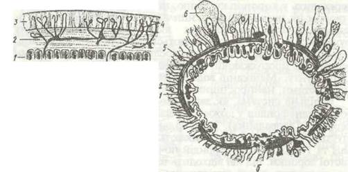
Fig. 24. Blood vessels of Lumbricus terrestris:
a - cutaneous capillaries; b - cross-section through the dorsal vessel; 1 - longitudinal musculature; 2 - circular musculature; 3 - epidermis; 4 - capillary branching; 5 - supporting lamella; 6 - chloragogen cells
A Respiratory system is generally absent: gas exchange occurs through the thin, moist skin and blood capillaries. However, certain aquatic forms possess external gills. For instance, in worms that bury their anterior ends in mud (genera Dero and Aulophorus), the caudal region features a broad lobe bearing paired, leaf-like gills. The gill surface is covered with cilia that generate Water currents, and the gills are richly supplied with blood capillaries. In bottom-dwelling species (e.g., from the families Tubificidae and Naididae), intestinal respiration supplements cutaneous respiration.
The Nervous system has a typical annelid structure, consisting of a paired supraoesophageal ganglion, circumoesophageal connectives, and a ventral nerve cord (Fig. 25). Only in a few species are the ventral trunks widely separated and the ganglia connected by long commissures (e.g., in Aelosoma).
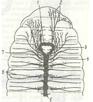
Fig. 25. Nervous system of Lumbricus terrestris:
1 - supraoesophageal ganglion; 2 - NERVES OF THE prostomium and segment I; 3 - circumoesophageal connective; 4 - segmental ganglia;
5 - ventral nerve cord; 6 - segmental nerves; 7 - suboesophageal ganglion
Sense Organs in microdriles are poorly developed. Eyes are generally absent, yet earthworms exhibit sensitivity to light. Scattered throughout their skin
epithelium are solitary cells or groups of sensory cells known as sensilla. These are of two types: sensory buds and photoreceptive sensilla (Fig. 26). Sensory buds are embedded in the cutaneous epithelium across the entire body, but are particularly numerous on the prostomium. These are clusters of sensory cells lying within the epidermal layer. The outer end of each cell bears a short sensory rod that passes through the cuticle and projects above its surface, while their basal ends possess processes that together form a nerve fibre. Sensory buds function as tactile and chemical receptors. Photoreceptive cells are either scattered singly in the epidermis or form groups—sensilla—on the fine branches of cutaneous nerves, and they are also most abundant on the prostomium. Each cell of a sensillum is connected to a sensory nerve fibre.
All oligochaetes are hermaphrodites; their Reproductive System is restricted to a few segments, the exact Location varying across families within the body trunk. The reproductive apparatus comprises Gonads, male and female genital ducts (coelomoducts), seminal and ovarian vesicles, and seminal receptacles. In addition, the reproductive system includes the skin Glands of the clitellum and genital setae.
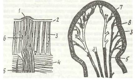
Fig. 26. Sensory sensilla of Lumbricus terrestris:
a - section of a sensory bud; b - cluster of photoreceptive cells on prostomial nerves; 1 - sensory rods; 2 - cuticle; 3 - epidermis; 4 - circular muscles; 5 - nerve fibres; 6 - sensory cells; 7 - prostomium; 8 - photoreceptive cells
In the earthworm Lumbricus terrestris, the Male Reproductive System is represented by two pairs of Testes located in segments X and XI (Fig. 27). Immature sperm pass from them into the seminal vesicles—sac-like outgrowths of the coelomic epithelium that enclose the testes. Here the sperm mature and accumulate. Opposite each Testis, a ciliated funnel of the coelomoduct opens into the coelom, leading into an efferent duct. Both ducts on each side unite into a single sperm duct (vas deferens), which opens on the ventral side of segment XV.
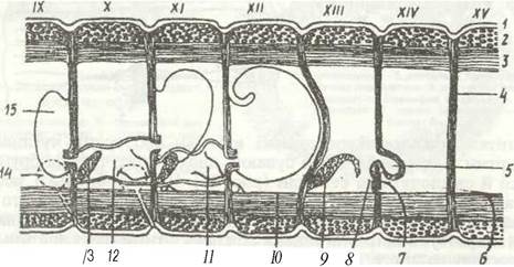
Fig. 27. Diagram of a sagittal section through the reproductive segments (IX-XV) of Lumbricus terrestris:
1 - epidermis; 2 - circular muscles; 3 - body wall muscles; 4 - ovisac; 5 - male genital pore;
6 - female genital pore; 8 - oviduct funnel; 9 - Ovary; 10 - sperm duct; 11 - sperm duct funnel; 12 - seminal capsule; 13 - testis; 14 - seminal receptacle; 15 - Seminal Vesicle
The FEMALE REPRODUCTIVE SYSTEM consists of a pair of Ovaries located in the XIII segment. Mature eggs are released into the coelom, where they undergo development and accumulate within the egg sacs; from there, they exit via the oviducts, which open into the coelom through funnel-shaped ostia and to the outside via pores on the XIV segment. The female reproductive system also includes seminal receptacles—two pairs of deep epidermal investments on the ventral side of the IX–X segments, which have no connection to the body cavity. Their function is to store the sperm of another individual during cross-Fertilization.
During copulation, two worms align their ventral surfaces with their anterior ends pointing in opposite directions, such that the clitellum of one worm lies opposite the seminal receptacle pores of the other (Fig. 28). The clitellums secrete abundant mucus that envelops the bodies of both specimens in the form of two sleeves. From the sperm duct openings, the worms discharge sperm, which is transported by muscular contractions toward the clitellum, where it enters the mucous sleeve and subsequently passes into the partner's seminal receptacles, which actively engulf the sperm through swallowing movements. Afterward, the worms separate. Thus, copulation involves solely an exchange of sperm, rather than fertilization. When a worm's eggs reach maturity, its clitellum secretes a mucous sleeve containing nutrients essential for the embryo. The worm begins to shift this sleeve forward. As it moves, it first picks up the eggs and then the sperm from the seminal receptacles, thereby achieving fertilization within the sleeve. Finally, the worm sheds the sleeve over its anterior end, its edges seal together, and a cocoon is formed, within which the eggs develop.
Self-fertilization is known to occur in certain oligochaete species, while others are capable of parthenogenesis. Very rarely, oligochaetes (such as those belonging to the family Eudrilidae) feature female reproductive ducts connected to their seminal receptacles, resulting in internal self-fertilization.
Development in microdriles proceeds without metamorphosis. The eggs develop entirely inside an egg cocoon, from which fully formed young worms emerge. In small aquatic oligochaetes, such as members of the family Naididae, the eggs are rich in yolk, which is consumed by the embryo during embryonic development. In larger worms, such as Lumbricidae, the eggs contain little yolk, but the cocoon is filled with a nutritious albuminous fluid. The developing embryo inside the cocoon (Fig. 29) possesses a mouth, pharynx, and midgut; it actively swallows the albumin and is therefore termed a "cryptic larva," which transforms into a young worm just prior to hatching. Juvenile earthworms emerge by rupturing the cocoon wall.
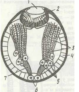
Fig. 29. Lumbricus embryo, ventral view:
1 - mouth, 2 - pharynx; 3 - ectodermal bands; 4 - endoderm; 5 - ectodermal teloblasts;
6 - mesodermal teloblasts; 7 - mesodermal bands
In addition to sexual reproduction, aquatic oligochaetes exhibit asexual reproduction through body division. In certain species (Lumbriculus, Enchytraeus), the body breaks apart into several fragments, each of which subsequently regenerates the missing parts. This phenomenon is known as architomy. In other instances, the body divides via constriction, yet both parts remain connected while the anterior segment regenerates its posterior end and the posterior segment regenerates its anterior end. Eventually, the young individuals separate, though sometimes prior to Separation each individual divides in turn, forming chains consisting of several specimens (e.g., in Aelosoma). This mode of asexual reproduction is termed paratomy.
A universally accepted division of the Class Oligochaeta into orders does not yet exist; therefore, we will examine the most important families.
Well-known representatives include the family Naididae, which predominantly inhabit freshwater bodies but can also be found in coastal marine zones or brackish areas. For instance, species of the genus Nais occur in great Abundance across various types of rivers and standing water bodies. These are small (10–15 mm), whitish, transparent animals bearing bundles of long, delicate setae, with a pair of eyes situated on the prostomium. They crawl along the substrate and algae, and are also capable of swimming. They feed on unicellular algae, protozoans, and detritus.
The family of sludge worms (Tubificidae) comprises over 300 species of marine and freshwater burrowing worms. The majority of tubificids inhabit freshwater environments. The most widespread genus is Tubifex. These are small worms reaching a body length of 2–5 cm. Approximately 40 species of this genus are known in Ukraine; they thrive in muddy sediments where oxygen is scarce. All of them possess haemocruorin in their blood to store oxygen. Tubificids anchor their anterior ends in the mud, while their posterior ends—rich in cutaneous blood capillaries—oscillate in the water to facilitate gas exchange. They feed on mud by assimilating organic residues from it. They can tolerate substantial aquatic pollution by various substances (petroleum products, pesticides, detergents, etc.). In organic-rich water bodies, their concentration can reach 100,000 individuals per 1 m2 of the bottom. Marine forms also exist among tubificids. The coastal zone of tropical seas is inhabited by species of the genus Phalodrilus, which are characterized by a unique Symbiosis with bacteria residing beneath the worms' cuticle. These bacteria are capable of utilizing hydrogen sulfide generated during the decomposition of organic remains. The gut is absent in Phalodrilus, and they derive Nutrition entirely from their symbionts.
Members of the family Branchiobdellidae live on the gills and body surface of freshwater crayfish. Their adaptation to ectoparasitism has drastically altered their Morphology: formerly mistaken for leeches, these oligochaetes are small (no more than 10–12 mm), stout, and consist of a HEAD region formed by the fusion of the prostomium and four segments, alongside 11 trunk segments terminating in a sucker. The mouth is equipped with chitinous upper and lower jaws. Juveniles feed on detritus, while adults consume crayfish blood.
Species of the family Enchytraeidae inhabit marine environments, freshwater bodies, and soils, numbering over 400 species. These are whitish worms typically 2–3 mm long, occasionally up to 45 mm, with well-developed setae. Certain species, particularly soil-dwellers, multiply in enormous numbers. They feed on humus. Aquarists frequently culture them in pots of garden soil as fish food, which is why they are commonly known as "potworms."
The most prominent are the so-called earthworms, which play a major role in soil formation. They belong to several families and do not constitute a single systematic group. The best-known among them—true earthworms (family Lumbricidae)—range in length from 20 mm to 1 m and in thickness from 1 to 20 mm. All of them are saprophages (feeding on organic detritus). Based on their feeding habits, they are divided into two ecological groups. Some species, including the well-known earthworm (Lumbricus terrestris), feed on plant litter and occasionally even green plant parts, pulling them from the soil surface into their burrows; their bodies are heavily pigmented, the head lobe is well developed, and they are capable of venturing onto the surface, living in the leaf litter, and even inhabiting decaying wood (Dendrobaena). Species of the second group feed on soil humus; their body pigmentation is poorly developed, the prostomium is small, the cuticular-muscular sac is thinner than in the previous group, and they inhabit the deeper soil layers (most species of Allolobophora), with some species maintaining permanent deep burrows (up to 1 m).
Earthworms perform vertical Migrations; the drier the soil or the lower its Temperature, the deeper they burrow into the earth. Certain species can form specialized capsules at depth to survive unfavorable seasons (winter, dry periods, etc.). During periods of intensive feeding, worms deposit mounds of processed soil—coprolites—onto the surface. The presence and quantity of these deposits can be used to estimate the population density of the worms themselves.
Earthworms are widely distributed across diverse soils, even in deserts; in some soils, their population density is immense, reaching up to 10 million per hectare in chernozems. More than 50 species of earthworms are known in Ukraine, among which the endemic species of the Carpathians (Helodrilus cemosvitovianus, Allolobophora carpathica) and the lowland regions of our country (Allolobophora leoni, etc.) are of particular interest. These species are potentially in need of conservation; some are listed in the Red Data Book of Ukraine. Interestingly, Eisenia submontana, a species known from the Alps and the Carpathians, is capable of Bioluminescence in the dark.
Resembling Lumbricidae in appearance, the worm Criodrilus lacuum of the family Criodrilidae reaches a length of 12–32 cm, leads an aquatic lifestyle, inhabits the mud of freshwater and brackish water bodies, and is widely distributed in Ukraine. Species of the family Megascolecidae vary in size, with the largest specimens attaining a length of 3 m.
They inhabit soils in South and East Asia, Australia, and the islands of Indonesia; a single species has been discovered in Transcaucasia, having been introduced there by humans. The coprolites of large species reach a height of 20–25 cm, resembling small towers.
Practical significance. Tubificids and enchytraeids serve as excellent fish food and are therefore harvested and cultured. Due to their high pollution tolerance, tubificids hold potential for the biological Treatment of wastewater. Certain giant megascolecids are consumed as food by indigenous peoples of Southeast Asia and South America.
Certain earthworm species are commercially bred to produce feed supplements for livestock and even humans. The species Eisenia foetida, common in our region (commercial name "red Californian"), lives in manure slurry. Through selective breeding, strains capable of breaking down various types of manure and organic waste have been developed. These worms are successfully cultured industrially and utilized—including in Ukraine—for Processing diverse biological waste. After passing through the worms' digestive tract, this waste is converted into a valuable complex fertilizer, while the worms themselves, processed into meal, can serve as high-value additives that significantly enhance the nutritional efficiency of livestock feed.
Nevertheless, the most celebrated role of earthworms lies in soil formation, a phenomenon first highlighted by Charles Darwin. In his work *The formation of Vegetable Mould, through the Action of Worms*, he demonstrated their positive impact on soil fertility. Subsequently, this topic attracted the attention of numerous researchers. Earthworms loosen and mix the soil, improving its aeration and the infiltration of water into deeper layers: the total length of worm burrows per 1 m2 can reach 1–8 km. Soil that has passed through their digestive tract exhibits an increased content of mineral nutrients essential for plants, alongside a reduction in soil acidity. Over the course of a year, worms pass a layer of soil ranging from one to seven meters in thickness through their bodies, and their biomass varies from 10–20 to 2,000–3,000 kg/ha depending on the soil type.
The NEGATIVE IMPACT OF oligochaetes is negligible. It is known that during their reproductive period (June–July), most earthworm species become toxic and may cause mortality in domestic poultry. Some microdriles serve as intermediate hosts for helminths. For instance, plerocercoids of the cyprinid parasite Caryophyllaeus (Cestoda) inhabit the coelom of Tubifex, while earthworms host nematode larvae responsible for severe diseases in pigs (Metastrongylus) and chickens (Syngamus).
Last update: 13/08/2026
Editorial and Educational Adaptation: This material has been compiled based on the primary/original source text. The project team performed an editorial review, corrected technical inaccuracies, structured sections, and adapted the content for an educational format.
What was processed:
- elimination of formatting defects (OCR errors, structural breaks, corrupted characters);
- editorial organization of content;
- standardization of terminology in accordance with academic sources;
- verification of factual statements against the original source text.
All mentions of the author, publication year, and origin of the primary text have been preserved in accordance with the source.