Tuberculosis Study Guide - M.M. Savula 2002
Extrapulmonary tuberculosis
Tuberculosis of the meninges and central nervous system
Tuberculosis of the Meninges (Tuberculous meningitis) is an inflammation of the leptomeninges caused by Mycobacterium tuberculosis. If, In addition to the meninges, the Brain parenchyma is also affected, meningoencephalitis develops.
People of all ages can be affected, but it predominantly occurs in the elderly and occasionally in children, most commonly during the winter-spring season. In 2000, 45 cases of tuberculous meningitis were diagnosed in Ukraine. This is one of the most severe forms of tuberculosis, which is fatal if left untreated.
Pathogenesis. Meningitis is a complication of Other forms of intrathoracic (most commonly primary forms or disseminated tuberculosis) or Extrapulmonary tuberculosis. It can also be one of the manifestations of Miliary tuberculosis. The Development of meningitis requires the dissemination of MTB from the primary focus of infection into the bloodstream and increased permeability of the Blood-brain barrier. Contributing factors may include hypothermia, hyperthermia, concomitant diseases (including childhood infections), trauma, etc.
Inflammatory changes are most pronounced at the Base of the brain, where a gelatinous exudate and tuberculous tubercles are found. The choroid plexuses of the brain are also affected. The localization of these changes at the Skull base explains the frequent involvement of the Cranial Nerves. The inflammatory process can spread to the spinal meninges (spinal meningitis) and the brain parenchyma (meningoencephalitis).
Clinical presentation. In 70% of cases, the onset of the disease is gradual: general malaise, loss of appetite, irritability, drowsiness, intermittent headache, and sometimes low-grade fever. This prodromal period lasts from 1 to 4 weeks. In infants, the disease may onset acutely with high fever, epileptiform seizures, and digestive disorders.
The subsequent period (clinical manifestations of meningeal and cranial nerve involvement) begins with a rise in body Temperature (meningitis almost never occurs without fever) to 38-39 °C and higher, severe headache, and frequent vomiting, which can occur suddenly upon changing body position or after taking medication. Drowsiness, lethargy, and irritability increase. Patients cannot tolerate noise or bright light. Constipation develops.
Physical examination may reveal relative bradycardia and often symptoms of cranial nerve involvement (Fig. 22). The oculomotor (CN III) and abducens (CN VI) nerves are most commonly affected, presenting as drooping of one or both eyelids (ptosis), pupillary constriction or dilation, and strabismus. In Facial Nerve (CN VII) palsy, the face becomes asymmetric, the nasolabial fold is flattened, and the corner of the Mouth droops. Symptoms of Vestibulocochlear nerve (CN VIII) involvement may include tinnitus, vertigo, and a sensation of "falling". Peripheral paralysis of the Hypoglossal nerve (CN XII) causes deviation of the Tongue to one side. In the later Stages of the disease, swallowing difficulties, choking, and hiccups develop.
Class="center">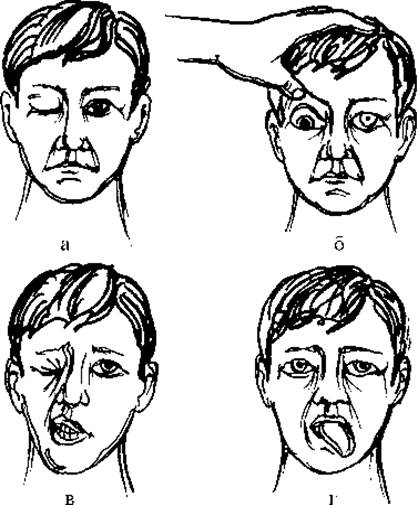
Fig. 22. Most common manifestations of cranial nerve involvement in tuberculous meningitis:
a - right Oculomotor nerve palsy (ptosis); b - right oculomotor nerve palsy (dilated pupil); c - peripheral facial nerve palsy; d - peripheral hypoglossal nerve palsy.
A specialized neurological examination reveals meningeal signs. The most important of these are: nuchal rigidity (when attempting to passively flex the HEAD to the chest, resistance and pain are felt, and the chin cannot reach the Sternum). Kernig's sign (it is very difficult to fully extend the leg at the knee joint when it is flexed at a right angle at the Hip and knee joints). Brudzinski's sign:
- upper - attempting to flex the head to the chest causes flexion of the lower limbs at the hip and knee joints;
- middle - pressure on the Pubic Symphysis is accompanied by flexion of the lower limbs at the hip and knee joints;
- lower - during the extension of one lower limb (while testing Kernig's sign), the other limb reflexively flexes.
In infants, diagnostic indicators include: bulging and tension of the fontanelle, pupillary dilation during rapid neck flexion, and Lesage's sign ("hanging" sign). It consists of the sick child flexing their legs at the hip and knee joints when lifted by the armpits and holding them in this position for a long time, whereas a healthy child quickly lowers their legs.
By the end of the second week of the illness, all these symptoms intensify. The patient lies with their head hyperextended, eyes closed, legs drawn up to the abdomen, abdomen retracted, and Abdominal Muscles tense.
If adequate Treatment is not provided, by the 3rd week of the illness, the process spreads to the brain parenchyma, developing into meningoencephalitis. The patient's consciousness becomes clouded, seizures may occur, and the temperature rises to high levels (40-41 °C) or drops below normal. Sensory disturbances, central paresis, and paralysis occur.
If the inflammatory process spreads to the spinal meninges (spinal meningitis), girdling pain in the spine and abdomen appears. Dysfunction of the pelvic Organs ensues, presenting as difficulty urinating and persistent constipation, followed by urinary and fecal incontinence. As a result of Nervous system dysfunction, pressure ulcers develop, and death occurs.
If tuberculous meningitis is suspected, the patient must be hospitalized immediately and a lumbar puncture performed. The CEREBROSPINAL FLUID flows out under increased pressure (normally no more than 60 drops per minute) and is clear. Its protein content is elevated to 0.6-3.0 g/L (normally no more than 0.43 g/L). An increase in Cell count from 30 to 400 Cells/mL (normally up to 10 cells/mL) with a predominance of lymphocytes (90% or more) is mandatory.
The glucose level in the cerebrospinal fluid is decreased (normally it is not lower than half of the Blood Glucose Level). When the CSF is left to stand in the cold, a delicate web-like clot forms after 12-24 hours. The cerebrospinal fluid is tested for MTB, but they are detected in no more than 10-15% of cases.
Lumbar puncture
A lumbar puncture is performed by a physician in the lumbar region between the L3 and L4 vertebrae. At this level, the Spinal Cord is no longer present, as it ends at the upper border of the L2 vertebra. The lumbar puncture is performed with the patient lying on their side, with the spine arched and the spinous processes maximally separated from each other, which facilitates needle insertion (Fig. 23). After local anesthesia, the spinal needle with a stylet is inserted horizontally, slightly angled cranially. The depth of the puncture is up to 3 cm in children and 4-6 cm in adults. Therefore, a spinal needle of appropriate length with a stylet must be prepared. After puncturing the dura mater, during which the physician feels a certain resistance followed by a "give" sensation, cerebrospinal fluid begins to drip from the needle. The physician carefully removes the stylet from the needle, and the CSF is collected in sterile containers. The tubes containing the cerebrospinal fluid, along with the appropriate referral form (patient's last name, initials, and department name), are delivered to the clinical, biochemical, and bacteriological laboratories.

Fig. 23. Lumbar puncture.
Below is The sequence of assistance provided by a junior medical worker to the physician during a lumbar puncture.
Assisting the physician in performing a lumbar puncture
|
Required Materials |
|
|
1. Ethyl alcohol 70° 2. 3 % alcoholic iodine solution 3. Anesthetic (0.5 % novocaine solution, 1 % lidocaine solution) 4. Sterile spinal needle with stylet 5. Disposable or sterile syringe with needle 6. Sterile tray 7. Rack with sterile test tubes 8. Forceps, clamp 9. Sterile gauze wipes, cotton balls 10. Adhesive plaster |
1. Before the puncture, it is advisable for the patient to empty their bowels (enema) and bladder 2. The patient is placed on their side on a firm couch, near its edge 3. The patient's head is flexed as much as possible until the chin touches the chest 4. The legs are bent at the hip and knee joints and drawn up to the abdomen 5. To prevent sudden movements during the puncture, a healthcare worker holds the patient in this position 6. The nurse hands the physician a clamp with a cotton ball soaked in iodine solution 7. The physician palpates the highest points of the iliac crests and connects them with a line using an iodine-soaked cotton swab. This line crosses the spine at the level between the III and IV lumbar vertebrae - the puncture site 8. The nurse thoroughly preps this area of the Skin with 70° alcohol and alcoholic iodine solution, removing excess iodine with a cotton ball soaked in alcohol 9. The nurse hands the physician a syringe with the anesthetic 10. After the physician performs infiltration anesthesia, the nurse hands them the spinal needle with the stylet 11. The physician performs the lumbar puncture 12. The nurse collects the first drops of cerebrospinal fluid into a sterile tray, and the subsequent ones into sterile test tubes 13. After the needle is removed, the nurse applies a sterile wipe to the puncture site and secures it with adhesive plaster 14. The patient is transported to the ward in a prone position without a pillow and transferred to the bed in the same position 15. After 2 hours, the patient is allowed to turn over. The patient must remain on bed rest for two days |
To establish a Diagnosis of tuberculous meningitis, a fundoscopic examination is required, which may reveal papilledema, signs of optic neuritis, and sometimes tuberculous tubercles.
The patient must undergo a comprehensive examination. X-ray Examination helps to identify respiratory tuberculosis, while urinalysis changes may suggest Renal tuberculosis. The complete blood count (CBC) reveals moderate leukocytosis or a normal WHITE BLOOD CELL count (in infants, high leukocytosis of 30- 109/L or more is sometimes observed), lymphopenia, and a normal or slightly elevated ESR.
The reaction to the Mantoux test is usually positive, although it becomes negative if the patient's condition is severe.
A clinical presentation similar to tuberculous meningitis can occur in meningitis of other etiologies (viral, bacterial) or Brain Tumors.
Treatment and outcomes. Treatment for patients with tuberculous meningitis must begin immediately at the facility where the disease is suspected, as the success of the outcome depends on this. Therapy is continued in a specialized hospital. The cornerstone of treatment is a combination of anti-tuberculosis drugs for 9-12 months. In addition to tuberculostatic agents, Vitamins B1, B6, and B12 are prescribed, along with detoxification therapy, including infusions of 5 % glucose solution, rheopolyglucin, and albumin. Concurrently, glucocorticoids and Diuretics are prescribed to reduce inflammatory changes and cerebral edema.
To monitor treatment efficacy and reduce elevated intracranial pressure, lumbar punctures are performed periodically.
The regimen and patient care are of great importance for recovery. Patients must remain on bed rest for 1.5-2 months, after which they are allowed to sit up, and later, to walk around the ward.
In severe cases or when swallowing is impaired, patients are tube-fed, prescribed intravenous infusions of glucose and protein preparations, and monitored for regular urination and bowel movements. In case of pelvic organ dysfunction, urinary catheterization and cleansing enemas are required. Prevention of pressure ulcers is crucial (clean bed linen, hygiene compliance, washing the most vulnerable areas with warm soapy Water followed by rubbing with camphor spirit, applying liquid paraffin, and using positioning bolsters and inflatable rubber rings).
The prognosis for tuberculous meningitis is relatively favorable if treatment is initiated no later than the 10th day of the illness. After 1-2 weeks of treatment, headaches subside and vomiting stops; however, a marked improvement in the general condition does not occur sooner than 2-3 months. Meningeal signs disappear during this time, but normalization of the cerebrospinal fluid composition is observed only after 5-6 months. Timely treatment leads to recovery in 95-98 % of patients. If treatment is delayed, complications may arise, the consequences of which can persist for a long time, sometimes for life. These include intellectual impairment, severe Vision loss, and motor disorders.
Questions
1. What is tuberculous meningitis? Meningoencephalitis?
2. What is the most common onset of tuberculous meningitis?
3. What are the primary Complaints of a patient with tuberculous meningitis?
4. What are the neurological manifestations of tuberculous meningitis?
5. Which symptoms of cranial nerve involvement are frequently detected in tuberculous meningitis?
6. What examinations should be performed on a patient to confirm the diagnosis of tuberculous meningitis?
7. What pathological changes are found in the cerebrospinal fluid in tuberculous meningitis (cell count, protein, glucose)?
8. Clinical Features and diagnosis of tuberculous meningitis in children.
9. Care and regimen for patients with tuberculous meningitis.
Last update: 10/08/2026
Editorial and Educational Adaptation: This material has been compiled based on the primary/original source text. The project team performed an editorial review, corrected technical inaccuracies, structured sections, and adapted the content for an educational format.
What was processed:
- elimination of formatting defects (OCR errors, structural breaks, corrupted characters);
- editorial organization of content;
- standardization of terminology in accordance with academic sources;
- verification of factual statements against the original source text.
All mentions of the author, publication year, and origin of the primary text have been preserved in accordance with the source.