Orthopedics - Oleksa A.P. 2006
Osteochondropathies
Osteochondropathies of the lower limb bones
Osteochondropathy of the phalanges of the hand
Osteochondropathy of the femoral HEAD was first described in 1910 independently by Legg (USA), Calvé (France), and Perthes (Germany).
The Etiology of the disease is multicausal and attributed to the same harmful factors that cause osteochondropathy in other anatomical sites, namely trauma, inflammation, endocrinopathies, and heredity (Harper P.S. et al., 1976; Wynne-Davies R., Gormley J., 1978).
Joseph B. (1991), and Neidei J., Zander D., Hackenbroch M. (1992) reported a marked increase in IgG and IgM levels alongside a decrease in IGF-1 and somatomedin C in this condition.
Toxic synovitis of the hip joint is associated with Legg-Calvé-Perthes disease in 1-12% of patients, although this finding has not been corroborated by other studies.
It is believed that in hemophilia, recurrent hemarthroses lead to increased intra-articular pressure, which impedes the progression of osteochondropathy.
The onset of Legg-Calvé-Perthes disease is attributed to venous Hypertension and arterial insufficiency by Sanshis M., 21ahir A., and Freeman M.A. (1973), as well as Liu S.L. and Ho T.C. (1991).
Using radioisotope, capillaroscopic, and thermometric studies, as well as the sweat test, biological and hydrophilic tests, Kryukova N.N. (1985) detected neurocirculatory and vascular disorders throughout the entire lower limb rather than being confined to the hip joint area. These disorders manifested as early as
The First stage of the disease and persisted through The final stage.
There are also recent reports (Glueck C.J., Crawford A., Roy D. et al., 1996) regarding hypofibrinolysis, protein C and protein S deficiencies, and elevated lipoprotein a levels, all of which contribute to venous thrombosis, hypertension, and consequently The Development of osteonecrosis.
Clinical manifestations of the disease. Among all osteochondropathies, Legg-Calvé-Perthes disease is the most thoroughly documented in medical literature. It predominantly affects children aged 5-12 years, with some potential age-related variations. Typically, a single hip joint is affected, occasionally both (in about 10% of cases), and the pathological process in bilateral cases may develop asynchronously. Boys are affected 4 to 5 times more frequently than girls.
The initial signs of the disease are pain and a limp during walking; children stop running and their overall physical activity declines. However, it should be noted that a small number of patients may experience no pain at the onset, or the pain may radiate to the knee, which can mislead the physician and hinder early Diagnosis.
According to Kapetanakis (1993), hip pain is reported by 65.9% of children, knee pain by 12.4%, and pain throughout the entire limb by 10.6%. Limping is caused both by the child's attempt to unload the affected leg during ambulation and by protective antalgic contracture of the hip joint. According to Limin O.L. and Khvysyuk M.I. (1970), flexion and external rotation contractures develop in 55% of children, while others exhibit various alternative patterns. Notably, 91.2% of children examined show restricted medial Rotation of the thigh, and 87.7% show restricted abduction. Flexion is also limited in one-third of patients. All of these factors lead to progressive Muscle atrophy in the limb, particularly in the thigh and gluteal regions.
Additionally, During the first stage of the disease, vascular and autonomic disorders can be detected in the affected limb: Skin pallor, increased moisture, and localized cooling due to impaired Blood Circulation, as demonstrated by Kryukova N.N. (1985) using radioisotope studies.
Radiological examination is the cornerstone of osteochondropathy diagnosis, revealing changes characteristic of each stage of the disease (Fig. 348). Nevertheless, radiographic diagnosis can be challenging at the onset of the disease, particularly when low-quality radiographs are obtained in the anteroposterior view alone.
Comparative radiography of the hip joints in the lateral projection is significantly more informative for the early diagnosis of Legg-Calvé-Perthes disease, clearly demonstrating widened joint space and broadening of the femoral neck in the subepiphyseal region.
Computed tomography undoubtedly allows for the assessment of Bone tissue density within the femoral head and the precise localization of the necrotic focus at a stage when they remain invisible on standard radiographs.
Magnetic Resonance imaging is particularly valuable for diagnostic purposes, as it reveals pathological alterations not only in the bone itself but also in the Joint Capsule and surrounding soft Tissues. The joint capsule is thickened and distended due to synovitis, Muscles are indurated, and edema may cause widening of the intermuscular fascial planes between the gluteus medius and minimus muscles.
Furthermore, bone scintigraphy (using 99mTc-polyphosphate) remains a definitive and precise diagnostic tool for evaluating the vascular status (Sutherland A.D. et al., 1980).
In addition, other authors utilize sonography to determine the shape of the femoral head and associated morphological changes depending on the stage of the disease (Danigelis J.A. et al., 1975). This method also successfully detects synovitis and thickening of the joint capsule (Futami T. et al., 1994; Eckerwall G. et al., 1994). Grebenyuk L.A., Menshchikova T.I., and Soldatov Yu.P. (2001) note that with the thigh abducted and medially rotated, sonography can reveal early, pre-radiographic signs of osteochondropathy: small ossification foci along the margins and at the apex of the epiphysis, rarefaction of the subchondral bone layer, capsular thickening, and widening of the joint space.
Direct signs of the initial stage of the disease include widening of the joint space, increased bone density at the upper pole of the femoral head presenting as "pseudosclerosis," heterogeneity and mottling of the paraepiphyseal Structure, and occasionally simulated widening of the femoral neck. Protopopov A.N. and Priyezzheva V.N. (1972), using arthropneumography, also found that the apparent widening of the joint space was not true widening, but rather the result of hyaline Cartilage thickening.
Class="center">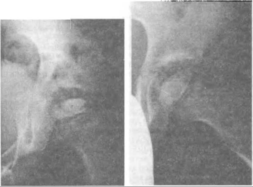
Fig. 348. Osteochondropathy of the femoral head in the fragmentation stage, radiographic views in two projections.
A characteristic radiological sign is also Cages' sign, which involves irregularity and undulation of the epiphyseal cartilage plate, along with marginal erosions on the adjacent surfaces of the Head and Neck epiphysis. Furthermore, Osteoporosis is observed not only in the BONES OF THE hip joint but also in the corresponding half of the pelvis, especially in the supra-acetabular region.
By the end of the first stage, standard plain radiographs clearly show a focus of bone tissue destruction within the femoral head while its anatomical shape remains preserved. The first stage lasts approximately six months, and occasionally longer.
The Second Stage of fragmentation (resorption) is radiographically characterized by the appearance of radiolucent clefts within the necrotic focus of the femoral head, although the overall anatomical shape of the head is maintained (Fig. 349). This stage involves revascularization of the epiphysis from the periosteum and the trabecular bone of the neck. Connective Tissue and Blood Vessels grow into the necrotic zone. Under the action of osteoclasts and phagocytes, necrotic masses—primarily those located along the vascular pathways—are resorbed, which is why the focus of necrosis in the femoral head appears fragmented on radiographs. Cysts of varying sizes may occasionally form in the femoral neck adjacent to the epiphysis.
During the fragmentation stage, complete unloading of the limb through traction and leg abduction helps preserve the delicate and fragile Water/140.html">Anatomical Structure of the fragmented upper femoral head. Any vertical single-load application to the head compresses the bone fragments, causing the head to lose its natural spherical shape and flattening its upper pole (Fig. 346). This results in irreversible deformation of the head, ultimately leading to persistent deforming osteoarthritis once the process concludes.

Fig. 349. Impression of the right femoral head in osteochondropathy.
The fragmentation stage is quite prolonged, lasting from one to one and a half or even two years.
Simultaneously with the "resorption" of necrotic bone, reparative processes take place, though they are initially uneven. As a result, central areas of necrosis may persist for a long time and undergo slow replacement. This marks the onset of the Third Stage of the process—repair (reossification).
At this stage, reossification of the fibrovascular structures formed in the previous stage takes place. Sequestrum-like shadows gradually disappear, and the epiphysis is completely replaced by newly formed bone, with possible areas of radiolucency in the center. Reossification proceeds from the margins of the epiphysis, indicating paraphyseal reossification. Subsequently, the metaphyseal and epiphyseal regions undergo ossification, completing transphyseal ossification.
The Fourth Stage is remodeling—the final phase, during which the shape and STRUCTURE OF THE femoral head are restored.
According to Studenikin and Yakovleva (1987), complete recovery, where the femoral head is indistinguishable from a healthy one, occurs in only 85% of patients treated for Legg-Calvé-Perthes disease. In less favorable cases, the head acquires an irregular shape (such as mushroom-like) and is not fully covered by the acetabular roof, which had grown and shaped itself around the femoral head during Treatment. Consequently, the acetabulum tends to be shallow, flat, and more inclined. This condition of the hip joint leads to deforming osteoarthritis with corresponding clinical symptoms, and the altered shape of the acetabulum may contribute to subluxation of the femoral head.
Overall, the course of Legg-Calvé-Perthes disease lasts three years or more. Medical literature describes cases of mild disease progression with a small focus of necrosis (so-called localized forms of osteochondropathy) and favorable treatment outcomes (Lymin A.L., Khvysyuk M.I., 1970; Krylova M.D., 1971).
As mentioned above, the diagnosis of hip osteochondropathy is based on patient Complaints, clinical findings, and X-ray data. Diagnostic challenges typically arise early in the disease before any radiographic changes are visible. When examining a child, the physician must keep osteochondropathy in mind, and in doubtful cases, recommend limb unloading, thermal Procedures, and mandatory follow-up with a lateral X-ray of the hip joint in three months.
Treatment. Management of Legg-Calvé-Perthes disease involves a comprehensive set of measures aimed at relieving pain, improving local blood circulation and metabolic processes, preventing femoral head compression, and preserving normal hip joint function.
Orthopedic treatment begins in an inpatient Setting with complete and absolute limb unloading in internal rotation using skin traction (adhesive or cuff traction) applied to the lower leg immediately after diagnosis.
Given the presence of autonomic-vascular disorders throughout the limb and to avoid compressing the legs with a cuff, some orthopedists suggest applying a plaster cast fixed at the FOOT and ankle region with damper traction, or a plaster bed with abducted and medially rotated thighs, or skeletal traction alternating every 3–4 months with functional-restorative treatment.
Clinical observations indicate that skin traction with a lightweight load, combined with abducted lower limbs using a Viennese splint and relaxed muscles in bed, ensures sufficient unloading of the femoral head and instills discipline in the child.
In the first stage of the disease, along with traction, cocarboxylase Electrophoresis is prescribed for the Scarpa's triangle area (20–30 sessions), along with Vitamins B1, B2, B6, C, PP, and glycerophosphates. Muscle massage is also administered.
Inpatient treatment is repeated every two months. The child is discharged wearing foot plaster casts connected by a spreader bar to keep the limbs abducted and slightly internally rotated. At home, muscle massage, ozokerite-paraffin Applications to the hip joint area (20–30 sessions) are performed, and A balanced diet is prescribed for the child.
During the second stage (fragmentation), strict adherence to bed rest and child compliance are crucial. In the hospital setting, a control X-ray of the hip joint is performed, and skin traction with leg abduction using a Viennese splint is applied. A course of cocarboxylase electrophoresis (15 sessions) is prescribed every other day, alternating with Trypsin electrophoresis applied to the hip joint region.
Trypsin or Chymotrypsin can be administered intramuscularly (3 mg for children under 5 years, and 5 mg as a single dose for children over 5). A course of multivitamins, glycerophosphates, and gluteal muscle massage is repeated.
If radiographs reveal residual fragments within the femoral head during subsequent inpatient treatment, trypsin electrophoresis is repeated along with previous prescriptions, including vitamin B12 injections (200 mcg), aloe or FIBS (30 injections) at 0.5–1.0 ml daily. At home, treatment with leg abduction, thermal procedures, and massage is continued.
During the recovery stage in a hospital setting, comprehensive treatment is continued with The addition of ATP (15–30 mg per 1 ml of saline), vitreous body (1 ml, 20–30 injections), Calcium and phosphorus electrophoresis on the hip joint area, mud therapy, etc. Magnetotherapy using the millimeter non-thermal range device Gch-142 or the portable "Porog-1" device is also beneficial.
At this stage, children with unilateral involvement are allowed to walk using crutches without bearing weight on the affected leg, but skin traction is applied at night. The reparative process lasts up to a year and a half. During convalescence, sending children to sanatorium-resort rehabilitation is recommended.
Thus, children must avoid any weight-bearing on the affected leg for 2.5–3 years, which is key to preserving the normal anatomical shape of the femoral head and restoring full range of motion in the hip joint. Minor, even single-instance weight-bearing on the head during the fragmentation stage can cause compression, deformation, and consequently, deforming osteoarthritis.
Timely, comprehensive conservative treatment is highly effective and leads to patient recovery, which, in Schwartz's words, represents the "ideal type of healing."
To accelerate the revascularization of the necrotic focus, pediatric patients with stage I–II disease at the Turner Institute undergo vascularized pedicle bone grafting of the femoral neck. The autograft is harvested from the proximal bone area along with a flap of the tensor fasciae latae (according to Shapiro) or the anterior portion of the gluteus medius muscle (according to Adrianov-Tikhonenkov), or even a bone graft on two muscular pedicles (Andrianov V.L., Tikhonenkov E.S., Veselovsky Yu.A., Kharlamov M.N., 1987) (Fig. 350). Postoperative treatment duration was thus reduced. For the same purpose, percutaneous drilling with a Kirschner wire is performed from the subtrochanteric region into the upper pole of the head, or an allogeneic pin is driven
in along a similar trajectory according to Zandi. Instead of a bone pin, Pitzen used a thin Smith-Petersen nail, which additionally prevented Varus deformity of the femoral neck. Drilling with wires can be considered a sparing method, as driving an allogeneic pin or nail damages the growth epiphyseal cartilage, potentially leading to adverse consequences. Occasionally, fenestration of the femoral neck is performed with excochleation of the head's osteonecrosis and its tamponade using allogeneic bone or kergan.
However, there are cases when the course of osteochondropathy is prolonged due to delayed reparative processes; although the femoral head has slowly restored its structure, its shape remains altered—it is somewhat flattened, which led Waldenström to coin the term "coxa plana".
During the remodeling stage, the femoral head forms in an oval rather than a spherical shape; the femoral neck is shorter and wider than normal, and varus-deviated at the epiphyseal level, indicating the development of typical coxa vara (Fig. 351) resulting in functional limb shortening. Radiographs are taken in two projections, as femoral neck anteversion may simultaneously develop during growth.
Andrianov et al. report that in cases of stage II–III osteochondropathy involving the proximal Femur without head deformation, they performed corrective intertrochanteric femoral osteotomies. Following the correction of neck angles, the pathological process typically halted, and The structure of the femoral head was restored within 6–8 months.
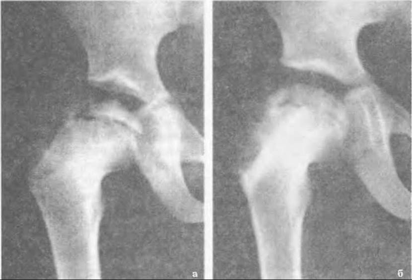
Fig. 350. Radiographs of the proximal femur in a 10-year-old patient with Perthes disease: a — before surgery, b — after impaction of a bone graft into the femoral neck.
In cases of femoral head flattening, subchondral remodeling is performed to achieve joint surface congruency and restore hip joint mobility. If cartilage is absent on the femoral head, arthroplasty using a demineralized osteochondral cap is applied.
To correct the axis of the femoral neck, an intertrochanteric wedge valgus osteotomy is most frequently performed. Prior to surgery, the wedge shape and base width are measured using skiagraphy.
Under general anesthesia via the Smith-Petersen approach, the intertrochanteric region of the femur is exposed. Occasionally, a smaller vertical incision along the anterior aspect of this region is used. The muscles are bluntly separated without damaging blood vessels. After determining the exact site for the intertrochanteric osteotomy, the bone wedge is removed. By abducting the femur, the fracture fragment surfaces are apposed and fixed percutaneously with two to three Kirschner wires. Postoperatively, the limb is immobilized in a hip spica cast extending to the other thigh. After 3–4 weeks, the pins are removed by their ends protruding above the skin. The plaster cast is removed once bone union is achieved.
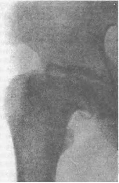
Fig. 351. Sequelae of untreated Perthes disease: flattened femoral head with a shortened, varus-deviated neck; the flattening of the acetabulum corresponds to the anatomical shape of the head.
Last update: 10/08/2026
Editorial and Educational Adaptation: This material has been compiled based on the primary/original source text. The project team performed an editorial review, corrected technical inaccuracies, structured sections, and adapted the content for an educational format.
What was processed:
- elimination of formatting defects (OCR errors, structural breaks, corrupted characters);
- editorial organization of content;
- standardization of terminology in accordance with academic sources;
- verification of factual statements against the original source text.
All mentions of the author, publication year, and origin of the primary text have been preserved in accordance with the source.