Orthopedics - Oleksa A.P. 2006
Osteochondropathies
Osteochondropathies of the Lower Extremity Bones
Osteochondritis dissecans of the femoral condyle (König's disease)
Osteochondritis dissecans was first identified by König, who reported it in 1887 under the name "osteochondritis dissecans". This condition typically localizes to the femoral condyle and occurs relatively frequently, accounting for 0.8% of orthopedic disorders and 2% of knee joint pathologies (Studenikin M.Ya., Yakovleva A.A., 1987). It is rarely seen in children (Diasemidze A.Kh., 1976), being more common in adolescents and adults (aged 20–40 years), with a twofold higher incidence in males, particularly those engaged in heavy physical labor. Bilateral involvement is observed in 68% of patients (Diasemidze A.Kh., 1976).
The Essence of the disease lies in localized subchondral necrosis, typically affecting the medial femoral condyle. Aichroth P. (1971) notes that in 75% of cases, the intercondylar area is also affected alongside the medial condyle.
Although several hypotheses regarding its Etiology exist, the exact cause remains unclear. It is widely believed to result from microtrauma caused by continuous mechanical overload of weight-bearing articular surfaces. Petrie P.W. (1977) and Mubarak S.J. and Carrol N.C. (1981) suggest that it stems from abnormal epiphyseal ossification.
The vascular theory of the origin and progression of femoral osteochondritis dissecans is currently the most widely accepted. Osteonecrosis develops as a consequence of local embolism in the artery supplying a restricted subchondral bone area. Because the deep, productive layers of the articular Cartilage in this region also lack a direct Blood supply, secondary degenerative changes develop within the cartilage.
During both radiographic examinations and surgical Procedures, we have frequently observed cone-shaped osteochondral sequestra with a broader cartilaginous base, which strongly Supports this theory.
Pappas proposed a Classification of femoral osteochondritis dissecans based on the patient's medical history and age (Pappas A.M., 1981). The first category includes children: girls under 11 years of age and boys under 13. The second category comprises girls aged 12 to 20 and boys aged 14 to 20, during the period when longitudinal bone growth is still possible. The third category includes patients over 20 years of age in whom the physis is closed; these individuals typically exhibit more extensive destruction and defects within the subchondral bone and cartilage. This classification has been instrumental in guiding Treatment strategies.
Class="center">
Fig. 352. Schematic diagram illustrating the progression of König's disease.
Mironova Z.S. and Badnin (1976) distinguish three distinct stages in the progression of the disease (Fig. 352).
In The First stage, the patient complains of joint pain triggered by weight-bearing, which subsides at rest. Children instinctively protect the affected limb, trying to avoid running, jumping, and similar activities. Palpation may occasionally reveal knee synovitis. Radiographs (including AP, lateral, and patellofemoral views recommended by Cherney S. (1997)) reveal a localized subchondral area of bone sclerosis bordered by a narrow radiolucent zone.
In the second stage, pain becomes more intense and occurs more frequently during walking and squatting, while synovitis may recur. Radiographs clearly show the dense necrotic subchondral focus surrounded by a wider radiolucent cleft within the bone.
In the Third Stage, persistent pain during walking and synovitis persist. Radiographs clearly demonstrate a detached sequestrum; if the fragment dislodges into the joint cavity, it causes locking and mechanical symptoms, severely impairing limb function.
Diagnosing osteochondritis dissecans at an early stage is crucial. When routine radiographs show no visible bone changes yet the clinical history and physical examination raise suspicion of osteochondritis dissecans, computed tomography (CT) or Magnetic Resonance imaging (MRI) of the knee is indicated (Fig. 353). This allows for the comparison of bone density in symmetrical subchondral regions, thereby clarifying the Diagnosis. Alternatively, radionuclide bone scintigraphy using osteotropic radioisotopes (technetium) can be employed.
For the clinical diagnosis of femoral osteochondritis dissecans, Wilson J.N. (1967) recommends an extension test: with the knee flexed at 90° and the Tibia internally rotated, extending the leg will exacerbate the pain.
Certain orthopedic specialists (such as Diasemidze A.Kh. et al.) distinguish atypical forms of femoral osteochondritis dissecans characterized by small areas of osteonecrosis appearing as thin plates or cyst-like changes. Furthermore, typical osteochondritis dissecans in children generally does not present with synovitis.

Fig. 353. Magnetic resonance imaging of the knee joint in König's disease.
What happens to the articular cartilage overlying the necrotic focus? Deprived of Blood supply to its deep layers from the underlying bone, the cartilage undergoes degeneration. During surgery or arthroscopy, the cartilage in this area exhibits altered coloration and appears depressed due to thinning, reflecting degenerative changes. In some patients, mechanical stress during ambulation leads to The formation of cartilage fissures. The severity of these pathological changes is directly proportional to the physical load placed on the limb.
Naturally, The Development of a cartilage fracture surrounding the zone of osteonecrosis leads to the complete detachment of the osteochondral sequestrum, facilitating its displacement into the joint cavity with the corresponding clinical and radiological manifestations.
Guhl J.F. (1984) proposed an arthroscopic classification of femoral osteochondritis dissecans: 1) intact and non-fissured articular cartilage, slightly depressed, measuring 1 to 3 cm in diameter; 2) mildly detached cartilage with fissures, held in place by fibrous tissue; 3) partial cartilage detachment palpable upon probing, or exhibiting slight mobility with interposition of fibrous tissue; 4) a crater covered with fibrous tissue at the site of a detached osteochondral sequestrum, accompanied by a loose body within the joint cavity (Fig. 354).
This arthroscopy-based classification assists in selecting the optimal treatment regimen for the patient.
Treatment. Conservative management alone is indicated for stages I and II in children (Pappas category I). The earlier osteochondritis dissecans is diagnosed, the more effective the treatment. Primarily, patients are advised to unload the affected limb, refrain from all athletic activities, running, and prolonged standing. A course of physical therapy and balneotherapy is prescribed in alternating cycles. In stage II, the limb is immobilized in a removable plaster splint worn during the day; at night, the splint is removed, and the knee is warmed using an electric heating pad or paraffin-ozokerite Applications. Patients are also prescribed vitreous body extracts, Rumalon, Nerobol, etc.
If synovitis is present, joint aspiration (arthrocentesis) is performed, followed by the intra-articular administration of Kenalog-40 (1 ml) combined with a 0.5% procaine solution (4 ml) and an antibiotic.
The efficacy of conservative treatment is monitored every three months via radiography, bone scanning, or, preferably, arthroscopy. This helps determine whether to continue conservative management or proceed with surgery. If a minor cartilage defect (up to 1 cm in diameter) is detected, conservative treatment is maintained.
Clinical observations confirm the effectiveness of conservative treatment provided the patient strictly adheres to a restricted activity regimen for two years or longer and complies with all prescribed therapies and medication schedules.
Follow-up examinations reveal progressive resorption and remodeling of the osteonecrosis, along with the disappearance of the radiolucent halo surrounding it, although the sclerotic rim at the boundary of healthy Bone tissue persists for quite some time. Thus, the patient achieves practical recovery.
At the age of 12-20 years (the second category of patients according to Pappas), treatment in all cases begins with Conservative Methods, accompanied by dynamic observation of the disease progression.
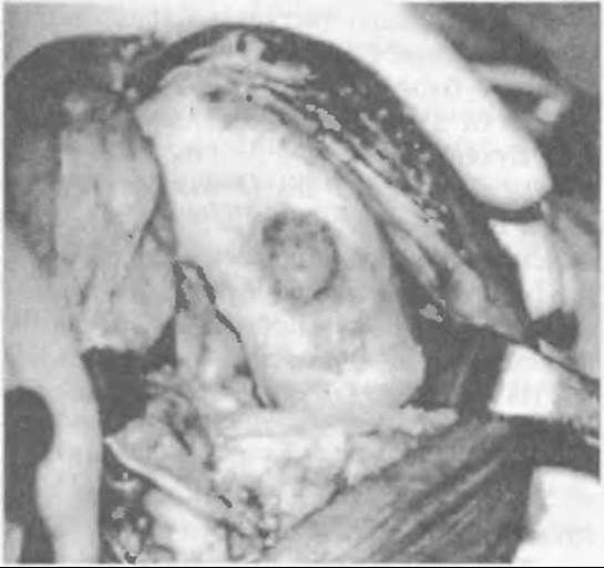
Fig. 354. Crater at the site of the osteochondral sequestrum detached in König's disease.
In non-progressive cases, conservative treatment proves effective, leading to patient recovery. An absolute indication for emergency surgery in these patients may be frequent entrapment or locking of the joint caused by a loose osteochondral sequestrum that has fallen into the joint cavity.
If treatment of patients in the second and third categories (according to Pappas) reveals widening of the radiolucent zone around the osteonecrosis, coupled with arthroscopic signs of articular cartilage depression and softness—indicating disease progression—surgical intervention is warranted. In cases where the cartilage has retained its density and remains undisturbed in its anatomical position, the optimal treatment modality is drilling channels through the chondro-osteonecrotic focus using a Kirschner wire to promote vascular ingrowth and accelerate the resorption and remodeling of subchondral osteonecrosis.
Nowadays, this Procedure is performed using arthroscopic techniques, which allow for visual inspection of the knee and execution of the drilling. In the absence of an arthroscope, a planned open arthrotomy is performed following routine radiological evaluation.
If softness and mobility of the undisturbed articular cartilage overlying the subchondral necrosis are detected, it is fixed with a bone peg driven into the drilled channel down to the healthy bleeding bone tissue, while the areas adjacent to the cartilage are drilled.
Postoperatively, the limb is immobilized with a plaster cast with the knee semi-flexed for 2.5-3 months. Afterwards, mobilization of the knee is initiated, along with paraffin-ozokerite applications, while avoiding weight-bearing on the leg for an additional two months.
If arthroscopy reveals a small crater (up to 1 cm2) covered with fibrous tissue, it is left untouched, but the loose sequestrum that has detached from the crater must be located and removed.
If a sequestrum with a preserved shape is found, the crater is cleaned of fibrous tissue via arthrotomy (curettage), the sequestrum is reduced into its bed and fixed with a thin bone peg inserted through a channel drilled within it. The bone peg is harvested from the epiphyseal region (through the same surgical incision) after opening the synovial membrane.
If arthroscopy reveals a larger crater (4-5 cm2) accompanied by fragmented loose sequestra within the knee joint, the sequestra are removed arthroscopically, while the closure of the cartilage defect is postponed to a Second Stage procedure.
Under general anesthesia, an open arthrotomy is performed, and the condition of the cartilage adjacent to the cartilage defect is assessed. The defect is cleared of the fibrous membrane down to the bleeding cancellous bone using curettage, and circular channels are drilled into it, into which osteochondral autografts (pegs) of matching diameter are inserted to fill the defect. The grafts are harvested from a non-weight-bearing area of the articular cartilage using a circular trephine with end-cutting Teeth. If the cartilage adjacent to the defect is soft, it is perforated with Kirschner wires. Postoperatively, the limb is immobilized in a plaster cast with the knee in a semi-flexed position. After 2.5-3 months, the cast is removed, and the condition of the knee is evaluated radiologically and arthroscopically. Subsequently, the limb joints are mobilized, and paraffin-ozokerite applications and Muscle massage are administered. Weight-bearing is permitted only after arthroscopic confirmation of graft incorporation.
The outcomes of various surgical procedures for Osteochondritis Dissecans of the knee joint have been reported by Lipscomb R. (1977). To enhance the contact between the detached cartilage fragment and the underlying bone layer, as well as to facilitate vascular ingrowth, drilling was performed, yielding good results in seven out of eight operated patients.
Gillespie N.F. and Day B. (1979) achieved similar outcomes in 17 out of 18 patients who underwent bone pegging and drilling of the adjacent cartilage in the presence of defects.
Furthermore, Lindholm S. and Pylkkänen P. (1974) observed good results over a five-year period in 73% of patients who underwent osteochondral pegging.
Minas T. and Spector M. (1996) reported on a novel method for filling articular cartilage defects using cultured cartilage Cell grafts, which they tested in animal experiments. This method was applied to an elderly patient in Sweden, but it is still premature to discuss its widespread clinical adoption.
Last update: 10/08/2026
Editorial and Educational Adaptation: This material has been compiled based on the primary/original source text. The project team performed an editorial review, corrected technical inaccuracies, structured sections, and adapted the content for an educational format.
What was processed:
- elimination of formatting defects (OCR errors, structural breaks, corrupted characters);
- editorial organization of content;
- standardization of terminology in accordance with academic sources;
- verification of factual statements against the original source text.
All mentions of the author, publication year, and origin of the primary text have been preserved in accordance with the source.