Cytology, General Histology and Embryology - V. K. Naphanyuk 2002
Tissues
Nervous Tissue
Cells of the Nervous Tissue
Cells of Nervous Tissue, or neurocytes, Neurons (neurocytus, neuronum), are the morphological and functional units of nervous tissue capable of perceiving stimuli, entering a state of excitation, generating, and transmitting impulses.
Due to their morphological features and functional purpose, neurocytes in different parts of The Nervous system vary significantly. There are two classifications of neurocytes: morphological and functional (Figs. 102, 103).
Class="center">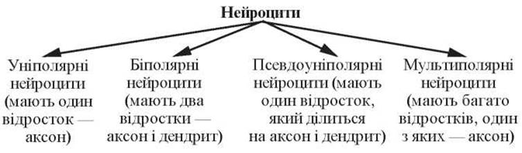
Fig. 102. Morphological Classification of neurocytes

Fig. 103. Structure/81.html">Functional Classification of neurocytes
Structure of Neurocytes
A neurocyte comprises a Cell body, or perikaryon (corpus neuro-cyti), processes, and nerve endings (terminationes neurocytum).
There are Two Types of processes:
— an axon, or neurite (axon, neuritum), a process that conducts nerve impulses away from The Cell body. It is a long process, measuring up to 1.5 m;
— a dendrite (dendritum), a process that receives nerve impulses and conducts them toward the cell body. It is a short, branching process.
The size of neurocytes varies widely, ranging from 4-6 µm in the granular layer of the Cerebellum up to 130 µm (giant pyramidal Cells of the cortex — Betz cells).
The shape of neurocytes can vary.
The Nucleus of a neurocyte is rounded, usually located centrally, and more rarely eccentrically. Neurocytes typically possess a single nucleus; binucleated and multinucleated neurocytes are very rare. Chromatin within the nuclei is dispersed, and there is one, occasionally 2–3, prominent nucleoli.
The Cytoplasm of neurocytes contains Three types of structured components: inclusions, general-purpose Organelles, and special-purpose organelles.
Inclusions include CARBOHYDRATES (Glycogen), pigment substances (lipofuscin, melanin), and various secretions.
General-purpose organelles: a well-developed Golgi apparatus, Mitochondria located throughout the cell body and all processes, and abundant Ribosomes and Lysosomes. The cell center is usually positioned between the nucleus and the dendrites.
Special-purpose organelles include chromatophilic substance and neurofibrils.
Chromatophilic substance (substantia chromatophilica) is revealed when nervous tissue is stained with aniline Dyes; in the cytoplasm of neurocytes, it appears as basophilic clumps of various SHAPES AND SIZES. These basophilic clumps are located in the cell bodies and dendrites of neurocytes, but are absent in axons and their cone-shaped hillocks. The basophilic clumps of the neurocyte cytoplasm are characterized by a high ribonucleoprotein content. Electron Microscopy studies have established that they correspond to areas of cytoplasm containing clusters of dense cisternae of the granular Endoplasmic reticulum. The degree of cisternae orientation varies across Different types of neurocytes, being most orderly in Spinal Cord neurocytes. In motor neurocytes of the spinal cord, the clumps of chromatophilic substance are large and arranged around the nucleus, whereas in sensory neurocytes of spinal ganglia, they appear as fine granules. The chromatophilic substance serves as an indicator of the neurocyte's functional state.
Because axons lack protein-synthesizing organelles, they exhibit a continuous cytoplasmic flow from the neurocyte body to the terminals at a rate of 1–3 mm per day (slow transport), which delivers Enzymes required for neurotransmitter synthesis at the axon terminals. There is also a fast transport mechanism (5–10 mm/h) that delivers components essential for synaptic function. Dendritic transport occurs at a rate of 3 mm/h, delivering the enzyme acetylcholinesterase, which breaks down the neurotransmitter acetylcholine. Retrograde transport carries cytoplasmic components back from the terminals to the neurocyte body.
Neurofibrils (neurofibrilla) are revealed by silver impregnation of nervous tissue, appearing as thin threads 0.3–0.5 µm in diameter that form a dense network within the neurocyte body and run parallel within dendrites and axons, including their smallest branches. Electron microscopy has established that neurofibrils correspond to bundles of neurofilaments 6–10 nm in diameter and neurotubules 20–30 nm in diameter. Neurofilaments and neurotubules belong to the neurocyte Cytoskeleton, located within the cell body and dendrites among the chromatophilic substance and oriented parallel to the axon.
Slides for Study
Slide 32. Chromatophilic substance in multipolar neurocytes of the spinal cord (Fig. 104).
Low magnification. Under this magnification, locate a large blue-stained multipolar neuron.
High magnification. Examine the large, pale Nucleus of the neurocyte with its intensely stained nucleolus. In the cytoplasm, blue clumps of basophilic substance are distributed throughout the cell body and dendrites, with the exception of the axon hillock and the axon. Make a drawing of the specimen.

Fig. 104. Chromatophilic substance in multipolar neurons of the spinal cord. Nissl staining. x 400:
1 — multipolar Nerve Cells (a — nucleus with a nucleolus; b — axon; c — dendrites; d — clumps of chromatophilic substance); 2 — glial cell nuclei
Label on the figure: 1) multipolar nerve cells: a — nucleus with a nucleolus; b — axon; c — dendrites; d — clumps of chromatophilic substance; 2) nuclei of glial cells.
Slide 33. Neurofibrils in neurons (Fig. 105).
Low magnification. At this magnification, locate a large multipolar neuron.
High magnification. A pale nucleus with a clearly visible brownish or black nucleolus is observed. Neurofibrils are clearly visible in the neuron body (as black or brown threads) forming a network, whereas in the processes, they run parallel to one another. Neurofibrils are present in both dendrites and the axon. Draw the slide.
Label on the figure: 1) neurons; 2) nucleus of the neuron; 3) nucleolus; 4) processes; 5) neurofibrils.
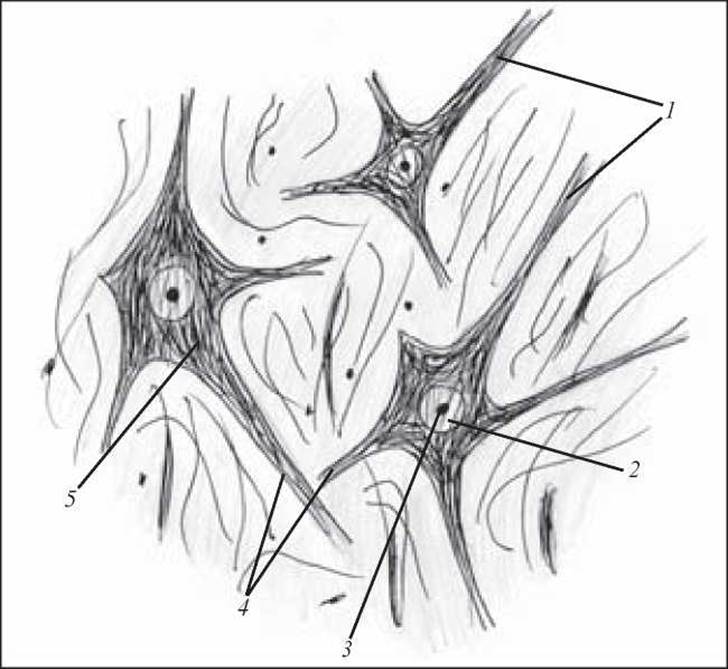
Fig. 105. Neurofibrils in neurons. Silver impregnation. x 600:
1 — neurons; 2 — nucleus of the neuron; 3 — nucleolus; 4 — processes; 5 — neurofibrils
Neuroglia
Neuroglia (neuroglia) is a collection of cellular elements of nervous tissue that surround neurons and perform supporting, demarcating, trophic, secretory, and protective Functions with respect to neurons.
All neuroglial cells are divided into two genetically and functionally distinct types: gliocytes (macroglia) and glial macrophages (microglia).
Gliocytes (gliocyti) are a group of cells that develop from the elements of the neural tube. Three types of cells are distinguished among gliocytes: ependymocytes, astrocytes, and oligodendrocytes.
Ependymocytes (ependymocyti) are cells that form a dense layer lining the spinal canal and all the Brain ventricles. During the Histogenesis of nervous tissue, ependymocytes differentiate first from neural tube glioblasts, performing demarcating and supporting functions at this stage of development. The elongated bodies of glioblasts on the inner surface of the neural tube form a layer of epithelium-like cells. On the surface facing the canal cavity, these cells bear cilia, the number of which in a single cell can reach 40. The beating of the cilia facilitates the movement of CEREBROSPINAL FLUID. The basal part of ependymocytes has long processes which, branching out, cross the entire neural tube and form its supporting apparatus. On the outer surface of the neural tube, these processes participate in forming the superficial glial limiting membrane, which separates the substance of the tube from other tissues.
After birth, ependymocytes gradually lose their cilia, which persist only in certain PARTS OF THE Central Nervous System (the cerebral aqueduct).
In the region of the posterior commissure of the cerebrum, ependymocytes perform a secretory function, forming a special "subcommissural organ" that secretes a substance presumably involved in the Regulation of Water METABOLISM.
Ependymocytes covering the choroid plexuses of the brain ventricles are cuboidal in shape. In newborns, cilia are located on their surface, which later degenerate. The cytoplasm of the basal pole forms numerous deep folds and contains large mitochondria, lipid droplets, and pigment inclusions.
Astrocytes (astrocyti) are small star-shaped cells with numerous processes extending in all directions to form the supporting apparatus of the central nervous system.
Two types of astrocytes are distinguished: protoplasmic and fibrous (fibrillar).
Protoplasmic astrocytes (astrocyti protoplasmatici) are located predominantly in the grey matter of the brain. These are cells 15-25 µm in size, possessing short, thick, highly branched processes and a large, oval, pale nucleus.
The cytoplasm contains a small number of endoplasmic reticulum cisternae, free ribosomes, microtubules, and numerous mitochondria.
Fibrous astrocytes (astrocyti fibrosi) are located mainly in the White matter OF the brain. These are cells up to 20 µm in size, with 20-40 smoothly contoured, long, sparsely branched processes that form glial fibers, creating a dense network—the supporting apparatus of the brain. The terminal expansions of astrocyte processes on Blood Vessels and on the brain surface form perivascular glial limiting membranes.
The cells have a large, pale nucleus; the nuclear envelope sometimes forms deep folds, and the karyoplasm is characterized by uniform electron density.
The cytoplasm contains few ribosomes and elements of the granular endoplasmic reticulum, and is packed with numerous 8-9 nm thick fibrils that extend into the processes as bundles.
Oligodendrocytes (oligodendrocyti) are the most numerous glial cell population, surrounding neuronal cell bodies in both the central and peripheral nervous systems. These cells are characteristically small across various Regions of the nervous system and exhibit diverse shapes (oval, polygonal). Each cell gives rise to several short, sparsely branched processes.
In terms of density, the cytoplasm closely resembles that of neurons and lacks neurofilaments.
Oligodendrocytes perform a trophic function and participate in the metabolic processes of nerve cells. They play a significant role in forming myelin sheaths around cellular processes, in which case they are referred to as neurolemmocytes (Schwann cells), and are involved in Water-Salt Metabolism, as well as degeneration and regeneration processes.
Microglia (microglia) comprise a population of small cells (glial macrophages) derived from Bone Marrow promonocytes. They typically feature 2-3 processes capable of giving off short secondary and tertiary branches.
The nuclei of these cells are elongated or triangular in shape and rich in chromatin.
When microglial cells are stimulated, they can alter their Morphology: their processes retract and the cells round up, at which point they are referred to as gitter cells (granular corpuscles). Recent studies have confirmed that microglia are capable of synthesizing immunoglobulin Proteins (Antibodies). These cells also exhibit amoeboid motility.

Fig. 106. Astrocytes of the cerebellum. Silver impregnation. x 400:
1 — astrocytes; 2 — glial fibers
Slides for Study
Slide 34. Astrocytes of the cerebral Gray matter (Fig. 106).
High magnification. Locate the body of an astrocyte—a small cell almost entirely occupied by the nucleus. Numerous branched processes radiate outward in all directions. Make a drawing of the slide.
Label the following on the drawing: 1) blood capillary: a) fibrous astrocyte; b) protoplasmic astrocyte.

Fig. 107. Nissl substance (tigroid bodies) within a nerve cell. Electron micrograph. x 30,000:
1 — endoplasmic reticulum membranes; 2 — ribosomes; 3 — cisternae (adapted from I.I. Glezer)
Electron micrographs provide a more detailed understanding of the ultrastructure of Nerve Tissue cells (Figs. 107-110).
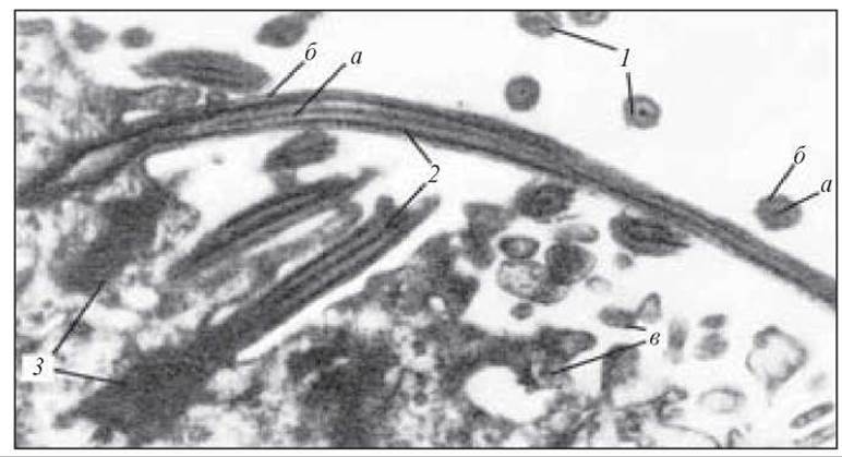
Fig. 108. Cellular cilia and microvilli of ependymal cells—distal region of ependymal cells from the Third ventricle of the rat Diencephalon. Electron micrograph. x 24,000:
1 — Cytology/practical/72.html">Cross section of cellular cilia; 2 — Longitudinal section of cellular cilia (a — central doublet microfibril; b — peripheral doublet microfibril; c — cellular microvilli); 3 — basal body — kinetosome (adapted from Brightman and Palay)

Fig. 109. Ependymal cells of the third ventricle of the rat diencephalon. Electron micrograph. x 17,000:
1 — lumen of the third ventricle; 2 — cross sections of cellular cilia with central and peripheral doublet microfibrils; 3 — cellular microvilli; 4 — desmosomes; 5 — nucleus; 6 — mitochondria (adapted from Brightman and Palay)

Fig. 110. Astroglial cell of the human Cerebral Cortex. Electron micrograph. x 24,000:
1 — nucleus of the astroglial cell; 2 — cytoplasm; 3 — mitochondria; 4 — processes of the astroglial cell; 5 — neurite (axon) (after De Robertis and Gerschenfeld)
1. Development of nervous tissue.
2. General structural plan of nervous tissue.
3. Functions of Nervous Tissue.
4. Cells of nervous tissue. Classification of nervous tissue cells.
5. Ultramicroscopic characteristics of neurocytes.
6. Special organelles of neurocytes and their functional purpose.
7. Axonal transport: types and functional significance.
8. Macroglia. Structure and origin of macroglial cells.
9. Microglia. Structure, functions, and origin.
10. Concept of reflex arcs and their cellular composition.
Situational Problems
1. There are two micropreparations of nervous tissue stained with Nissl stain. In the first one, large clumps of chromatophilic substance are visible in the neurocytes, while In the second one, they appear small, resembling dust-like granularity. To which functional types do the neurocytes in these preparations belong?
2. When examining two brain micropreparations from healthy individuals, one reveals A large number of lipofuscin inclusion granules in the cytoplasm of the neurocytes, whereas the other shows no lipofuscin. To which age groups do these micropreparations belong?
Sample Examination Questions
1. Histogenesis of nervous tissue.
2. Morphofunctional characteristics of neurocytes.
3. Morphofunctional characteristics and sources of development of macroglia and microglia.
Last update: 10/08/2026
Editorial and Educational Adaptation: This material has been compiled based on the primary/original source text. The project team performed an editorial review, corrected technical inaccuracies, structured sections, and adapted the content for an educational format.
What was processed:
- elimination of formatting defects (OCR errors, structural breaks, corrupted characters);
- editorial organization of content;
- standardization of terminology in accordance with academic sources;
- verification of factual statements against the original source text.
All mentions of the author, publication year, and origin of the primary text have been preserved in accordance with the source.