MEDICAL BIOLOGY, ANATOMY, HUMAN PHYSIOLOGY AND PATHOLOGY - Y.I. Fedoniuk 2010
BIOLOGY
CHAPTER 1. BIOLOGICAL FOUNDATIONS OF HUMAN VITAL ACTIVITY
1.3. MOLECULAR-GENETIC AND CELLULAR LEVELS OF LIFE ORGANIZATION
1.3.2. Structural, chemical, and functional organization of eukaryotic cells
Morphofunctional characteristics and Classification of Human Chromosomes
Chromosomes are nuclear structures of The Cell that contain genes (DNA). They are the physical carriers of heredity. Together with extrachromosomal structures, chromosomes ensure the preservation, Replication, expression, and transmission of Genetic information across generations. Chromosomes are capable of self-replication. Before every Cell Division, they duplicate and are then precisely and evenly distributed between the daughter Cells. They were named by the German histologist W. Waldeyer (1888) for their ability to stain intensely with basic Dyes (from the Greek chroma meaning color and soma meaning body).
The main Chemical Components of chromosomes are DNA, basic (histone) and acidic (non-histone) Proteins, which account for approximately 40%, 40%, and about 20%, respectively. DNA does not exist in a free state, but rather in a complex with proteins. Proteins spanning the entire length of the chromosome are called Histones. In the sperm chromosomes of certain species (such as salmon and herring), DNA is bound to protamines instead of histones. Histone proteins are represented by five main fractions (H1, H2A, H2B, H3, H4) and perform structural and regulatory Functions. The number of non-histone protein fractions exceeds 100. Among them are Enzymes involved in RNA Synthesis and Processing, as well as DNA Replication and Repair.
Chromosomes can exist in two Structural and functional states: condensed (spiralized) and uncondensed (despiralized), which are mutually convertible. During this process, the linear dimensions (length and thickness) of the chromosomes change. The spiralized state is characteristic of chromosomes during mitosis (mitotic chromosomes), whereas the despiralized state occurs between divisions (interphase chromosomes). Mitotic chromosomes are visible under a Light Microscope as thread-like or rod-like bodies that stain well with basic dyes. The function of mitotic chromosomes is transport: the precise distribution and transfer of genetic material into daughter nuclei. Interphase chromosomes are maximally despiralized, are not individually distinguishable, and occupy the entire volume of The Nucleus, forming Chromatin. The function of interphase chromosomes is synthetic: DNA Synthesis (replication) and RNA synthesis (METABOLISM/31.html">Transcription).
The Structure of chromosomes is studied during metaphase, when they are most condensed and clearly visible under a light microscope. A metaphase chromosome consists of two sister chromatids that lie side by side and are interconnected at the primary constriction (centromere). The centromere is a narrowed, unstained, and least spiralized region of the chromosome. It contains the kinetochore (from the Greek kinesis meaning motion and phoros meaning bearing), to which the microtubules of the spindle apparatus attach. The centromere divides the chromosome into two arms. The ends of the arms are called telomeres. These are regions containing tightly packed DNA that prevent chromosomes from fusing with one another or with their fragments. A chromosome end that has lost its telomere becomes "sticky" and readily joins with other chromosome fragments.
Depending on THE POSITION OF the centromere, chromosomes are classified into metacentric, submetacentric, and acrocentric types (Fig. 1.34).
Class="center">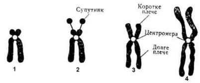
Fig. 1.34. Types of human chromosomes according to centromere position (metaphase stage):
1 - acrocentric; 2 - acrocentric with a satellite; 3 - submetacentric; 4 - metacentric.
In metacentric chromosomes, the centromere is located in the middle, resulting in arms of equal or nearly equal length (metacentric chromosomes). Submetacentric chromosomes are unequal-armed chromosomes in which the centromere is displaced from the center, so that one arm is long and the other is short. Acrocentric chromosomes are rod-like chromosomes in which the centromere is located close to one end, resulting in one long arm and a very short, often inconspicuous second arm. Telocentric chromosomes may also arise As a result of the loss of one arm, leaving only a single arm with the centromere at the tip; such chromosomes do not occur under normal conditions. Some chromosomes have secondary constrictions located in the pericentromeric Regions of the long arms (chromosomes 1, 9, 16) and at the terminal regions of the short arms (chromosomes 13-15 and 21, 22). In chromosomes 13-15 and 21, 22, the secondary constrictions separate small terminal segments, called satellites, from the main body via a thin thread, and such chromosomes are termed satellite chromosomes. The nucleolar organizers—the sites where nucleoli are formed—are located in the region of the secondary constrictions of certain chromosomes.
Ultrastructural Organization of chromosomes. A single-chromatid chromosome contains one hyperspiralized DNA molecule. There are several levels of coiling. The elementary chromosomal structure distinguishable under an Electron microscope is a fiber with a diameter of 10-13 nm (1 nm = 10~3 µm = 10~6 mm), which represents a complex of DNA and histone proteins (nucleohistone). Nucleosomes are arranged along its length like beads on a string. A nucleosome has a core consisting of 8 histone molecules of four classes (histone octamer, histone core), with two molecules of each class: H2A, H2B, H3, and H4 (Fig. 1.35). The DNA double helix (146 nucleotide pairs) makes 1.75 turns around it before extending to the core of the next nucleosome. The internucleosomal DNA regions (linkers) are connected by histone H1. Wrapping the DNA molecule around the histone bodies reduces the length of its double helix approximately 7-fold. At the next level of coiling, a fiber with a diameter of 20-25 nm is formed. The total length of DNA at this level is reduced 40-fold. Through further hyperspiralization involving loop formation and repeated longitudinal folding, mitotic chromosomes are formed. Bundles of fibrils form chromonemata. Along the chromonemata are regions (nodules) of denser DNA compaction called chromomeres. Super-spiralization achieves a tight Introduction/28.html">Packaging of Genetic material, which is crucial during chromosome movements in mitosis. The degree of compaction is illustrated by the following figures: the length of the largest human chromosome (chromosome 1) is 11 µm, whereas its fully extended DNA molecule is about 7 cm long. In this hypercompacted state, chromosomes are inactive, making it difficult for enzymes or regulatory proteins to access active DNA groups.
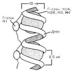
Fig. 1.35. Nucleosomal ORGANIZATION OF THE elementary chromosome fiber.
A prerequisite for the expression of any chromosomal region is its decondensation.
Last update: 08/08/2026
Editorial and Educational Adaptation: This material has been compiled based on the primary/original source text. The project team performed an editorial review, corrected technical inaccuracies, structured sections, and adapted the content for an educational format.
What was processed:
- elimination of formatting defects (OCR errors, structural breaks, corrupted characters);
- editorial organization of content;
- standardization of terminology in accordance with academic sources;
- verification of factual statements against the original source text.
All mentions of the author, publication year, and origin of the primary text have been preserved in accordance with the source.