Medical Radiology - Lazar A.P. 2008
Radiation examination of the digestive system
Diseases of the pancreas
The Pancreas is an exocrine and endocrine gland and one of the largest glands in The Human Body. It is 15–16 cm long, 4–5 cm wide, 2–3 cm thick, and weighs 80–100 g. Located retroperitoneally behind The Stomach at the level of the I-II lumbar vertebrae, the Peritoneum covers only the anterior surface of the organ body. The pancreas consists of a HEAD, neck, body, and tail. The main pancreatic duct lies closer to the posterior surface of the gland. Because the duct can have a straight or tortuous course, its length may exceed that of the gland itself, measuring 10–28 cm. The diameter of the duct is 2–4 mm in the head region, 1–3 mm in the body, and 0.5–2.5 mm in the tail.
Class="center">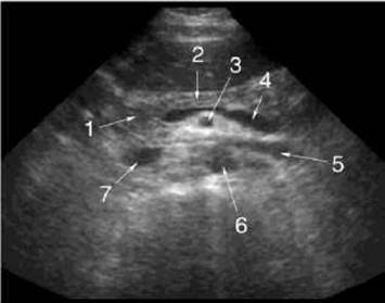
Fig. 223. Pancreas on a sonogram.
1 - head of the pancreas;
2 - body of the pancreas;
3 - superior mesenteric artery;
4 - splenic vein;
5 - left renal vein;
6 - aorta;
7 - INFERIOR VENA CAVA.
During plain radiography and on roentgenograms, the pancreas is not visualized; only calcifications within the glandular tissue in Chronic Pancreatitis may be noticeable. Pathological Changes in the pancreas can be inferred indirectly from the shape and position of the contrast-filled stomach and duodenum. For instance, tumors of the pancreatic head result in widening of the C-loop of the duodenum. Since a malignant tumor causes uneven enlargement of the gland, findings may include irregularity of the medial contour of the descending duodenum, luminal narrowing, and deformation of mucosal folds due to extrinsic pressure.
To study radiation anatomy and assess pancreatic function, modern Diagnostic Imaging Modalities are utilized: ultrasound (US), computed tomography (CT), Magnetic Resonance imaging (MRI), radionuclide Diagnostics (RND), endoscopic retrograde cholangiopancreatography (ERCP), selective angiography, and splenoportography.
On CT scans, pancreatic abnormalities can be detected in the majority of cases. The densitometric density of the pancreas on computed tomography ranges from +20 to +45 HU. The intravenous contrast enhancement technique increases glandular density by 20–40 HU.
On MRI scans, the signal from the pancreas has a medium intensity, generally matching or slightly lower than that of the Liver. Adipose tissue produces a high-intensity signal, which increases the signal from the pancreas.
On sonograms, the pancreas is comma-shaped (Fig. 223). An increase in the anteroposterior dimension of the pancreatic head exceeding 35 mm, the body exceeding 25 mm, or the tail exceeding 20 mm indicates The Development of a pathological process. General enlargement of the pancreas is most commonly caused by edema and is observed in pancreatitis, whereas focal enlargement is characteristic of malignant tumors, hematomas, and cysts. Normally, the echogenicity of the pancreas matches or is slightly higher than that of the liver; the Structure is homogeneous, fine-grained, and uniform throughout the gland. In elderly individuals, the echogenicity of the pancreatic structure increases due to fat deposition and Connective Tissue proliferation.
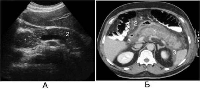
Fig. 224. Acute pancreatitis.
A - sonogram showing decreased echogenicity of the body (2) and enlargement of the head (1) of the pancreas;
B - computed tomography scan showing enlargement of the edematous pancreas (1), peripancreatic tissue edema (2), and fluid accumulation in the perirenal space (3).
The primary radionuclide imaging method is pancreatic scintigraphy, THE PRINCIPLE OF which involves obtaining an image of the pancreas using 75Se-Methionine. Pancreatic imaging can be achieved as early as 10 minutes after radiopharmaceutical administration. The highest radioactivity is observed in the head of the gland.
Radioimmunoassay is used to study the exocrine and endocrine Functions of the pancreas. This method assesses the levels of Trypsin, Insulin, and C-peptide. Determining C-peptide (an inactive form of insulin) is particularly important when a patient has been receiving long-term insulin therapy.
Acute pancreatitis typically develops As a result of biliary tract inflammation. The patient experiences acute girdle pain radiating to the lower back, left hypochondrium, and periumbilical region, along with nausea, dyspeptic symptoms, and vomiting. Leukocytosis and elevated serum amylase levels are observed.
Edematous, hemorrhagic, and necrotic forms of acute pancreatitis are distinguished. The goal of radiological evaluation is to differentiate these forms, which dictates the Treatment strategy—conservative for the edematous form, and surgical for the hemorrhagic and necrotic forms.
Plain radiography reveals a distended duodenal loop, high standing of the left dome of the Diaphragm, and reactive left-sided pleuropulmonary changes (Pleurisy).
In patients with acute edematous pancreatitis, ultrasound reveals generalized enlargement of the gland, indistinct contours, low echogenicity, and increased sound transmission of the edematous parenchyma (Fig. 224). In patients with hemorrhagic and necrotic pancreatitis, it shows alternating areas of parenchyma with increased and decreased echogenicity, with the latter predominating.
On CT, acute edematous pancreatitis typically manifests as a diffuse enlargement of the pancreas with blurred contours. A uniform or patchy decrease in glandular density is observed, dropping to +12 HU in certain areas. Hemorrhagic pancreatitis is characterized by structural inhomogeneity of the gland and the presence of areas whose density exceeds that of the unaffected portion of the organ by +10 to +20 HU. Bolus contrast enhancement is used to differentiate the necrotic form from the edematous one: necrotic areas do not enhance, unlike the edematous glandular tissue. This approach allows the detection of 90% of severe necroses and nearly 80% of small necrotic foci.
Magnetic resonance imaging reveals enlargement, structural inhomogeneity, and blurred contours of the pancreas. Hemorrhagic pancreatitis is distinguished by high signal intensity in areas of Hemorrhage, whereas areas of edema and necrosis produce low-to-medium signal intensity that differs little from that of the normal parenchyma.
Chronic pancreatitis may occur as a primary condition or develop as a consequence of acute recurrent pancreatitis.
During an Exacerbation of chronic pancreatitis, radiological signs of an enlarged pancreas include the Displacement of the stomach anteriorly and superiorly. Relaxation duodenography reveals straightening of the duodenal loop and leveling of its medial contour. Ultrasound in chronic pancreatitis demonstrates pancreatic enlargement, dilation of the pancreatic duct, and fluid collections in the form of pseudocysts. CT refines the pathomorphology of the lesion, particularly one of the most critical diagnostic signs—pancreatic calcification (Fig. 225). On MRI, the pancreatic tissue exhibits heterogeneous signal intensity.
It is generally recommended that retrograde pancreatocholangiography be performed in every patient who has experienced at least one episode of pancreatitis, though only 2 to 4 weeks after the acute symptoms have subsided. The Procedure involves locating the major duodenal papilla with a catheter probe, inserting a catheter into its orifice, and injecting 2 ml of a 50% cardiotrast or diodone solution, followed by radiography. In chronic pancreatitis, deformation of the pancreatic duct and its tributaries is observed, along with partial obliteration and alternating stenoses and dilatations. The contrast medium flows slowly out of the ducts, remaining visible for 10 minutes or more.
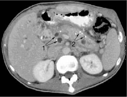
Fig. 225. Chronic pancreatitis on a computed tomography scan.
1 - pseudocysts;
2 - calcifications.
The scintigraphic pattern of chronic pancreatitis is characterized by poor uptake of 75Se-methionine throughout the entire gland or in its individual segments, depending on the duration and severity of the disease. Additionally, there is an increased level of extrapancreatic radiopharmaceutical accumulation in the PROJECTION OF THE Spleen, the splenic flexure of the colon, the Small Intestine, and the pyloric region of the stomach as a compensatory response to pancreatic exocrine insufficiency.
Pancreatic cysts are classified into congenital, parasitic, and pseudocysts. Congenital and parasitic pancreatic cysts are rare. Pseudocysts frequently form in chronic pancreatitis or during the terminal stage of acute pancreatitis. They can reach significant sizes (10-15 cm) and extend beyond the BOUNDARIES OF THE pancreas. On imaging, pseudocysts appear as rounded, cyst-like areas of uneven fluid accumulation (exudate, Blood, secretion) within the pancreatic parenchyma; they lack a true epithelial lining and result from tissue necrosis, duct rupture, or ductal occlusion with impaired secretory outflow (so-called retention cysts). Retrograde pancreatocholangiography can establish the communication between these cysts and the pancreatic duct system. Angiographically, a pancreatic cyst appears as a well-defined avascular zone. Both true cysts and pseudocysts on CT and MRI are typically solitary, regular and round in shape, with distinct margins, presenting low densitometric density ranging from -5 to +10 HU on CT, low signal intensity on T1-weighted images, and high signal intensity on T2-weighted MR images; true cysts additionally feature a thin, uniform wall. Ultrasonography detects cysts measuring 1 cm or more as echonegative areas with enhanced acoustic transmission and clear borders. Scintigraphically, a pancreatic cyst presents as a rounded radiopharmaceutical uptake defect with well-defined borders, causing compression and displacement of the organ.
Pancreatic tumors are divided into benign and malignant. Benign pancreatic tumors are extremely rare. Among malignant tumors of the pancreas, carcinoma is the most common.
Pancreatic Cancer is mostly observed in individuals aged 50 to 70 years, predominantly males. It is characterized by early metastasis to adjacent Organs, particularly the liver. In 50-70% of patients, the tumor is localized in the head of the pancreas, while in 25-50% it affects the body, tail, or the entire organ. Tumor dimensions range from 1 to 15 cm. Clinical manifestations include dyspeptic symptoms such as nausea, vomiting, loss of appetite, weight loss, and persistent dull aching pain. 3 to 4 months after the onset of these symptoms, jaundice, Skin pruritus, and a dull pain in the right upper quadrant appear.
Radiologically, secondary signs of a space-occupying lesion in the pancreas are identified through changes in adjacent organs: the stomach, duodenum, Gallbladder, Bile duct, and Kidneys. Occasionally, calcified inclusions within the tumor can be detected, which are best visualized in the lateral projection directly anterior to the spine.
Angiograms reveal enlargement of a specific part of the gland, The formation of pathological vessels, erosion of the walls of major vessels, and the presence of an avascular zone due to thrombosis and tumor compression of pancreatic Arteries. Splenoportography involves the puncture injection of 20-30 ml of a 70% cardiotrast solution into the spleen, followed by 3-4 radiographs taken at 1.5-2 second intervals. Lesions in the head of the pancreas are accompanied by displacement, compression, and deformation of the splenic vein and the portal vein. Involvement of major arteries and Veins indicates that the tumor is unresectable.
On CT and MRI, pancreatic cancer presents with alterations in the size and shape of the pancreas and blurred contours (Fig. 226). When a cancerous tumor is located in the head of the gland, signs of obstructive jaundice and post-stenotic dilation of the pancreatic duct are typically found. At the same time, carcinoma of the pancreatic head cannot be differentiated from Cancer of the major duodenal papilla or cancer of the intrapancreatic portion of the bile duct. With small tumors of the body and tail of the pancreas, only a slight enlargement of the gland may be detected. Invasion of adjacent organs by the cancerous tumor and metastasis to peripancreatic, para-aortic, paracaval Lymph Nodes, the liver, and the porta hepatis lymph nodes are clearly visualized.
Sonography reveals segmental enlargement of the pancreas, which occurs in 94% of patients with this localization of cancer. In most cases, the cancerous region is characterized by low echogenicity with small, unevenly distributed echoes; higher echogenicity, which may be homogeneous or heterogeneous, is observed less frequently. Indirect signs of pancreatic cancer include pre-stenotic dilation of the pancreatic duct and fluid accumulation in the Abdominal cavity (ascites), which is easily detected during sonography. Furthermore, in cancer patients, the pancreas loses its elasticity in the affected area and does not deform during Ultrasound examination.
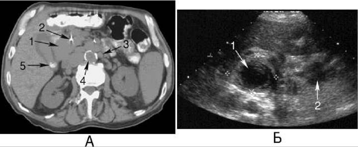
Fig. 226. Carcinoma of the pancreatic head.
A - computed tomography scan: 1 - enlarged pancreatic head; 2 - stent in the bile duct; 3 - enlarged para-aortic lymph node; 4 - sclerotic aorta; 5 - Gallbladder stone.
B - sonogram: 1 - hypoechoic enlarged pancreatic head; 2 - hypoechoic hyperplastic lymph node.
The scintigraphic pattern of pancreatic cancer is characterized by radiopharmaceutical uptake defects and alteration of the organ's shape, as only functioning areas of the parenchyma are visualized.
The Differential Diagnosis between pancreatic cancer and pancreatitis is one of the most challenging problems in abdominal imaging, especially since both conditions can coexist. Key considerations include: 1) loss of fat planes around peripancreatic vessels, which is more characteristic of cancer; 2) diffuse enlargement of the gland and peripancreatic inflammation with thickening of the renal fascia, which strongly favor pancreatitis; 3) the specific pattern of pancreatic duct dilation and the transition from the dilated segment to the narrowed one; 4) on scintigraphy, despite reduced radiopharmaceutical uptake in pancreatitis, the type and shape of the pancreas can always be determined. Interventional ultrasound- or CT-guided tumor biopsy is of paramount importance in diagnosing pancreatic cancer.
Last update: 08/08/2026
Editorial and Educational Adaptation: This material has been compiled based on the primary/original source text. The project team performed an editorial review, corrected technical inaccuracies, structured sections, and adapted the content for an educational format.
What was processed:
- elimination of formatting defects (OCR errors, structural breaks, corrupted characters);
- editorial organization of content;
- standardization of terminology in accordance with academic sources;
- verification of factual statements against the original source text.
All mentions of the author, publication year, and origin of the primary text have been preserved in accordance with the source.