Medical Radiology - Lazar A.P. 2008
Radiation Examination of the Digestive System
Diseases of the Major Salivary Glands
The major Salivary Glands are paired parenchymatous Organs whose excretory ducts open into the Oral Cavity. They include the parotid, submandibular, and sublingual glands.
The parotid gland is located in the posterior buccal region, posterior and lateral to the ramus of the Mandible. Its dimensions are: vertical 4–6 cm, sagittal 3–5 cm, and transverse 2–3.8 cm. The parotid duct, 5–7 cm in length, opens into the oral vestibule via a papilla.
The submandibular gland measures 3–4.5 cm in length, 1.5–2.5 cm in width, and 1.2–2 cm in thickness. It lies within the submandibular triangle, almost entirely on the Inferior surface of the mylohyoid Muscle. The posterior part of the gland wraps around the posterior border of the mylohyoid muscle onto its superior surface and gives rise to the submandibular duct, 4–6 cm long, which runs anteriorly along The surface of the sublingual gland and opens on the sublingual caruncle.
The sublingual gland measures 3.5×1.5 cm and rests on the superior surface of the mylohyoid muscle beneath the mucous membrane of the sublingual fold. The major sublingual duct opens on the sublingual caruncle adjacent to the submandibular duct (sometimes the ducts open together). The minor sublingual ducts, numbering about 10–12, open along the sublingual fold up to the sublingual caruncle inclusive.
Diagnostic Imaging Modalities for the major salivary glands include sialography, pneumosubmandibulography, CT, ultrasound, MRI, and sialoscintigraphy.
To perform sialography, 1–2 ml of a Water-soluble radiopaque contrast agent (verografin, urotrast, omnipaque) is warmed to body Temperature. A cannula with an attached syringe is inserted into the excretory duct, the contrast is injected, and a series of radiographs is acquired: at 15 s, 60 s, and 2 min. Duct patency and glandular angioarchitecture are evaluated during the parenchymal and resorption phases.
Radionuclide imaging evaluates the parenchymal distribution of 99mTc-pertechnetate in the salivary glands following intravenous administration. Gland massage is performed 2 min prior to the Procedure to clear saliva. Following radiopharmaceutical administration, scintigraphy is acquired at a rate of 1 frame per min for 40 min. Normally, the glands show somewhat higher radiopharmaceutical uptake in their central regions, exhibit an oval shape, and have smooth contours.
Pathological Changes in the salivary glands are accompanied by alterations in Symmetry, size, shape, densitometric density on CT, signal intensity on MRI, and echogenicity on sonograms.
Salivary gland and duct stones are referred to as sialolithiasis. Radiopaque salivary stones can be detected on conventional radiographs and CT scans, whereas radiolucent stones are identified using ultrasound, CT, MRI, and sialography.
Inflammation of a salivary gland is termed sialadenitis, and inflammation of its ducts is called sialodochitis. Acute sialadenitis is a contraindication for sialography due to the risk of retrograde infection during this diagnostic procedure.
In acute-phase sialadenitis, CT, MRI, and ultrasound reveal glandular enlargement and duct narrowing caused by compression from the edematous parenchyma and mucosal Swelling. Duct narrowing is also visualized via sialography. In the chronic phase, salivary ducts dilate due to paresis of the muscular layer (Fig. 227). Scintigraphy demonstrates relatively high but delayed accumulation of 99mTc-pertechnetate. Progression of chronic sialadenitis is accompanied by decreased radiopharmaceutical uptake.
Class="center">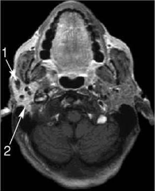
Fig. 227. Chronic sialadenitis of the right parotid gland on an MRI scan.
1 – dilation of the parotid duct;
2 – hyperintensity of the glandular parenchyma.
Chronic sialodochitis is characterized by alternating stenotic and dilated segments of the ducts, which is visualized using sialography.
Salivary gland tumors most commonly occur in the parotid gland.
Benign tumors (adenoma, adenolymphoma, mixed tumor) are more frequent, accounting for 80% of cases. They appear on CT, MRI, and ultrasound as a well-circumscribed mass with distinct margins and some organ distortion. Sialography reveals a filling defect with smooth, regular contours.
Malignant tumors (Sarcoma, carcinoma) more frequently affect the deep lobe of the parotid gland. They manifest with pain and Facial Nerve paresis. Sialography demonstrates cutoff of the salivary ducts adjacent to the tumor and pooling of the contrast agent. On CT, MRI, ultrasound, and scintigrams, the affected portion of the gland is enlarged, heterogeneous, and poorly marginated. Scintigraphically, decreased radiopharmaceutical uptake in the tumor region is most common, while increased uptake due to tumor obstruction of the glandular excretory duct is less frequent.
Last update: 08/08/2026
Editorial and Educational Adaptation: This material has been compiled based on the primary/original source text. The project team performed an editorial review, corrected technical inaccuracies, structured sections, and adapted the content for an educational format.
What was processed:
- elimination of formatting defects (OCR errors, structural breaks, corrupted characters);
- editorial organization of content;
- standardization of terminology in accordance with academic sources;
- verification of factual statements against the original source text.
All mentions of the author, publication year, and origin of the primary text have been preserved in accordance with the source.