Human Anatomy - H. I. Koliadenko 2009
Locomotor System
Skull Skeleton
Structure and Joints of the Skull Bones
The Skull (cranium) forms the skeletal framework of the HEAD and consists of 23 bones, which are divided into two main sections: the neurocranium (braincase) and the facial Skeleton (viscerocranium). The neurocranium serves a protective function, enclosing and safeguarding the Brain. The BONES OF THE facial skeleton form fossae and cavities that house the Organs of Hearing, Vision, and smell, as well as the initial segments of the respiratory and digestive tracts.
The neurocranium comprises a base and a vault (roof). The bones of the vault feature a specialized Structure. Each bone consists of an outer and an inner compact plate of Bone tissue, between which lies spongy bone (diploe) containing Cells filled with Cytology/practical/86.html">Red Bone Marrow. The inner compact plate, which lies directly adjacent to the brain, is known as the glassy plate (lamina vitrea). In the event of head trauma, it shatters into small fragments and can potentially lacerate cerebral Blood Vessels.
The neurocranium. The neurocranium is composed of 8 bones — 2 paired (2 parietal bones, 2 temporal bones) and 4 unpaired (frontal, ethmoid, sphenoid, and occipital bones).
The Parietal bone (os parietale; Fig. 47) is a paired, broad bone featuring two surfaces (external and internal) and four borders. The serrated upper borders of both parietal bones articulate with each other along the sagittal suture.
Class="center">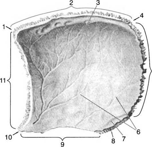
Fig. 47. Right parietal bone (internal view):
1 — frontal angle; 2 — sagittal border; 3 — sulcus for superior sagittal sinus; 4 — occipital angle; 5 — occipital border; 6 — arterial sulci; 7 — sulcus for sigmoid sinus; 8 — mastoid angle; 9 — squamous border; 10 — sphenoid angle; 11 — frontal border
The anterior borders articulate with the Frontal bone via the coronal suture. The posterior borders of the parietal bones join the Occipital bone to form the lambdoid suture. The lateral borders, together with the temporal bones, form the squamous suture. The parietal bone possesses four angles: the anterosuperior angle, or frontal angle; the posterosuperior angle, or occipital angle; the anteroinferior angle, or sphenoid angle; and the posteroinferior angle, or mastoid angle. The cerebral surface of the bone features digital impressions and arterial sulci. The external surface of the parietal bone is convex and displays an arcuate temporal line, to which the masticatory Muscles attach. Near the center of the bone lies the parietal eminence; it is prominent in infants, whereas in adults it becomes smoothed out and appears as a slight elevation.
The Temporal bone (os temporale; Fig. 48) is a paired bone located in the lateral wall and Base of the skull. It consists of three parts: the tympanic part, the squamous part, and the petrous part (pyramid).
The tympanic part is situated in the center of the bone, surrounding the external acoustic meatus anteriorly, inferiorly, and posteriorly. This meatus leads to the Middle ear (the tympanic cavity), which is located within the substance of the petrous part.
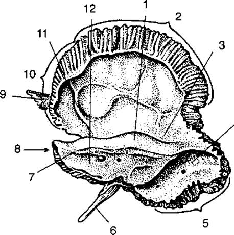
Fig. 48. Temporal bone (internal view):
1 — arcuate eminence; 2 — parietal border; 3 — tegmen tympani; 4 — sulcus for sigmoid sinus; 5 — occipital border; 6 — styloid process; 7 — apex of petrous part; 8 — petrous part; 9 — zygomatic process; 10 — basal border of squamous part; 11 — arterial sulcus; 12 — posterior surface of petrous part; 13 — internal acoustic meatus
The squamous part (squama temporalis) partially forms the lateral surface of the cranial vault; its internal surface exhibits digital impressions and vascular grooves. The upper border of the squamous part articulates with the parietal bone to form the squamous suture, while its anterior border articulates with the greater wing of the Sphenoid bone. Projecting horizontally from the squamous part is the zygomatic process, which joins the Zygomatic bone to form the zygomatic arch. Inferior to the zygomatic process lies the oval mandibular fossa for articulation with the condylar process of the Mandible. Anterior to the articular surface of the squamous part is the articular tubercle.
The petrous part resembles a three-sided pyramid, with its apex directed forward and medially. This portion of the temporal bone presents anterior, posterior, and inferior surfaces. The interior of the pyramid houses the middle and Inner ear. The anterior surface of the pyramid features the trigeminal impression, and posterior to it lies the arcuate eminence. The posterior surface contains the internal acoustic meatus, which leads into the internal acoustic canal, transmitting the facial and vestibulocochlear nerves.
The Inferior surface of the petrous part features the external opening of the carotid canal, through which the Internal Carotid Artery passes. Adjacent to this opening on the inferior surface of the pyramid is a deep jugular fossa, bounded by the jugular notch.
Between the petrous and squamous parts lies the musculotubal canal, which leads into the tympanic cavity. This canal houses the cartilaginous part of the pharyngotympanic (auditory) tube, connecting the middle ear cavity with the nasopharynx.
Externally, the petrous part features the mastoid process, its base directed upward and its apex downward. On the inner surface of the mastoid process lies the sigmoid sulcus, which continues into the transverse sulcus, curving around the jugular process of the occipital bone and extending to the jugular foramen of the skull. Inside the mastoid process, enclosed by the trabeculae of the spongy bone, are numerous air-filled mastoid air cells (mastoid sinuses). These communicate with the middle ear cavity. In young children, these cells are entirely absent and develop progressively with age. In elderly individuals, the cells enlarge As a result of Osteoporosis. Inferior to the mastoid process is the styloid process. Between the mastoid and styloid processes lies the stylomastoid foramen, through which the Facial Nerve exits the cranial cavity.
The frontal bone (os frontale; Figs. 49, 50) is an unpaired bone comprising a nasal part (or body), two orbital parts, and the squamous part (frontal squama).
The nasal part is located medially in the frontal bone and contains an air-filled cavity divided by a septum into two parts. This cavity communicates with the middle nasal meatuses; consequently, the frontal bone is classified as a pneumatic bone. The inferior border of the frontal bone's body features nasal notches that articulate with the nasal bones. The external surface of the nasal part lies within a depression situated between the lateral surfaces and the squama.
The frontal squama occupies the anterosuperior region of The Skull and articulates via a serrated suture with the parietal bone. On its lateral surfaces, the squama bears the frontal eminences, which vary in prominence among individuals. Inferior to the frontal eminences are the superciliary arches, between which lies the glabella.
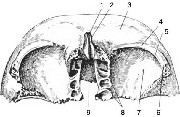
Fig. 49. Frontal bone (inferior view):
1 — nasal part; 2 — glabella; 3 — superciliary arches; 4 — supraorbital margin; 5 — fossa for lacrimal gland; 6 — zygomatic process; 7 — orbital surface; 8 — ethmoidal foramina; 9 — ethmoidal notch

Fig. 50. Frontal bone (internal view):
1 — groove for superior sagittal sinus; 2 — frontal crest; 3 — zygomatic process; 4 — digital impressions; 5 — orbital part; 6 — cerebral juga (eminences); 7 — arterial grooves; 8 — parietal margin
The orbital PARTS OF THE frontal bone, situated beneath the supraorbital margins, form the roof of the Orbit. In the lateral part of the orbital wall, near the base of the zygomatic process, lies the fossa for the lacrimal gland, lateral to which is the trochlear fossa where a small Cartilage is attached, serving as a pulley for the tendon of the superior oblique Muscle. Between the orbital parts lies a deep notch filled by the cribriform plate of the Ethmoid bone.
On the internal surface of the squama, there are digital impressions and eminences, as well as the frontal crest, which bifurcates superiorly to bound the groove for the sagittal sinus.
The occipital bone (os occipitale; Figs. 51, 52) is unpaired and consists of four parts: the basilar part, two lateral parts, and the occipital squama.
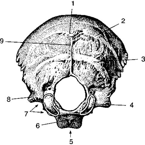
Fig. 51. Occipital bone (external view):
1 — internal occipital protuberance; 2 — superior nuchal line; 3 — inferior nuchal line; 4 — occipital condyle; 5 — basilar part; 6 — pharyngeal tubercle; 7 — jugular notch; 8 — jugular process; 9 — external occipital crest
The basilar part, or clivus of the occipital bone, lies anterior to the foramen magnum. The internal surface of the clivus is smooth, whereas the external surface is uneven and bears the pharyngeal tubercle in the midline, to which one of the pharyngeal muscles attaches. The clivus articulates via a flat suture with the body of the sphenoid bone. Posteriorly, the foramen magnum is enclosed by the occipital squama, which features a serrated margin, as well as external and internal surfaces. In the middle of the external surface of the squama is the external occipital protuberance, which extends inferiorly as the crest. Extending laterally from the occipital crest are the superior and inferior nuchal lines, to which the occipital muscles attach. On the internal, or cerebral, surface of the squama, There is a cruciform eminence along which run the grooves for the transverse sinus, while superiorly ascends the groove for the superior sagittal sinus, representing the impression of the venous sinuses of the dura mater. From the cruciform eminence down to the foramen magnum runs the internal occipital crest. Laterally, the foramen magnum is flanked by the lateral parts of the bone, on the inferior surface of each of which is an articular condyle by means of which the occipital bone articulates with the first cervical vertebra, the atlas. Above the condyles are the hypoglossal canals. Along the lateral margin of the lateral parts lies a deep jugular notch, bounded by the jugular process. The jugular notch, together with the jugular fossa of the petrous part of the temporal bone, forms the jugular foramen.
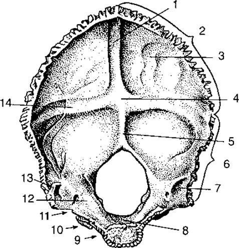
Fig. 52. Occipital bone (internal view):
1 — groove for superior sagittal sinus; 2 — lambdoid margin; 3 — occipital squama; 4, 14 — internal occipital protuberance; 5 — internal occipital crest; 6 — mastoid margin; 7 — groove for sigmoid sinus; 8 — clivus; 9 — basilar part; 10 — lateral part; 11 — jugular notch; 12 — jugular process; 13 — groove for transverse sinus
The ethmoid bone (os ethmoidale; Fig. 53) is unpaired and contributes to both the neurocranium and the viscerocranium. Its neurocranial component is the cribriform plate, which lies horizontally between the orbital parts of the frontal bone. Numerous olfactory nerves pass through its foramina into the cranial cavity. Projecting upward from the plate is the crista galli, while downward extends the perpendicular plate, which divides the upper Nasal cavity into right and left halves, thus forming the upper part of the nasal septum. Flanking the perpendicular plate are the ethmoidal labyrinths, composed of small, air-filled ethmoidal cells. The lateral walls of the labyrinth are enclosed by the orbital lamina, which forms the Medial surface of the orbit. Projecting from the medial surface of the ethmoidal labyrinths are thin plates that form the superior and middle nasal conchae.
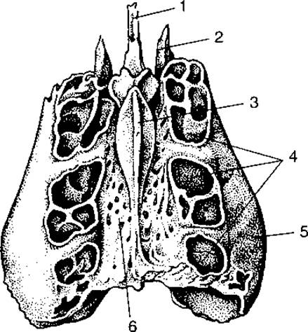
Fig. 53. Ethmoid bone (superior view):
1 — perpendicular plate; 2 — middle nasal concha; 3 — crista galli; 4 — ethmoidal labyrinth; 5 — orbital lamina; 6 — cribriform plate

Fig. 54. Sphenoid bone:
a — anterior view; b — posterior view; 1 — sella turcica; 2 — optic canal; 3 — foramen rotundum; 4 — foramen ovale; 5 — foramen spinosum; 6 — superior orbital fissure; 7 — opening of sphenoidal sinus; 8 — lateral plate of pterygoid process; 9 — medial plate of pterygoid process; 10 — pterygospinous process; 11 — pterygoid process; 12 — greater wing; 13 — lesser wing
The sphenoid bone (os sphenoidale; Fig. 54) is unpaired and occupies a central position at the base of the skull. It consists of a body and three pairs of processes, and is classified as a pneumatic bone. Attached to the body are the lesser wings anteriorly, the greater wings laterally, and the pterygoid processes inferiorly. The body of the sphenoid bone contains a cavity — the sphenoidal sinus — filled with air that enters through two openings on the anterior surface of the body. The sphenoidal sinus is divided into two compartments that communicate with the nasal cavity. The superior surface of the body features a depression known as the sella turcica, at the bottom of which is the hypophysial fossa lodging the Pituitary Gland. Anterior to the fossa is the tuberculum sellae, and posteriorly rises a plate-like elevation termed the dorsum sellae. Extending anterolaterally from the body are the lesser wings, which partly form the roof of the orbits. At the base of the wings lie the optic canals. The greater wings project from both sides of the body. At their bases are the foramina rotundum, ovale, and spinosum, through which Branches of the Trigeminal nerve and the middle meningeal artery pass. The anterior surface of the greater wing forms the lateral wall of the orbit. The external temporal and infratemporal surfaces contribute to the temporal and infratemporal fossae of the skull. Between the greater and lesser wings lies the superior orbital fissure, which connects the cranial cavity with the orbit and transmits the ophthalmic artery and branches of the trigeminal nerve. The pterygoid process consists of lateral and medial plates, between which lies the pterygoid fossa. Running anteroposteriorly through the base of the pterygoid process is the pterygoid canal. Anteriorly, the pterygoid processes articulate with the body of the Maxilla. The masticatory muscles attach to the pterygoid processes.
Facial skeleton. The facial skeleton comprises 15 bones, of which 6 are paired and 3 are unpaired. Paired bones: nasal, maxilla, zygomatic, palatine, Inferior nasal concha, and lacrimal; unpaired bones: Vomer, mandible, and Hyoid bone.
The maxilla (maxilla; Fig. 55) consists of a body from which four processes extend: the frontal, zygomatic, palatine, and alveolar. Within the body lies the paranasal maxillary sinus, commonly known as the antrum of Highmore. Through the maxillary hiatus, the sinus communicates with the middle nasal meatus. The body of the maxilla presents four surfaces: orbital, anterior, nasal, and infratemporal. The orbital surface forms the floor of the orbit and is marked by the infraorbital groove, which continues into the infraorbital canal opening onto the anterior surface of the bone via the infraorbital foramen. Below this foramen lies a depression termed the canine fossa. The posterior surface, facing the infratemporal region, bears the maxillary tuberosity. The medial surface of the bone features the nasal notch, above which is located the frontal process articulating with the nasal part of the frontal bone. The nasal notches together with the nasal bones form the piriform aperture of the nasal cavity on the skull. Medially from the nasal surface of the maxilla extends the palatine process; the palatine processes of both maxillae together form the anterior part of the hard palate. Laterally, the zygomatic process projects from the body to articulate via a flat suture with the zygomatic bone. From the inferior part of the body of the maxilla, the alveolar process arches inferiorly, containing 8 to 9 dental alveoli.

Fig. 55. Maxilla:
a — external view; b — internal view; 1 — orbital surface; 2 — frontal process; 3 — infraorbital foramen; 4 — canine fossa; 5 — anterior surface of body; 6 — maxillary sinus; 7 — infratemporal surface; 8 — palatine process; 9 — alveolar process; 10 — lacrimal groove; 11 — maxillary tuberosity; 12 — zygomatic process
The Palatine bone (os palatinum) consists of two plates: horizontal and perpendicular. The horizontal plates of the palatine bones join together to form the posterior part of the median palatine suture, and articulate with the palatine processes of the maxilla to form the transverse palatine suture. The perpendicular plate of the palatine bone contributes to The formation of the lateral wall of the nasal cavity and lies anterior to the medial plate of the pterygoid process of the sphenoid bone. On its superior border, the palatine bone features the sphenopalatine notch.
The zygomatic bone (os zygomaticum; Fig. 56) is a paired bone located between the maxilla, temporal, and frontal bones, featuring two processes: the frontal and temporal processes. Together with the maxilla, the zygomatic bone forms the inferior margin of the orbit. The temporal process of the bone, along with the zygomatic process of the temporal bone, forms the zygomatic arch.
The Lacrimal bone (os lacrimale) is a paired bone, the thinnest of all cranial bones, situated on the medial surface of the orbit. Its lateral surface bears a lacrimal crest that bounds the lacrimal groove posteriorly; this groove, together with the corresponding groove of the maxilla, forms the nasolacrimal canal leading from the orbital cavity into the inferior nasal meatus.
The Nasal bone (os nasale) is a paired, small, elongated plate that helps form the bridge of the Nose. Superiorly, the bone articulates with the nasal notch of the frontal bone; laterally, it articulates with the frontal processes of the maxilla. The inferior border of the nasal bone is free, forming the upper boundary of the anterior nasal aperture and connecting with the nasal cartilage.
The inferior nasal concha (concha nasalis inferior) is a paired bone, the smallest and thinnest of all facial bones, attached to the lateral wall of the nasal cavity, specifically to the maxilla and the perpendicular plate of the palatine bone.
The vomer (vomer) is an unpaired quadrilateral plate-like bone whose anterior border articulates with the inferior border of the perpendicular plate of the ethmoid bone. Its inferior border connects with the cartilaginous part of the nasal septum. The posterior border separates the posterior nasal apertures (choanae) from one another.

Fig. 56. Zygomatic bone:
1 — frontal process; 2 — temporal surface; 3 — temporal process; 4 — orbital surface
The mandible (mandibula; Fig. 57) is an unpaired and the only movable bone of the skull. It consists of a body and two rami. In the center of the outer surface of the body is the mental protuberance, and on its lateral surfaces are the mental foramina, through which branches of the trigeminal nerve emerge. In the center of the inner surface of the bone lies the mental spine. Below the spine is the digastric fossa, to which the tendons of the digastric Muscles of the neck attach. The body, joining with the rami, forms the right and left angles. On the outer surface of each angle is the masseteric tuberosity. On the inner surface of the ramus is the mandibular foramen, which leads into the mandibular canal. This canal opens onto the outer surface of the mandibular body. The rami terminate in anterior coronoid processes and posterior articular (condylar) processes. The articular process features a head and a neck. On the anterior surface of the neck is a small depression, the pterygoid fossa, to which the lateral pterygoid muscle attaches. The temporalis muscle attaches to the coronoid process. Between the processes lies the mandibular notch, below which is the opening leading to the mandibular canal. The upper part of the jaw terminates in the alveolar process containing 16 tooth sockets.

Fig. 57. Mandible:
1 — articular process; 2 — coronoid process; 3 — mandibular canal; 4 — mental foramen; 5 — body of mandible; 6 — angle of mandible; 7 — masseteric tuberosity; 8 — ramus of mandible
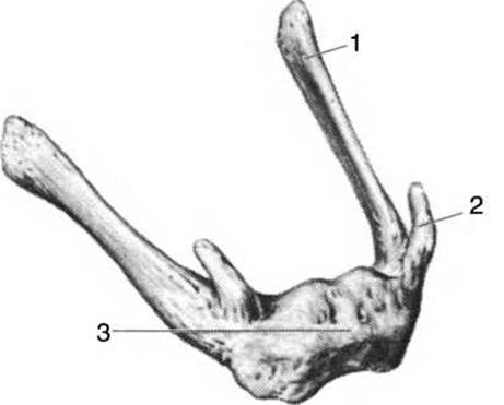
Fig. 58. Hyoid bone:
1 — greater horn; 2 — lesser horn; 3 — body
The hyoid bone (os hyoideum; Fig. 58) is an unpaired horseshoe-shaped bone located below the mandible, consisting of a body and two pairs of horns: greater and lesser horns. The hyoid bone does not articulate directly with other bones of the skeleton, but is connected to the Larynx by a broad ligament, and to the skull by ligaments extending from the styloid processes.
Last update: 08/08/2026
Editorial and Educational Adaptation: This material has been compiled based on the primary/original source text. The project team performed an editorial review, corrected technical inaccuracies, structured sections, and adapted the content for an educational format.
What was processed:
- elimination of formatting defects (OCR errors, structural breaks, corrupted characters);
- editorial organization of content;
- standardization of terminology in accordance with academic sources;
- verification of factual statements against the original source text.
All mentions of the author, publication year, and origin of the primary text have been preserved in accordance with the source.