MEDICAL BIOLOGY, HUMAN ANATOMY, PHYSIOLOGY AND PATHOLOGY - Ya.I. Fedonyuk 2010
BIOLOGY
CHAPTER 2. BIOGEOCENOTIC LEVEL OF LIFE ORGANIZATION AND HUMAN'S PLACE IN IT
2.4. MEDICAL HELMINTHOLOGY
Type Flatworms (Plathelminthes)
Class Tapeworms (Cestoidea)
Tapeworms are obligate endoparasites that inhabit the intestines of humans and vertebrates in their adult stage, causing diseases known as cestodiases. Their body is dorsoventrally flattened and ribbon-like. The anterior end bears a HEAD (scolex), followed by a neck and a strobila consisting of segments (proglottids). The head is equipped with Organs of attachment: suckers (usually 4) or bothria (slit-like attachment grooves). In many species, In addition to suckers, the apex of the scolex features a rostellum armed with cuticular hooks. The neck serves as the growth zone of the helminth, where new segments are continuously formed. These newly formed segments gradually push the older ones backward. Consequently, the youngest segments are located in the anterior part of the body, while the oldest, or mature, segments are situated at the rear and eventually detach from the strobila. The body is externally covered by a tegumentary-muscular sac, the Superficial layer of which—the tegument—contains various digestive Enzymes and an anti-proteolytic enzyme that prevents the parasite from being digested in the host's intestine. Beneath the tegument lie three layers of Muscles: an outer circular layer, an inner longitudinal layer, and a diagonal layer situated between them.
The Digestive System is completely absent. Nutrients present in the host's intestine are absorbed osmotically across the entire body surface. The tegument Cells secrete digestive enzymes that facilitate food Digestion and assimilation. The excretory system is of the protonephridial type. There are two main excretory canals running along the lateral sides of the strobila over its entire length, opening via excretory Pores in the terminal segment. In each proglottid, these longitudinal canals are connected by a transverse canal. The Nervous system is of the orthogenous (ladder-like) type. Circulatory and respiratory systems are lacking. Compared to other Organ Systems, the Reproductive System is extraordinarily developed and highly complex. Cestodes are hermaphroditic. Each segment contains repeating complexes of male and FEMALE REPRODUCTIVE ORGANS, which endows tapeworms with immense fecundity. In the youngest segments, located immediately behind the neck, the reproductive system is not yet developed; next, the Male reproductive organs appear, and as these proglottids are pushed further back, the female reproductive organs develop as well, rendering the segment hermaphroditic (or immature). Subsequently, in many species, part of the reproductive organs within the segments regresses, leaving only the Uterus filled with eggs containing onchospheres. Such a segment is termed mature; it can detach from the strobila and be expelled into the external environment. The Male Reproductive System consists of numerous Testes whose efferent ducts merge into a common vas deferens that opens through the male genital pore into the genital atrium. The distal end of the vas deferens Functions as a copulatory organ, the cirrus. The FEMALE REPRODUCTIVE SYSTEM comprises an Ovary, vitelline glands (vitellaria), Vagina, ootype, and uterus. The vagina is a specialized canal for receiving Male Germ Cells; one end connects to the ootype, while the other opens into the genital atrium adjacent to the male genital pore. In many cestode species, the uterus lacks an external opening. In such cases, as eggs accumulate, it enlarges progressively, eventually filling the entire segment, while other PARTS OF THE hermaphroditic reproductive system partially atrophy. The specific cestode species is often identified by The Structure of the uterus. Fertilization in tapeworms typically occurs between different proglottids of the same individual or between different individuals.
The life cycle is characterized by alternation of hosts and the presence of several larval stages. The definitive host is a vertebrate animal or human, whereas the intermediate host is predominantly vertebrate, though occasionally invertebrate. The developmental cycle of all cestodes invariably includes two larval stages: the onchosphere and the bladder worm (fin/cysticercus). The onchosphere, or first larval stage, develops within the egg while it is still inside the segment; it is spherical and bears six hooks. In the intermediate host's intestine, the onchosphere hatches from the egg, uses its hooks to penetrate the Blood Vessels, and is passively carried by the blood to various organs, where it transforms into the second larval stage—the metacestode (bladder worm).
Various types of metacestodes (larval forms) are recognized: cysticercus, coenurus, cysticercoid, echinococcus, and plerocercoid. As a rule, a metacestode is a fluid-filled bladder with one or more inverted scoleces inside.
The Development of metacestodes into the adult stage takes place in the intestine of the definitive host, where the scolex everts under the action of digestive juices and
attaches to the intestinal wall, while the bladder disintegrates.
Medical significance. Several species of tapeworms are causative agents of human cestodiases.
Pork tapeworm, or armed tapeworm (Taenia solium). In its adult stage, it causes taeniasis; in its larval stage (cysticercus), it causes cysticercosis.
Localization. The adult form parasitizes the Small Intestine, whereas the larval forms inhabit various organs, most frequently the muscles and the Central Nervous System.
Geographic distribution: ubiquitous wherever pig farming is practiced.
Morphophysiological characteristics. The body is ribbon-like, white, and 2-3 m in length.
The scolex is microscopic (2-3 mm) and bears 4 suckers and a double crown of hooks (hence the name "armed tapeworm"). An unsegmented neck is located immediately behind the scolex (Fig. 2.20).
Class="center">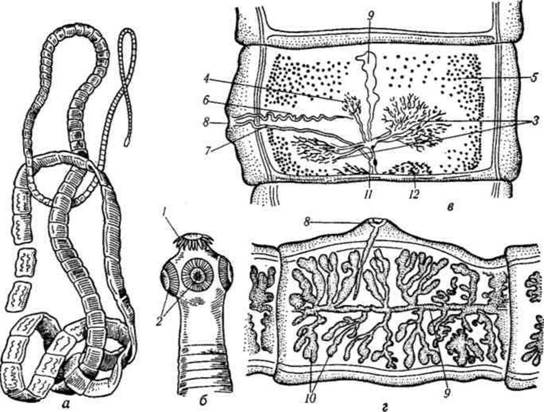
Fig. 2.20. Pork tapeworm (Taenia solium):
a - strobila; b - scolex; c - hermaphroditic proglottid; d - mature proglottid; 1 - hooks on the scolex; 2 - suckers; 3 - ovary; 4 - third (accessory) lobe of the ovary; 5 - testes; 6 - vas deferens; 7 - vagina; 8 - cirrus pouch; 9 - main uterine stem; 10 - lateral uterine branches;
The strobila contains up to 1,000 or more segments.
Hermaphroditic segments are square-shaped. The uterus is blind-ending (sac-like). The ovary has three lobes—two large ones and a small third (accessory) lobe, which serves as a diagnostic species feature. Situated beneath the ovary is the vitelline gland.
The testes are located in the lateral parts of the proglottid. The genital atrium is situated on the left side in one segment and on the right side in the next. In mature segments, the uterus forms 7-12 lateral branches on each side (a key diagnostic feature).
Life cycle. The pork tapeworm is a biohelminth. Its development involves a change of hosts (Fig. 2.21). The definitive host is exclusively human, while the intermediate host is a pig, and rarely, a human. A person suffering from taeniasis excretes mature segments containing eggs with onchospheres in their feces. Pigs rooting in sewage or garbage may ingest these segments or individual eggs. In the pig's Stomach, the hexacanth larva (onchosphere) hatches from the egg. Using its hooks, the onchosphere penetrates the blood vessels and is transported by the blood to various Internal Organs (Liver, muscles, Brain), where it transforms into a cysticercus (bladder worm) within 2 months.

Fig. 2.21. Life Cycle of the pork tapeworm (*Taenia solium*).
A bladderworm (cysticercus) can be seen with the naked eye. It is a small fluid-filled vesicle (up to 10 mm in diameter) with a single inverted scolex inside. Humans become infected by eating raw or insufficiently cooked pork contaminated with cysticerci. Under the action of digestive juices, the scolex everts from the cysticercus, attaches to the wall of the small intestine, and then proglottids begin to grow from the neck. After 2–3 months, the helminth reaches sexual maturity. Its lifespan in The Human Body is 10 years or more. Thus, the invasive stage for the intermediate host is the egg with an oncosphere, and for the definitive host, it is the cysticercus (bladderworm).
Pathogenic effect. Taeniasis is a biohelminthiasis and a zoonosis. The pathogenic impact is caused by mechanical damage, the consumption of the host's digested nutrients, and the Toxic effects of the parasite's Metabolic waste products. Digestive disorders, anemia, and general weakness are observed. The pork tapeworm is also dangerous because humans can act as an intermediate host upon accidental ingestion of eggs or As a result of autoinfection, when proglottids in patients with taeniasis enter The Stomach during vomiting. Here, oncospheres emerge from them and are carried by the bloodstream to various organs, where they develop into cysticerci. The localization of cysticerci in the brain is dangerous and can sometimes cause death, as well as in the visual organ, which can lead to blindness. The Treatment of cysticercosis is exclusively surgical.
Laboratory Diagnosis of taeniasis. Detection of mature segments in the feces of an infected person. The presence of 7–12 lateral uterine branches indicates taeniasis. The uterus has no excretory pore, which is why eggs are rarely found in the feces.
Laboratory diagnosis of cysticercosis. X-ray examinations, immunological reactions.
Prevention of taeniasis. Personal: do not consume pork that has not passed veterinary and sanitary inspection. Public: detection and treatment of infected individuals, especially those working in animal husbandry; veterinary and sanitary inspection of pork; protection of soil from contamination with human feces; indoor pig farming.
Prevention of cysticercosis. Personal: follow personal hygiene rules; public: health education work.
The beef tapeworm, or unarmed tapeworm (*Taeniarhynchus saginatus*), is the CAUSATIVE AGENT OF taeniorhynchiasis.
Localization: small intestine.
Morphophysiological characteristics. One of the largest human helminths, with a length of 4–7 m (some specimens reach up to 10 or even 18 m). Structurally, it is similar to the pork tapeworm (Fig. 2.22). It differs by the absence of hooks on the scolex (hence the name "unarmed") and the lack of a third (accessory) ovary lobe in the hermaphroditic proglottid (the ovary has only two lobes). In addition, the uterus in the mature proglottid has significantly more lateral branches (17–35). Mature proglottids, detaching from the strobila, can independently crawl out of the anus and move over the patient's body and underwear. Mature proglottids of the pork tapeworm do not have this ability.
Life cycle. The beef tapeworm is a biohelminth. Its development involves a change of hosts. The definitive host is exclusively humans, while the intermediate host is cattle. A person infected with taeniorhynchiasis excretes mature proglottids containing eggs with oncospheres in their feces.
If proglottids or eggs enter the digestive tract of cattle, the egg shell is digested, and the hexacanth

Fig. 2.22. Beef tapeworm (*Taeniarhynchus saginatus*):
a - general view; b - scolex; c - hermaphroditic proglottid; d - mature proglottid; 1 - ovary; 2 - vitelline gland; 3 - Mehlis' gland; 4 - uterus; 5 - Testis; 6 - vas deferens; 7 - vagina; 8 - cirrus pouch; 9 - genital atrium; 10 - excretory canal; 11 - nerve cord.
oncosphere penetrates the intestinal wall into blood vessels and is carried by the blood to various Organs of the host. Here, the oncosphere loses its hooks and transforms into a cysticercus. Cysticerci can live in muscles for years without developing further. Further development is possible only in the intestine of the definitive host, which is humans. Humans become infected by eating insufficiently cooked beef infected with cysticerci. In the human intestine, the scolex of the cysticercus everts, attaches to the wall of the small intestine, after which strobilation begins from the neck. Thus, the invasive stage for the intermediate host (cattle) is the egg with an oncosphere, and for the definitive host (human) is the cysticercus.
Pathogenic effect. In its clinical course, taeniorhynchiasis is similar to taeniasis.
Laboratory diagnosis. Detection of mature proglottids in the feces of an infected person. The uterus in a mature proglottid has 17–35 branches on each side. The uterus in the beef tapeworm is also blind-ended, so eggs are frequently not found in the patient's feces. If they are found (in scrapings from perianal folds), they are morphologically indistinguishable from the eggs of the pork tapeworm.
Prevention. Personal: do not consume meat that has not passed veterinary and sanitary inspection. Public: detection and treatment of patients; veterinary and sanitary inspection of beef; protection of soil from contamination with human feces; health education work.
The dwarf tapeworm (*Hymenolepis nana*) is the causative agent of hymenolepiasis.
Localization: human small intestine.
Geographical distribution: worldwide.
Morphophysiological characteristics. Its size is small compared to other cestodes: length is 1.5–2 cm, rarely up to 5 cm (hence the name "dwarf"). The scolex is pear-shaped, bearing four suckers and a retractable rostellum with hooks. The neck is long and thin (Fig. 2.23). The strobila consists of 100–200 proglottids. The genital pores of all proglottids are located on the same side. The posterior proglottids with a sac-like uterus are very fragile. Detaching from the strobila, they quickly disintegrate, and the eggs with oncospheres enter the lumen of the small intestine.
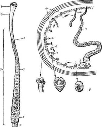
Fig. 2.23. Dwarf tapeworm (Hymenolepis nana):
a — general view; 1 — scolex; 2 — neck; 3 — body; 4 — immature proglottid; 5 — mature proglottid; 6 — gravid proglottid; b — life cycle in the human intestine: 1 — body of the adult worm; 2 — eggs; 3 — oncospheres emerged from eggs within the mucous membrane villi;
Life cycle. In the life cycle of the dwarf tapeworm, humans serve as both definitive and intermediate hosts, representing the sole source of infection. The parasite's eggs enter the environment along with feces and are immediately infective. Infection occurs via the oral route. Unwashed hands serve as the primary vehicle for transmitting the causative agent of hymenolepiasis. Once in the intestine, the eggs hatch into oncospheres, which penetrate the villi of the small intestine and develop into a cysticercus (cysticercoid). The latter is a small bladder-like structure with an invaginated head and a tail-like appendage.
The cysticercoid grows, destroys the villus, and drops into the intestinal lumen. Under METABOLISM/18.html">The Influence of digestive juices, the head of the cysticercoid everts, attaches to the wall of the small intestine, and proglottid growth begins.
An adult, sexually mature specimen forms within 14–15 days.
It is believed that autoinfection (the development of parasite eggs within the intestine without passing into the external environment) can occur in hymenolepiasis. Under such conditions, the infection persists for a prolonged period. A patient with hymenolepiasis who fails to observe personal hygiene rules may reintroduce their own parasites' eggs into the Mouth (autoinfection).
Pathogenic effect. Hymenolepiasis is a contagious biohelminthiasis and anthroponosis. The source of infection is an infected individual. The transmission mechanism is fecal-oral, occurring through the ingestion of eggs. Preschool-aged children are most commonly infected. The parasites can occur in large numbers (up to 1,500 specimens), which exerts a significant pathogenic effect on the host Organism, consisting of the destruction of intestinal villi and the toxic action of helminth metabolic products. Hymenolepiasis is characterized by headaches, abdominal pain, digestive and nervous system disorders, general weakness, and rapid fatigue. Children become fussy and irritable.
Laboratory Diagnostics. Detection of eggs in the feces of an infected person.
Prevention. Personal prevention involves strict adherence to personal hygiene rules, instilling hygienic habits in children, and washing hands before meals and after using the restroom. Public Prevention entails identifying and deworming infected individuals; isolating sick individuals from healthy ones in children's collectives; health education campaigns among parents and staff of childcare facilities; and rodent control, as it is believed humans may become infected with dwarf tapeworms from mice and rats.
Echinococcus (Echinococcus granulosus) is the causative agent of echinococcosis.
Localization. In humans, unlike other cestodes, it parasitizes in the larval (cysticercus) stage, affecting the liver, Lungs, brain, and tubular bones, though it can potentially occur in any organ.
Geographical distribution is ubiquitous, yet uneven. In Ukraine, it is predominantly found in the southern regions.
Morphophysiological characteristics. The body of the adult parasite is 3–6 mm in length. The scolex bears 4 suckers and a rostellum with a double crown of hooks. The neck is short. The strobila consists of 3–4 proglottids (Fig. 2.24, a). The penultimate proglottid is hermaphroditic, while the last one is gravid, containing a uterus with lateral branches filled with eggs containing oncospheres.

Fig. 2.24. Echinococcus and Alveococcus: a — Echinococcus; b — Alveococcus; 1 — scolex; 2 — neck;
3 — hermaphroditic proglottids; 4 — gravid proglottid; 5 — uterus filled with eggs.
Life cycle. Echinococcus is a biohelminth. Its development involves alternating hosts. Adult specimens parasitize the small intestine of dogs, wolves, and jackals, which serve as definitive hosts. Intermediate hosts include humans, cattle, small ruminants, pigs, camels, rabbits, and many other herbivorous mammals. The terminal proglottid detaches from the strobila and is eliminated with feces or actively crawls out of the anal opening of the definitive host (predominantly dogs), moving actively through the fur while releasing eggs. Proglottids shed with feces crawl onto grass. Domestic herbivores consume the grass and concurrently ingest Echinococcus eggs (Fig. 2.25).
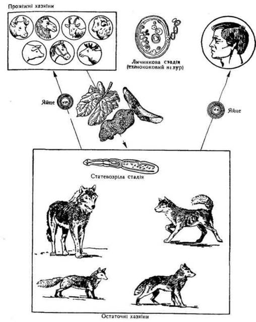
Fig. 2.25. Life cycle of Echinococcus.
In the intestine of intermediate hosts, the egg hatches an oncosphere, which penetrates blood vessels. Via the portal vein, it reaches the liver (the most frequent site of localization). Oncospheres that bypass the liver are carried through the right side of The Heart into the lungs. Frequently, oncospheres are carried by the bloodstream to the brain and other organs. Here, the oncosphere develops into a unilocular, or hydatid, cyst (larval stage). The latter represents a complexly structured bladder. The bladder wall consists of two layers: an inner (germinal) layer and an outer (cuticular) layer.
A dense fibrous capsule forms over the outer layer. The cyst cavity is filled with fluid. Brood capsules containing scolices develop from the germinal layer. Daughter cysts, which also possess brood capsules with scolices, may be present within the primary cyst cavity. The larva grows slowly and can reach large sizes—larvae weighing up to 64 kg have been described in animals, while in humans they can grow to the size of a newborn's head. Larvae persist in the intermediate host's body for years. For further development, they must reach the definitive host's intestine. Infection of dogs and other definitive hosts occurs through the ingestion of organs of intermediate hosts affected by echinococcosis. In the intestine of the definitive host, a single larva gives rise to A large number of tapeworm forms. Infective stages: for definitive hosts — the larva (cysticercus); for intermediate hosts — eggs containing oncospheres.
Humans most frequently contract echinococcosis due to poor personal hygiene when interacting with infected dogs whose fur carries eggs. Unwashed hands transfer the eggs to the mouth. Infection can also occur from sheep, to whose wool Echinococcus eggs adhere from infected guard dogs. The causative agent of echinococcosis is likewise transmitted via unwashed vegetables and garden produce contaminated with the feces of infected dogs. Humans act as a biological dead end in the Echinococcus life cycle, because after human death, the larva is generally not transmitted to animals and simply perishes.
Echinococcosis is a biohelminthiasis, a naturally focal infection, and a zoonosis. The initial signs of the disease appear long after infection, once the hydatid cysts have attained a certain size. Disease manifestations depend on the cyst's localization and the parasite's developmental phase. The pathogenic effect is driven by the toxicity of the cyst fluid and its mechanical impact on Tissues, which impairs organ function. Treatment is exclusively surgical. Rupture of a hydatid cyst is extremely hazardous, as the toxic fluid it contains can induce Shock and instant death. Furthermore, such a rupture can seed the cavity with scolices, leading to multiple echinococcosis. Occasionally, dissemination results from the spilling of cyst fluid containing scolices during the surgical management of echinococcosis.
Laboratory diagnostics. The diagnosis is established based on immunological reactions. X-ray examinations are also utilized. To identify infected dogs, their feces are examined for the presence of Echinococcus eggs.
Prevention. Personal prevention — washing hands after contact with dogs and observing personal hygiene rules. Public prevention — control of stray dogs; detection and deworming of infected working dogs, particularly those guarding sheep; veterinary and sanitary inspection of cattle, small ruminant, and pig carcasses at slaughter sites; prohibiting the feeding of echinococcosis-affected animal organs to dogs, as such organs must be destroyed.
Alveococcus (Alveococcus multilocularis) is the causative agent of alveolar echinococcosis (alveococcosis).
Localization. The localization is largely similar to that of echinococcosis, though the liver is primarily and predominantly affected, while involvement of other organs is rare.
Geographical distribution. Foci of alveolar echinococcosis are reported significantly less frequently than echinococcosis. Morphophysiological characteristics. The strobila form of Alveococcus resembles the tapeworm Echinococcus (Fig. 2.24, b). They differ in the number of hooks on the scolex, the shape of the uterus (in Alveococcus it is globular, lacking lateral branches; in Echinococcus, it features lateral branches), and THE POSITION OF the genital pore, which is located in the anterior part of the lateral margin of the proglottid in Alveococcus and in the posterior part in Echinococcus. Structurally, the larval stage (metacestode or bladder worm) of Alveococcus differs sharply from that of Echinococcus. The Alveococcus bladder worm is a cluster of small vesicles united within a common Connective Tissue capsule (stroma). The vesicles contain no fluid; they undergo exogenous budding and progressively infiltrate surrounding tissues like malignant tumors, forming metastases.
Life cycle. Alveococcus is a biohelminth. Its development involves a change of hosts. Definitive hosts include foxes, arctic foxes, wolves, dogs, and occasionally cats; intermediate hosts are murid rodents and, rarely, humans. The life cycle is similar to that of Echinococcus. Eggs containing oncospheres are released into the environment with the feces of definitive hosts. Humans become infected by consuming forest berries contaminated with Alveococcus eggs; by handling the pelts of infected foxes, arctic foxes, or dogs; or through contact with infected dogs or objects contaminated with dog feces containing Alveococcus eggs.
Pathogenic effect. Alveococcosis is a biohelminthosis, a natural focal disease, and a zoonosis. In humans, the parasite's primary localization is predominantly the liver. Hepatic alveococcosis remains asymptomatic for a long time (sometimes years). Pain emerges at later stages. The disease is characterized by a malignant clinical course. Metastatic nodes may develop in the lungs and brain. Treatment is exclusively surgical.
Laboratory diagnostics. Immunological tests are used to establish the diagnosis. Diagnosis is usually made at advanced stages, when surgical intervention is complicated or impossible.
Prevention. Personal prevention involves adhering to personal hygiene rules when handling dogs and Processing the pelts of foxes, arctic foxes, and dogs. Public prevention includes Setting up specialized facilities for skinning pelts and conducting sanitary and educational outreach.
Broad fish tapeworm (Diphyllobothrium latum) is the causative agent of diphyllobothriasis.
Localization. Small intestine.
Geographical distribution. The causative agent of diphyllobothriasis occurs among populations engaged in fishing, particularly in Karelia, the Baltic states, and along the banks of the Volga and Dniester rivers.
Morphophysiological characteristics. This is the largest human helminth (8–10 m in length, with individual specimens reaching up to 20 m). The scolex is elongated and bears two longitudinal sucking grooves, known as bothria, instead of suckers. The proglottids have a characteristic shape: their width is several times greater than their length. The structure and arrangement of the reproductive organs differ from those of tapeworms of the genus Taenia, which is of diagnostic significance. The genital pore is located not laterally, but on the ventral surface of the proglottid near its anterior margin. The vitelline glands (vitellaria) are situated in the lateral parts of the proglottid, ventral to the testes. The uterus has its own opening through which eggs are expelled as they are produced, and it lacks lateral branches. It is coiled into loops resembling a rosette (Fig. 2.26).

Fig. 2.26. Broad fish tapeworm (Diphyllobothrium latum):
a — body; b — scolex (showing bothria - 1); c — hermaphroditic proglottid; d — mature proglottids; 2 — uterus; 3 — vitelline gland; 4 — uterine loops; 5 — cirrus sac; 6 — ovary; 7 — Mehlis' gland; 8 — testes.
Life cycle. The broad fish tapeworm is a biohelminth. Its development involves intermediate hosts (Fig. 2.27).
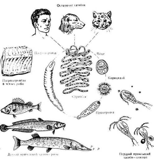
Fig. 2.27. Life cycle of the broad fish tapeworm.
Definitive hosts include humans and carnivorous mammals (cats, foxes, arctic foxes, dogs, bears). There are two intermediate hosts: the first is the cyclops copepod, and the second comprises freshwater fish (pike, burbot, pike-perch, etc.). Definitive hosts excrete eggs with their feces, which must reach Water. A ciliated larva, known as a coracidium, hatches from the egg and swims freely in the water for a time until swallowed by a cyclops. Inside the cyclops, the oncosphere emerges from the coracidium, penetrates the intestinal wall into the body cavity using its hooks, and develops into a procercoid larva. The cyclops is then eaten by a fish. In the stomach of the second intermediate host, the cyclops is digested, and the procercoid penetrates the muscles, transforming into a plerocercoid. The plerocercoid is a white, worm-like larva about 6 mm long, bearing two sucking grooves on its anterior end. If an infected fish is consumed by another predatory fish, the plerocercoid remains viable. Humans become infected by eating raw, sun-dried, or insufficiently thermally processed fish containing plerocercoids. In the definitive host's body, the plerocercoid attaches to the intestinal mucosa via its bothria and develops into the sexually mature stage. The broad fish tapeworm can live in the human body for up to 28 years. Infective stages: for the cyclops, the coracidium; for the fish, the procercoid; for the definitive host (humans, etc.), the plerocercoid.
Pathogenic effect. Diphyllobothriasis is a biohelminthosis and a natural focal disease.
The parasite damages the intestinal mucosa with its bothria, leading to tissue necrosis. Accumulations of parasites can cause intestinal obstruction. Patients frequently develop severe anemia associated with a deficiency of cyanocobalamin (vitamin B12), which the parasite selectively adsorbs from the intestinal contents onto its surface.
Laboratory diagnostics. Detection of eggs in the feces of an infected person. The eggs are broad, oval, with a smooth, transparent, double-contoured shell, yellowish or gray in color, featuring an operculum (lid), and measuring up to 70 µm. The diagnosis is frequently confirmed by finding strobila fragments of varying lengths in the feces. A broad strobila with short proglottids and a centrally located
rosette-shaped uterus is indicative of diphyllobothriasis.
Prevention. Personal prevention involves avoiding raw or undercooked fish and caviar. Public prevention entails the detection and dehelminthization of infected individuals; sanitary improvement of watercraft and settlements; protection of water bodies from contamination with human feces; sanitary control over compliance with fish processing regulations; and sanitary and educational outreach.
Last update: 08/08/2026
Editorial and Educational Adaptation: This material has been compiled based on the primary/original source text. The project team performed an editorial review, corrected technical inaccuracies, structured sections, and adapted the content for an educational format.
What was processed:
- elimination of formatting defects (OCR errors, structural breaks, corrupted characters);
- editorial organization of content;
- standardization of terminology in accordance with academic sources;
- verification of factual statements against the original source text.
All mentions of the author, publication year, and origin of the primary text have been preserved in accordance with the source.