HUMAN MEDICAL BIOLOGY, ANATOMY, PHYSIOLOGY AND PATHOLOGY - Ya.I.Fedoniuk 2010
BIOLOGY
CHAPTER 2. BIOGEOCENOTIC LEVEL OF ORGANIZATION OF LIFE AND HUMAN PLACE IN IT
2.4. MEDICAL HELMINTHOLOGY
Phylum Flatworms (Plathelminthes)
Class Trematodes (Trematoda)
The Class comprises about 3,000 species. All trematodes are parasitic organisms. The body is unsegmented, mostly leaf-shaped, and flattened in the dorsoventral direction. A characteristic feature is the presence of suckers, typically two: oral and ventral (hence the name of the class). The suckers serve as Organs of attachment of the parasite to the host's body. The body wall is a Skin-muscular sac consisting of a tegument (external covering) and Muscles fused with it. The musculature is represented by three layers: circular, diagonal, and longitudinal. The Digestive System has an ectodermal foregut (Mouth, Pharynx, Esophagus) and two branches (branched or unbranched) of the endodermal midgut that end blindly. There is no anus. Undigested food residues are expelled through the mouth. The excretory system is of the protonephridial type. The Nervous system has a typical Structure for Flatworms (orthogon). It consists of a circumpharyngeal nerve ring from which three pairs of nerve trunks extend, with the lateral ones being the most developed. The nerve trunks are interconnected by commissures. Overall, the nervous system resembles a lattice.
Almost all trematodes are hermaphrodites. The Male Reproductive System consists of two Testes, two vas deferens merging into an ejaculatory duct, and a copulatory organ (cirrus). The cirrus is enclosed in a cirrus sac located above the ventral sucker. The FEMALE REPRODUCTIVE SYSTEM is complex. The Ovary, vitellaria, and seminal receptacle open into the ootype, where Fertilization and the final formation of eggs take place. Nutrient material for the eggs, produced by the vitellaria and specialized glands (Mehlis' glands), also flows here. From the ootype, the eggs move into the Uterus and are expelled outward through the genital pore located next to the male opening. In some trematodes, fertilization occurs in the seminal receptacle. Fertilization is usually cross-fertilization. Self-fertilization is observed less frequently.
The life cycle involves alternation of hosts and several larval generations. The definitive hosts are vertebrates and humans, while the intermediate host is obligatorily a mollusk. Some trematodes additionally have a second intermediate host. A characteristic feature of the life cycle is the reproduction of larval stages via parthenogenesis (without fertilization). The sexually mature form of trematodes is called the marita.
Medical significance. Several species of trematodes parasitize humans. Diseases caused by trematodes are called trematodiases.
The Liver fluke (Fasciola hepatica) is the CAUSATIVE AGENT OF fascioliasis.
Localization: Bile ducts of the liver, Gallbladder, Pancreas.
Morphophysiological characteristics. Size 3-5 cm, anterior end elongated into a conical shape (Fig. 2.13).
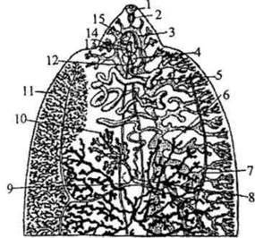
Fig. 2.13. Liver fluke (anterior third of the body from the ventral side):
1 - oral sucker; 2 - pharynx; 3 - intestine; 4 - ventral sucker; 5 - uterus; 6 - testes; 7 - vitelline duct; 8 - Mehlis' gland; 9 - Laurer's canal; 10 - ovary; 11 - vitelline ducts; 12 - vas deferens; 13 - Seminal Vesicle; 14 - cirrus sac; 15 - cirrus. The vitelline ducts are shown only on the left side, the intestine on the right.
The ventral sucker is located close to the oral one.
Directly behind the ventral sucker lies the multilobed uterus, behind which is the ovary, and the entire middle part of the body is occupied by the testes. The two intestinal branches have numerous ramifications. The ovary and testes are also branched.
Numerous vitellaria are located along the sides of the body. An ootype and Mehlis' gland are present.
Life cycle. The liver fluke is a biohelminth.
Development involves alternation of hosts (Fig. 2.14). Definitive hosts are herbivorous mammals, large and small ruminants, horses, pigs, rabbits, and occasionally humans. There is a single intermediate host—the mud snail Limnea (Galba) truncatula. With the feces of the definitive host, the eggs are released into the external environment and must reach Water. Here, a ciliated larva—the miracidium—develops inside the egg. The egg operculum opens in the light, and the miracidium leaves the egg membranes. For some time, it swims freely in the water, then actively penetrates the body of the mollusk, enters the liver, and transforms into a sporocyst, inside of which rediae develop parthenogenetically (without fertilization) from Germ Cells. The rediae emerge from the sporocyst and continue development. From the germ Cells of the rediae, the next generation of larvae—cercariae—is formed parthenogenetically. The cercaria has a long muscular tail that provides forward movement. The cercaria leaves the mollusk, swims actively in the water, then attaches to aquatic plants or other objects, discards its tail, becomes encysted, and transforms into an adolescaria. Livestock eat the plants and thereby swallow the adolescariae. Humans become infected by drinking raw water from open, especially stagnant, bodies of water or by consuming unwashed vegetables and greens (most commonly lettuce) irrigated with water from a river or lake where adolescariae are present. In the human intestine, the membrane of the adolescariae dissolves, and young flukes emerge, migrating into the host's liver.
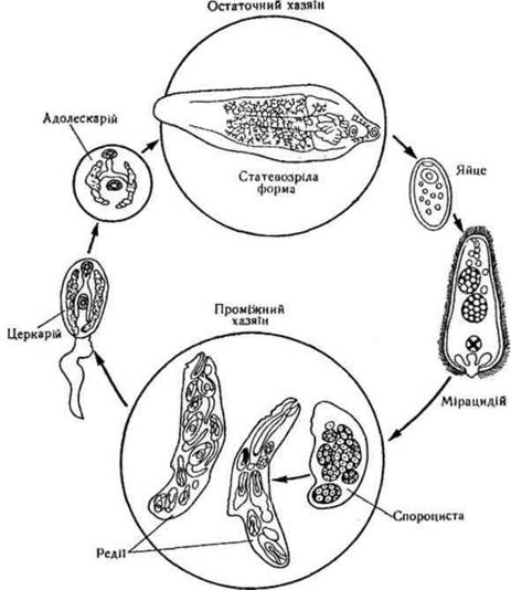
Fig. 2.14. Life Cycle of the liver fluke (Fasciola hepatica).
Thus, in the life cycle of Fasciola, the invasive stage for the intermediate host (mollusk) is the miracidium, and for the definitive hosts, it is the adolescaria. Pathogenic action. Fascioliasis is a biohelminthiasis, a natural focal disease, and a zoonosis.
Flukes exert a mechanical effect on the host; their metabolic products are toxic and have an allergic effect. With prolonged infection
- inflammation of the bile ducts accompanied by fibrous tissue proliferation, and liver cirrhosis.
Laboratory Diagnostics involves identifying eggs in the feces of an infected person. The eggs are large (135 x 80 µm), oval-shaped, yellowish-brown, and feature an operculum at one of the poles. Eggs may also be detected in healthy individuals if they have consumed the liver of fasciola-infected animals (transit eggs). To avoid misdiagnosis, liver should be eliminated from the patient's diet and the analysis repeated.
Prevention. Personal measures: avoid drinking raw water from open water bodies, especially stagnant ones, and thoroughly wash vegetables consumed raw. Public and veterinary measures include the Treatment of infected animals (deworming), pasture rotation, and health education.
Lancet liver fluke (Dicrocoelium lanceatum) is the causative agent of dicrocoeliasis.
Localization: bile ducts of the liver, gallbladder, and pancreas.
Geographic distribution: ubiquitous.
Morphophysiological characteristics: The body is lanceolate in shape and measures 8-10 mm in length. It possesses two suckers—oral and ventral. Two unbranched intestinal branches extend along the sides of the body to the posterior end, ending blindly. Two rounded testes are located behind the ventral sucker. The female reproductive system consists of a small, rounded ovary situated posterior to the testes, paired vitelline glands lying along the sides of the body, and a well-developed uterus occupying the posterior part of the body (Fig. 2.15, a).

Fig. 2.15. STRUCTURE OF THE lanceolate liver fluke (Dicrocoelium lanceatum) and the cat liver fluke (Opisthorchis felineus)
Life cycle. The lancet liver fluke is a biohelminth. Its life cycle involves a change of hosts.
Definitive hosts include small and large ruminants, other herbivorous mammals, and occasionally humans. There are two intermediate hosts: the first consists of land snails of the genera Helicella and Zebrina, and the second is the ant. Eggs are shed into the environment with the feces of the definitive host and must be ingested by a snail. The egg already contains a miracidium. Inside the snail, the miracidium escapes from the egg membranes, penetrates the liver, and transforms into a first-generation sporocyst, which gives rise to second-generation sporocysts, and subsequently cercariae. The redia stage is absent. Cercariae leave the sporocysts and migrate to the Lungs of the snail, where they encyst, adhering together in groups to form collective cysts.
The latter are discharged to the exterior along with mucus and deposit on plants. When eaten by the second intermediate host
(ants of the genus Formica), each cercaria sheds its membrane within the ant and develops into the next larval stage—the metacercaria.
Definitive hosts become infected by ingesting ants containing metacercariae. Humans become infected accidentally, also through the ingestion of ants infested with metacercariae. Invasive stages: for the first intermediate host (snail) — an egg containing a miracidium; for the second intermediate host (ant) — a cercaria; for the definitive hosts — a metacercaria.
Pathogenic effect. Dicrocoeliasis is a biohelminthiasis, a natural focal disease, and a zoonosis. It is accompanied by liver dysfunction and inflammatory processes that may lead to cirrhosis. The liver is painful and enlarged. Obstruction of the bile ducts can cause jaundice.
Laboratory diagnostics: Detection of eggs in the feces of an infected person. The eggs are elongated, asymmetric, with an operculum at the narrowed end, and range in color from yellowish to dark brown. Transit eggs are possible.
Prevention. The main measures are veterinary: treatment of infected animals (deworming) and pasture rotation.
Cat liver fluke, or feline liver fluke (Opisthorchis felineus), is the causative agent of opisthorchiasis. The disease was first described in Siberia in 1891 by K.N. Vinogradov.
Localization: bile ducts of the liver, gallbladder, and pancreas.
Geographic distribution: along the riverbanks of Western Siberia, particularly the Ob and Irtysh river basins. Isolated foci have been recorded in the Baltic states and the basins of the Kama, Volga, Dnieper, and Southern Bug rivers.
Morphophysiological characteristics: The anterior end of the body is narrower than the posterior. Size is 8-13 mm. The intestinal branches are unbranched and terminate short of the posterior end of the body. The uterus occupies the middle part of the body. Posterior to it lie the rounded ovary and bean-shaped seminal receptacle. In the posterior part of the body are two rosette-shaped testes, between which the S-shaped canal of the excretory system is visible. The vitelline glands are located between the intestinal canals and the body margin (Fig. 2.15, b).
Life cycle (Fig. 2.16).
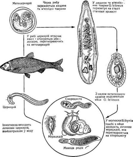
Fig. 2.16. Life cycle of the cat liver fluke.
The cat liver fluke is a biohelminth with development involving host alternation. Definitive hosts include humans, cats, dogs, foxes, otters, muskrats, and other fish-eating mammals. There are two intermediate hosts: the first is the freshwater snail Bithynia leachi, and the second consists of cyprinid fish (ide, roach, common carp, Caspian roach, etc.).
The parasite's eggs are excreted with the definitive host's feces into the external environment, where they must enter water and be ingested by a mollusk. Inside the mollusk, the miracidium hatches from the egg, penetrates the liver, and transforms into a sporocyst. Rediae develop parthenogenetically within the sporocyst, and cercariae subsequently develop inside the rediae, also parthenogenetically.
The latter emerge from the mollusk into the water, swim freely, and then attach to a fish or are swallowed by it, actively penetrating its subcutaneous tissue and muscles. Here they encyst and develop into metacercariae. Humans become infected by consuming insufficiently cooked, raw, or dried fish containing metacercariae. Once inside the definitive host, the larvae reach the digestive tract, shed their membranes under the action of digestive juices, and migrate to the gallbladder and liver, where they mature into adults.
Infective stages: for the first intermediate host (mollusk) - the egg containing the miracidium; for the second intermediate host (cyprinid fish) - the cercaria; for the definitive host (humans, cats, etc.) - the metacercaria.
Pathogenic effect. Opisthorchiasis is a biohelminthiasis, a natural focal disease, and a zoonosis. The causative agent is particularly prevalent in regions where eating raw, flash-frozen fish is a culinary tradition (e.g., Siberia). Early-stage opisthorchiasis manifests as allergies, fever, liver pain, and eosinophilia. Heavy parasite burdens lead to bile stasis and liver cirrhosis. Complications include biliary Peritonitis, primary liver Cancer, or Pancreatic Cancer. In severe cases, death may occur.
Laboratory diagnostics. Detection of eggs in feces or duodenal contents of an infected person. The eggs are very small (23-34 x 10-19 µm), oval-shaped, slightly asymmetrical, tapered at the anterior pole, and yellowish in color. The tapered pole features an operculum, while the opposite pole has a distinct knob; the shell is thick and double-contoured, with a finely granular content and a developing miracidium inside.
Prevention. Personal: avoid consuming raw, undercooked, or underfried freshwater fish. Public: supervision of fish Processing and cooking technologies, identification and treatment (deworming) of infected individuals, protection of water bodies from fecal contamination, avoiding feeding raw fish to domestic dogs and cats, and health education campaigns.
Lung fluke (Paragonimus ringeri, syn. P. westermani) is the causative agent of paragonimiasis.
Localization - lungs.
Geographical distribution - East Asia, Russian Far East.
Morphophysiological characteristics. The body is ovoid, reddish-brown, covered with tiny spines, and measures 7.5-12 x 4-6 mm (Fig. 2.17).
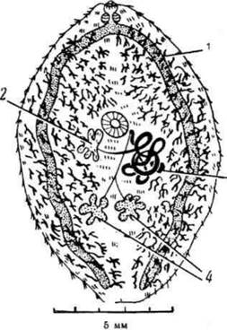
Fig. 2.17. Lung fluke (Paragonimus ringeri):
1 - vitellaria; 2 - ovary; 3 - uterus; 4 - testes.
Life cycle. The lung fluke is a biohelminth. Its development involves a change of hosts. Definitive hosts include humans, cats, dogs, and pigs. There are two intermediate hosts: the first comprises freshwater
Mollusks of the genus Melania, and the second comprises freshwater crayfish and crabs. The parasite's eggs are released into the environment with the definitive host's sputum and feces. For further development, they must reach water. A miracidium hatches from the egg and actively penetrates the mollusk, where larval stages successively develop: sporocysts, rediae, and cercariae. The cercaria leaves the mollusk and actively penetrates the second intermediate host (freshwater crayfish and crabs), where it encysts into a metacercaria. Humans become infected by eating insufficiently cooked freshwater crayfish and crabs infected with metacercariae.
Infective stages: for the first intermediate host (mollusk) - the miracidium; for the second intermediate hosts (freshwater crayfish and crabs) - the cercaria; for the definitive hosts - the metacercaria.
Pathogenic effect. Paragonimiasis is a biohelminthiasis, a natural focal disease, and a zoonosis. In the lungs, paragonimuses cause inflammation, hemorrhages, and The formation of cystic cavities. This leads to a cough with sputum and Blood-tinged streaks, which may resemble tuberculosis. Extrapulmonary localization of the parasite also occurs. Cerebral paragonimiasis often results in blindness, paralysis, seizures, and mental disorders.
Laboratory diagnostics. Detection of eggs in sputum or feces. The eggs are relatively large (0.1 mm), golden-brown, and operculated.
Prevention. Personal: avoid consuming raw freshwater crayfish and crabs. Public: protection of water bodies from fecal contamination, and health education. Clonorchis sinensis is the causative agent of clonorchiasis. Localization - intrahepatic bile ducts, gallbladder, and pancreas. Geographical distribution - Far East, China, Japan, Korea.
Morphophysiological characteristics and life cycle are very similar to those of the cat liver fluke. Definitive hosts: humans, dogs, cats, pigs, and other mammals. First intermediate host: freshwater mollusks. Second intermediate host: fish (primarily cyprinids) and freshwater crustaceans, consumption of which infects definitive hosts. Humans contract the infection by eating insufficiently cooked fish and freshwater crustaceans infected with Clonorchis metacercariae.
Pathogenic effect. Clonorchiasis is a biohelminthiasis, a natural focal disease, and a zoonosis. Parasite accumulations cause bile stasis and liver cirrhosis. The lifespan in The Human Body can reach 25-40 years.
Laboratory diagnostics. Detection of eggs in the feces of an infected person. The eggs are virtually indistinguishable from those of the cat liver fluke.
Prevention. Personal: avoid eating insufficiently cooked fish and freshwater crustaceans. Public: identification and treatment of infected individuals, protection of water bodies from fecal contamination, quality control in fish product preparation, avoiding feeding raw fish to domestic dogs and cats, and health education campaigns.
Nanophyetus salmincola schikhobalowi is the causative agent of nanophyetiasis.
Localization - Small Intestine.
Geographical distribution: The Far East (Amur River basin), northern part of Sakhalin Island. Morphophysiological characteristics: The smallest human trematode, measuring 0.6 × 0.4 mm, with a nearly round shape (Fig. 2.18, a).
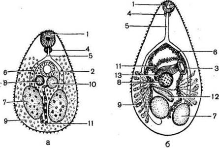
Fig. 2.18. Nanophyetus salmincola schikhobalowi, Metagonimus yokogawai
a) Nanophyetus salmincola schikhobalowi; 6) Metagonimus yokogawai: 1
- oral sucker; 2 - ventral sucker; 3 - genital sucker; 4 - pharynx; 5
- esophagus; 6 - intestine; 7 - testes; 8 - ovary; 9 - vitellaria; 10
- genital bursa; 11 - uterus with eggs; 12 - seminal receptacle; 13 - seminal vesicle.
Life cycle: Definitive hosts include humans, cats, dogs, and various wild mammals. Eggs are excreted with the definitive host's feces. There are two intermediate hosts: the first is freshwater snails, and the second is freshwater fish. Humans become infected by consuming insufficiently cooked fish infected with nanophyetid larvae.
Pathogenic effect: Nanophyetiasis is one of the most common helminthiases in the Far East. The disease manifests with digestive disorders.
Laboratory Diagnosis: Detection of eggs in the patient's feces. The eggs are oval, gray or yellowish, operculated, and very similar to the eggs of the broad fish tapeworm.
Prevention: Same as for opisthorchiasis.
Metagonimus yokogawai is the causative agent of metagonimiasis.
Localization: Small intestine.
Geographical distribution: Japan, China, South Korea, Indonesia, and the Amur River region. In Ukraine, it is found in the basins of the Dnipro, Dniester, and Danube rivers.
Morphophysiological characteristics: Length 1-2.5 mm, width 0.4-0.7 mm. The body is covered with minute spines. The oral sucker is significantly smaller than the ventral sucker. The intestinal branches extend to the posterior end of the body, where two round testes are located. The vitellaria are situated laterally in the posterior third of the fluke (Fig. 2.18, b).
Life cycle.
Metagonimus is a biohelminth.
Definitive hosts include humans, dogs, cats, foxes, pelicans, cormorants, and others. There are two intermediate hosts. With the definitive host's feces, the egg is released into the environment and must reach water. Here, a miracidium hatches and penetrates the first intermediate host—a snail of the genus Melania—where it develops to the cercaria stage. Cercariae emerge into the water and actively penetrate the second intermediate host—various species of cyprinid and salmonid fish—transforming into metacercariae in their muscles, scales, and fins. Humans become infected by consuming undercooked, insufficiently fried, raw, or dried fish infected with Metagonimus metacercariae. Invasive stages: miracidium for the snail, cercaria for fish, and metacercaria for humans and other definitive hosts.
Pathogenic effect: Metagonimiasis is a biohelminthiasis, a natural focal disease, and a zoonosis. It is accompanied by intestinal disorders. The larvae burrow into the intestinal mucosa, causing inflammatory processes.
Laboratory diagnosis: Detection of eggs in the patient's feces. The eggs are light brown, lemon-shaped, with an operculum at one pole and a small knob at the opposite pole; dimensions are 26-28 × 15-17 µm.
Prevention: Individual measures include avoiding raw, dried, and insufficiently cooked fish. Public measures include protecting water bodies from fecal contamination and conducting health education.
Blood flukes, or schistosomes, are the causative agents of Schistosomiasis.
Localization: Lumen of Blood Vessels, typically Veins.
Geographical distribution: Countries with tropical and subtropical climates.
Morphophysiological characteristics: Unlike other trematodes, schistosomes are dioecious. The body is narrow and cylindrical. The male is significantly shorter and broader than the female. Along his ventral side runs a groove—the gynaecophoric canal—in which the female resides during fertilization. The two intestinal caeca unite at the middle of the body into a single unpaired trunk that ends blindly near the posterior end of the parasite.
Life cycle. The definitive hosts are humans and various mammals, while the intermediate hosts are freshwater mollusks. The life cycle includes two generations of sporocysts (First and Second order); rediae are absent. The invasive stage for definitive hosts is the cercaria. Infection occurs through the active penetration of cercariae via the skin or mucous membranes upon contact with water. The eggs feature a spine. Parasitic species affecting humans include Schistosoma haematobium, Schistosoma mansoni, and Schistosoma japonicum (Fig. 2.19).
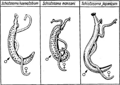
Fig. 2.19. Schistosomes. The male possesses a specialized canal containing a long, slender female.
Schistosoma haematobium is the causative agent of urogenital schistosomiasis (bilharziasis).
Localization: VEINS OF THE intestine and the Urogenital System.
Geographical distribution: Africa, the Middle East, and Central and South America.
Morphophysiological characteristics. The male measures 10-15 mm in length, and the female up to 20 mm. The body is covered with spines (Fig. 2.19).
Life cycle. Definitive hosts include humans and monkeys. Intermediate hosts are snails of the genera Bulinus and Planorbis. Mature individuals reproduce primarily in the veins of the Urinary Bladder. The fertilized female leaves the gynaecophoric canal of the male and deposits eggs in small venules. The egg, bearing a sharp terminal spine, punctures the vessel wall, enters the cavity of the urinary bladder, and is excreted with urine. For further development, the egg must reach water, where it hatches a miracidium that penetrates the intermediate host—a snail—within which two generations of sporocysts and cercariae develop. Humans become infected through exposure to water. Cercariae actively penetrate the skin during swimming, washing clothes, or working in rice paddies. Via the lymphatic and blood vessels, cercariae pass through the right ventricle—along with the long, slender female—into the lungs and subsequently into the hepatic portal veins. Adult forms migrate to the mesenteric veins, intestinal walls, and urinary bladder. Infection can also occur by drinking water containing cercariae. Within the host Organism, they can live for up to 40 years.
Pathogenic effects. Punctate hemorrhages and papules appear at the site of cercarial penetration, accompanied by skin itching and tissue edema. The eggs, armed with sharp spines, damage the Tissues of infected organs, leading to inflammatory processes and ulceration.
Laboratory diagnosis. Detection of eggs in urine. Parasite eggs appear in the urine predominantly during the hottest hours of the day when the probability of entering water is highest. This must be considered during diagnosis: schistosome eggs are generally not found in a patient's morning urine.
Prevention. Personal: in schistosomiasis endemic foci, avoid swimming in infected water bodies, drinking raw water, and washing with unfiltered water. Public: protecting water bodies from urine contamination, eradicating snails, and conducting health education campaigns.
Schistosoma mansoni is the causative agent of intestinal schistosomiasis.
Localization: mesenteric veins, intestine, and the portal Venous system.
Geographical distribution: Africa and South America (especially Brazil).
Morphophysiological characteristics. Structurally similar to Sch. haematobium, but slightly smaller: the male is up to 10 mm in length, and the female up to 15 mm. The body is covered with somewhat larger spines (Fig. 2.19). The egg features a prominent spine located laterally rather than at the pole.
Life cycle. The definitive host is humans, and intermediate hosts are snails of the genera Planorbis and Bulinus. Fertilized females lay eggs in the small venules of the intestine, from where they pass into the intestinal lumen and are expelled with feces.
Pathogenic effects. The eggs damage the intestinal wall with their spines, leading to ulceration and polypoid growths. A severe complication in the late Stages of the disease is liver Damage caused by schistosome eggs carried to the organ.
Laboratory diagnosis. Detection of eggs in feces.
Prevention. Similar to that of urogenital schistosomiasis.
Schistosoma japonicum is the causative agent of Asiatic (Japanese) schistosomiasis.
Localization: intestinal veins, mesenteric veins, and the portal system.
Geographical distribution: China, southern Japan, and the Philippine Islands.
Morphophysiological characteristics. It differs from other schistosomes by having a smooth body surface devoid of spines. The male measures up to 20 mm, and the female up to 26 mm (Fig. 2.19). The eggs are rounded, with a rudimentary, knob-like spine located laterally.
Life cycle. Definitive hosts include humans, as well as domestic and wild animals (cattle, horses, pigs, dogs, rats, etc.); however, humans remain the primary reservoir of infection. The intermediate host is the snail of the genus Oncomelania. Development does not differ significantly from that of other schistosomes.
Pathogenic effects. Compared to Other forms of schistosomiasis, Japanese schistosomiasis is characterized by a malignant course and high mortality (Katayama fever).
Laboratory diagnostics. Detection of eggs in feces.
Prevention. The same as for other schistosomiases.
Last update: 08/08/2026
Editorial and Educational Adaptation: This material has been compiled based on the primary/original source text. The project team performed an editorial review, corrected technical inaccuracies, structured sections, and adapted the content for an educational format.
What was processed:
- elimination of formatting defects (OCR errors, structural breaks, corrupted characters);
- editorial organization of content;
- standardization of terminology in accordance with academic sources;
- verification of factual statements against the original source text.
All mentions of the author, publication year, and origin of the primary text have been preserved in accordance with the source.