MEDICAL BIOLOGY, HUMAN ANATOMY, PHYSIOLOGY AND PATHOLOGY - Ya.I. Fedonyuk 2010
BIOLOGY
CHAPTER 1. BIOLOGICAL BASES OF HUMAN VITAL ACTIVITY
1.4. ONTOGENETIC LEVEL OF LIFE ORGANIZATION
1.4.2. Fundamentals of human genetics
Chromosomal diseases
Chromosomal diseases are hereditary disorders caused by genomic (changes in chromosome number) and chromosomal (changes in Chromosome Structure) Mutations. As a rule, they are not transmitted to offspring and occur sporadically in families. Chromosomal diseases arise de novo As a result of mutations in the Gametes of one of the healthy parents or in the zygote during the early Cleavage stages. If the mutation occurs in the gametes, it is the full form of the disease; if it occurs at the zygote cleavage stage, it is the mosaic form. In the full form, chromosomal alterations are present in all Cells of the offspring. Unlike Gene Mutations, chromosomal mutations involve a much larger amount of genetic material and are characterized by multiple impairments manifested as lethality and Congenital Malformations. Patients with chromosomal diseases account for nearly 25% of hospital beds worldwide. Cytogenetic Methods are used to diagnose chromosomal diseases. Quantitative and structural chromosomal abnormalities are visible under a Microscope.
Genomic mutations associated with an increase or decrease in haploid chromosome sets are incompatible with human life. In clinical practice, only heteroploidies are encountered: trisomies, less commonly tetra- and pentasomies, and a single variant of monosomy; nullisomy is incompatible with life. Chromosomal diseases are divided into those caused by changes in autosome number and those associated with abnormalities in sex chromosome number. Chromosomal diseases caused by changes in autosome number include Down syndrome, Edwards syndrome, and Patau syndrome.
Down syndrome (trisomy 21). The Clinical presentation of the syndrome was first described in 1866 by the English physician J. Down. In 1859 [sic], the French scientist J. Lejeune discovered an extra chromosome 21 in the karyotype of patients. The karyotype of patients is 47, XX, +21 or 47, XY, +21. The incidence is 1:1100, and in some regions 1:700–1:800 newborns. The risk of having a child with Down syndrome increases with maternal age. Their birth rate is unaffected by sex, racial, geographic, or population differences. The complex of congenital malformations characteristic of Down syndrome gives rise to the clinical picture of "all children look like they are from the same family."
Clinical diagnostic features: short stature, varying degrees of intellectual disability, craniofacial anomalies: upslanting palpebral fissures, short neck, epicanthus (a Skin fold covering the inner corner of the eye), flat face, small short Nose, large Tongue, small deformed ears (Fig. 1.81). Muscle hypotonia, joint laxity, a single transverse palmar crease, and clinodactyly (curving) of the fifth finger are also characteristic. Congenital malformations of Internal Organs (The Heart) and reduced Immunity are frequently the cause of death in these children.
Class="center">
Fig. 1.81. Down syndrome (trisomy 21) and patient karyogram.
Cytogenetic Variants of the syndrome are diverse. The majority (94%) consist of cases of complete trisomy 21 resulting from chromosomal nondisjunction during Meiosis. Of these, maternal nondisjunction accounts for 80% and paternal for 20%. Approximately 4% of patients have a translocation form (translocation of chromosome 21 most commonly to chromosome 13 or 22) and 2% have mosaicism due to mitotic nondisjunction, where one group of cells has a normal chromosome count (46) and another has an aneuploid count (47). The translocation form does not depend on maternal age, meaning There is a high risk of recurrence of an affected child in the family.
Patau syndrome (trisomy 13). Karyotype 47, XX, +13 or 47, XY, +13. Incidence: 1:5000–1:7000 newborns. Clinical diagnostic features: cleft lip and palate, microcephaly (reduced Skull volume), sloping low forehead, microphthalmia (small Eyeball size), anophthalmia (absence of one or both eyeballs), depressed nasal bridge, deformed auricles, polydactyly, Congenital heart defects, and anomalies of other internal organs. Most children die in the first weeks or months of life. Cytogenetic examination is decisive for Diagnosis.
Edwards syndrome (trisomy 18). Karyotype 47, XX, +18 or 47, XY, +18. Incidence: 1:5000–1:7000. The male-to-female ratio is 1:3. The reasons for the predominance of female patients remain unknown. Clinical diagnostic features: dolichocephalic skull (longitudinal HEAD diameter exceeding the transverse), small Mouth and lower jaw, narrow palpebral fissures, deformed auricles, flexed positioning of the hands, and abnormal feet ("rocker-bottom feet"). The syndrome is characterized by congenital Malformations of the heart, Skeletal System, Kidneys, and reproductive organs. Children predominantly die by 2 months of age. Diagnosis is confirmed by cytogenetic examination.
Chromosomal diseases caused by changes in sex chromosome number include Turner syndrome, Klinefelter syndrome, Triple-X syndrome, and Y-chromosome disomy syndrome.
Turner syndrome (monosomy X). Karyotype 45, XO. Sex Chromatin bodies (Barr bodies) are absent in the cells. Incidence: 1:2000–1:5000. The syndrome was described by the Russian clinician M.A. Shereshevsky (1925) and H. Turner (1938). Clinical diagnostic features: occurs in females; short stature, short neck with excess skin and webbed folds (sphinx neck), low posterior hairline, shield-shaped chest with widely spaced nipples, and ovarian hypoplasia (Fig. 1.82).
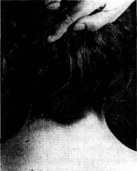
Fig. 1.82. Turner syndrome (45, X0): low hairline in X-linked dominant, X-linked recessive, Y-linked disorders.
Klinefelter syndrome. Karyotype 47, XXY. Incidence: 1:400. The syndrome manifests exclusively in males, predominantly during Puberty. Clinical diagnostic features: tall stature, long limbs, eunuchoidism, gynecomastia (enlargement of the Mammary Glands), absence of Spermatogenesis, and underdeveloped Gonads. Sex chromatin bodies are detected in 80% of cases. Occasionally, patients with Klinefelter syndrome have 48 and 49 Chromosomes (48, XXXY; 49, XXXXY). The greater the number of X chromosomes in the karyotype, the higher the likelihood of developing intellectual disability.
Triple-X syndrome (trisomy X). Karyotype 47, XXX.
The vast majority of such women have normal physical and mental development and are identified incidentally during examinations. Only a few of them experience impaired reproductive function. Most women exhibit normal fertility, although there is an increased risk of spontaneous miscarriages and Chromosomal aberrations in offspring. The cells contain two sex chromatin bodies (Fig. 1.75). As the number of X chromosomes increases, the degree of deviation from normal becomes more pronounced. Intellectual disability, craniofacial anomalies, occipital defects, dental, skeletal, and genital abnormalities have been described in women with tetra- and pentasomy. However, women even with X-chromosome tetrasomy can have offspring.
Y-chromosome disomy syndrome. Karyotype 47, XYY. Incidence: 1:1000. The syndrome occurs in males. In terms of their mental and physical development, such men do not differ from normal individuals. No noticeable deviations in sexual and hormonal status have been revealed. However, some clinicians have pointed to an increased degree of aggressiveness in certain individuals.
Last update: 08/08/2026
Editorial and Educational Adaptation: This material has been compiled based on the primary/original source text. The project team performed an editorial review, corrected technical inaccuracies, structured sections, and adapted the content for an educational format.
What was processed:
- elimination of formatting defects (OCR errors, structural breaks, corrupted characters);
- editorial organization of content;
- standardization of terminology in accordance with academic sources;
- verification of factual statements against the original source text.
All mentions of the author, publication year, and origin of the primary text have been preserved in accordance with the source.