BOTANY, VOLUME 1: CELL BIOLOGY. ANATOMY. MORPHOLOGY — 2007
2. CELL STRUCTURE AND ULTRASTRUCTURE
2.3. Structure of the Prokaryotic Cell
Prokaryotes are exceptionally heterogeneous in ecological, physiological, and structural terms. Below is a Brief Overview of the General Structural Features of Prokaryotic Cells, which nevertheless clearly demonstrates just how vast the differences are between prokaryotic and Eukaryotic cells. No transitional forms between these two Cell types are known in the modern biological world. Only sequence similarities in informational macromolecules (DNA, RNA, Proteins) indicate a common evolutionary origin for pro- and eukaryotes.
Even outwardly, the fundamental difference between pro- and eukaryotes is manifested in vastly different cell sizes (see Fig. 2). The dimensions of an Escherichia coli cell are 2–4 × 1 µm, corresponding to a volume of about 2.5 µm3. By contrast, the cytoplasmic volume of an average Introduction/5.html">Eukaryotic Cell without vacuoles is roughly 1,500 – 3,000 µm3, making it approximately 3 orders of magnitude larger. This corresponds to a significantly lower DNA content in
the Prokaryotic Cell. While the total length of nuclear DNA in a human haploid cell approaches 1 m, in E. coli it barely exceeds 1 mm. The small size of prokaryotic cells also means that generation times can be extremely short; in E. coli, for instance, it is just 20 min. Eukaryotic cells in meristematic Tissues, by contrast, typically divide no more than once a day. A single bacterial cell can thus give rise to over a billion cells in just 10 hours — a fact that, alongside other factors, explains the immense ecological importance of Bacteria. There are no true Multicellular Organisms among prokaryotes.
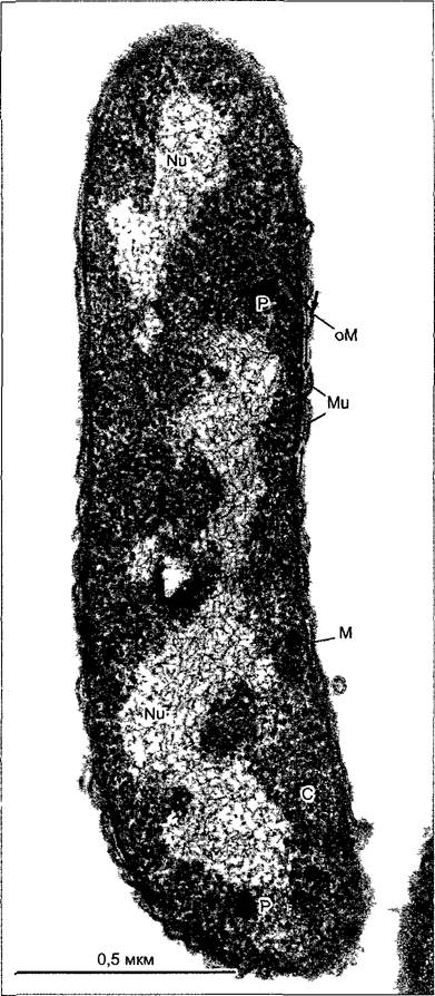
The small size of prokaryotes results in an especially simple intracellular Differentiation of the cell. Such a small cell would lack the space for an extensive internal membrane system (Fig. 2.93). In most prokaryotic cells, The Plasma Membrane is the sole biomembrane, meaning The Cell constitutes a single, undivided compartment. Intracellular non-plasmic compartments rarely occur in bacteria, and in no case are there ER, dictyosomes, vesicles, true vacuoles, or membrane-bounded compartments such as Plastids or Mitochondria.
Class="center">Fig. 2.93. Ultrastructure of a typical bacterial (Gram-negative) cell, Rhodospirillum rubrum (preparation by R. Ladwig, electron micrograph by R. Marx).
The amorphous nucleoid, in which DNA strands are clearly distinguishable, is surrounded by a ribosome-rich Cytoplasm containing polyphosphate granules. The cell is separated from The Cell wall by the plasma membrane. The cell wall comprises a thin murein sac (peptidoglycan layer, thin arrows) and a membrane-like layer known as the outer membrane; this is absent in Gram-positive bacteria, where the murein portion of the wall is considerably thicker and multi-layered (cf. Fig. 2.98). C — cytoplasm; M — plasma membrane; Mu — murein layer; Nu — nucleoid; oM — outer membrane; P — polyphosphate granules
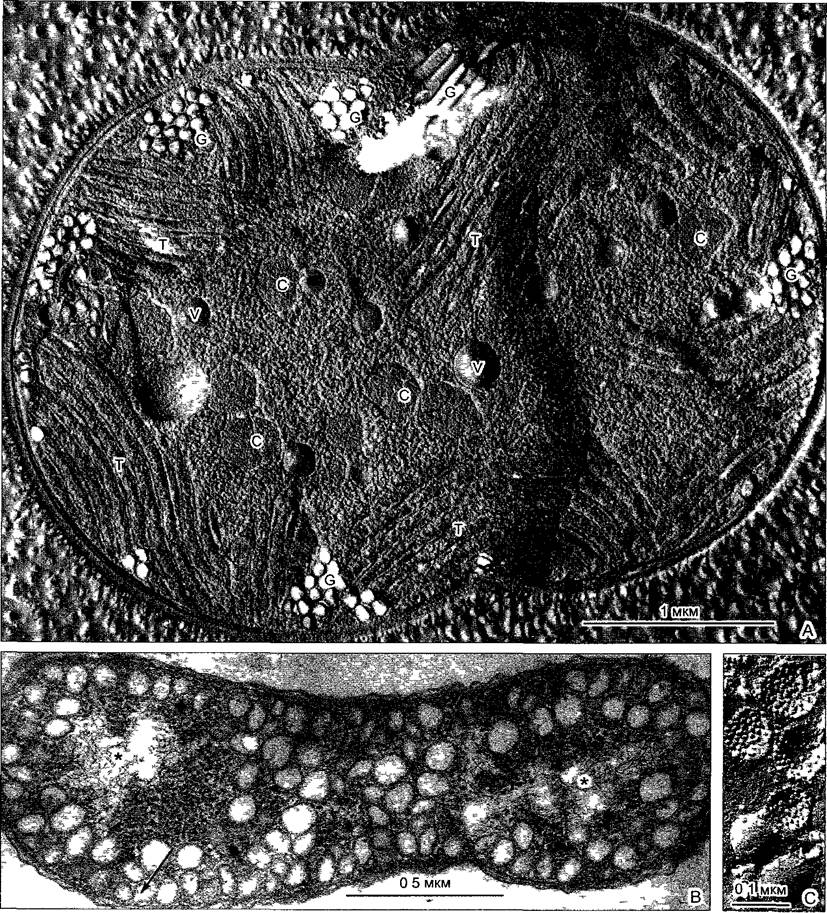
The so-called thylakoids of cyanobacteria (Fig. 2.94, A) are not compartments bounded by plastid membranes, as seen in eukaryotic Algae and higher plants. Instead, they consist of flat double membranes within the cytoplasm that bear Photosynthetic Pigments and carry out light-dependent reactions coupled with Water splitting. They arise via invaginations of the plasma membrane. In some bacteria, there are Various Forms of plasma membrane invaginations (Fig. 2.94, B, C) that remain continuously connected to the membrane for long periods; these are referred to as intracytoplasmic membranes (ICMs). These membrane vesicles, pockets, or tubules also harbor photosynthetic pigments.
Fig. 2.94. Intracytoplasmic membranes (ICMs) of prokaryotes (preparations and electron micrographs by J. R. Golecki)
A — The cyanobacterium Microcystis aeruginosa (shown here following freeze-fracturing) contains several types of intracytoplasmic membranes: thylakoids (T); storage vacuoles (V); carboxysomes (C), which serve as storage structures for the photosynthetic enzyme RubisCO (see 6.5.1); and gas vacuoles (G) — gas-filled cylindrical cavities that provide cell buoyancy in the water Column. These are bounded not by lipoprotein membranes, but by protein shells that can be synthesized de novo in the cytoplasm. The cell is at the onset of division. B — In the Gram-negative bacterium Rhodospirillum rubrum, exposure to light under anaerobic conditions induces a system of vesicular intracytoplasmic membranes resembling chromatophores, which perform Photosynthesis using bacteriochlorophyll (naturally without water splitting). These "chromatophores" originate from cell membrane invaginations (arrow) and are connected partly to the membrane and partly to one another. Asterisks (*) denote nucleoids. C — Corresponding "chromatophores" after freeze-fracturing in Rhodobacter capsulatus; their surfaces appear smooth (cf. Box 2.3, Fig. A), whereas fracture faces reveal numerous intramembrane particles corresponding to pigment-Structure/178.html">Protein Complexes involved in photosynthesis.
The immense and still incompletely understood diversity of prokaryotes is reflected not only in numerous unusual metabolic pathways unknown in eukaryotes, but also in striking departures from the typical characteristics listed above. For instance, bacterial cells occasionally reach sizes comparable to those of eukaryotic cells. The maximum dimensions have been recorded in Epulopiscium fishelsoni, a Gram-positive gut bacterium from tropical marine fish, whose rod-shaped cells measure 600 × 80 µm. Even larger is Thiomargarita namibiensis, a spherical sulfur bacterium recently discovered in marine sediments off the coast of Namibia, with a diameter of up to 750 µm; its vacuolated cells accumulate significant amounts of sulfur and nitrate. This bacterium forms chains comprising up to 50 individual cells. Similar cell aggregates are the rule in cyanobacteria (see Figs. 5.1, 11.15), while in myxobacteria they even culminate in The formation of complex "fruiting bodies."
2.3.1. Reproduction and the Genetic Apparatus
Prokaryotic DNA is circular and is not organized into distinct linear structures analogous to eukaryotic Chromosomes. Nevertheless, bacterial DNA rings are commonly referred to as bacterial chromosomes. These DNA rings possess a specific membrane-attachment locus and a single site of Replication origin; they are monorepliconic. The proportion of non-coding sequence regions is small. Despite its modest length (ranging from 0.2 mm in Mycoplasmas to 37 mm in certain cyanobacteria), the DNA must be tightly folded to fit within the central region of the cell, known as the nucleoid. Nucleoids are not separated by single or double membranes from the ribosome-containing cytoplasm, yet they are distinctly demarcated from it. Nucleoids lack nucleolus-like structures. In cyanobacterial cells (blue-green algae), which are larger than other prokaryotes, the centrally located nucleoid can be discerned even under light Microscopy and was formerly described as a chromidial apparatus. The nucleoid lies within the centroplasm, which is surrounded by a chromatoplasm colored by "thylakoids" (see Fig. 2.94, A).
Histones are absent from the nucleoids of prokaryotic cells (with the exception of methanogenic archaea). Neutralization and packaging of the genetic material are instead carried out by other basic proteins, amines, and inorganic cations. METABOLISM/31.html">Transcription and Translation clearly illustrate that nucleoids are bounded by no membranes: even before the transcription of a single Gene or a group of adjacent genes (an Operon) is complete, translation begins at the initially synthesized 5'-end of the mRNA. RNA Processing does not occur. Co-transcriptional translation takes place on 70S Ribosomes (50S and 30S subunits; see Fig. 2.42), whose activity is inhibited by Antibiotics distinct from those affecting eukaryotic 80S ribosomes (see 2.2.4). 70S ribosomes are smaller and structurally simpler than 80S ribosomes, with an RNA-to-protein ratio of approximately 1.5:1 (compared to 1:1 in eukaryotic ribosomes).
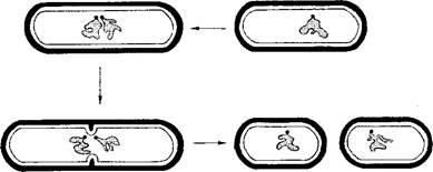
Processes analogous to mitosis or Meiosis are absent in prokaryotes. They possess neither microtubules, Actin, nor Myosin, and lack anything directly comparable to the eukaryotic spindle apparatus. The distribution of genetic material to daughter cells is achieved because, following the replication of the circular DNA molecule, the replication start sites within the nucleoid move as far apart as possible, while membrane attachment sites are similarly displaced by cell membrane growth. The formation of a septum (cross-wall) then initiates between them (Fig. 2.95). For Cell Division, the Cell Cytoplasm is constricted in the plane of the septum by a contractile ring. The FtsZ protein, a tubulin homolog that forms filaments and ring-like structures under appropriate conditions, plays a central role in this ring.
Despite the absence of syngamy and meiosis, bacteria exhibit sexual-like processes — namely, The transfer of Genetic information from one cell to another, as well as recombination (parasexuality). This is primarily mediated by Plasmids, which are (typically) small, circular DNA molecules capable of autonomous replication within a host cell. They do not encode central metabolic housekeeping genes, but rather carry so-called adaptive genes that confer, for example, Antibiotic Resistance (resistance genes on R-plasmids), mediate conjugation (F-plasmids), or encode toxins.
Fig. 2.95. Genome segregation and cell division in a bacterium. Diagram (circular DNA and cell membrane attachment complexes are shown in gray)
2.3.2. Bacterial Flagella
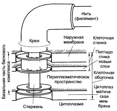
Many eubacteria bear flagella, which are structurally entirely different from the complex flagella or cilia of eukaryotes. A bacterial flagellum (Fig. 2.96) is only 20 nm thick, meaning it does not even reach the diameter of a single microtubule. It is constructed from a single structural protein, flagellin. Bacterial flagella are helical and rigid. At the base, they feature a structure composed of four coaxial rings embedded in the plasma membrane and cell wall (Fig. 2.97). The flagellum itself lies entirely outside the cell. Unlike eukaryotic flagella, which are ten times thicker and capable of changing shape, prokaryotic flagella are not enclosed by a membrane. When a bacterial cell swims forward or backward (these movement directions constantly alternate), the entire flagellum rotates clockwise or counterclockwise without changing its shape, functioning like a ship's propeller. The motor driving this rotational movement is located at the Base of the flagellum. It is powered not by ATP, but directly by the proton gradient across the plasma membrane. As the bacterial flagellum moves, this gradient is dissipated by the inward translocation of protons into the cell.
Fig. 2.96. Bacterial flagella (Agrobacterium tumefaciens, negative staining) (electron micrograph by H. Falk)
The arrow in the inset (higher magnification) points to the flagellar "hook" housing the rotary motor (Fig. 2.97)
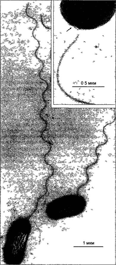
Fig. 2.97. Diagram of the flagellar base in Escherichia coli (after J. Adler)
The four rings of the basal protein complex, which Functions as the motor apparatus, have a diameter of approximately 20 nm. Both outer rings are absent in Gram-positive bacteria
2.3.3. Wall Structures
The cell walls of prokaryotes can vary significantly in appearance. In exceptionally small and simply structured mycoplasma cells—representing the lowest level of Cellular Organization—cell walls are entirely absent. By contrast, most other prokaryotic cells are enclosed by a cell wall that serves not only for protection, but also for osmotic stabilization, maintenance of shape, and controlled interaction with the environment. The wall functions as an exoskeleton. If prokaryotic cells are artificially deprived of their walls, they round up (spheroplasts and protoplasts), become osmotically labile, and can resume division only after the wall has regenerated.
Fig. 2.98 illustrates the layered Structure of Bacterial Cell Walls. (The cell walls of archaebacteria differ markedly in both structure and molecular composition.) The structural backbone of the wall is the peptidoglycan, or murein, layer. It is constructed from unbranched polysaccharide chains cross-linked by oligopeptide chains. Because the entire murein layer constitutes a single giant molecule, it is also referred to as the murein sacculus. Through the localized incorporation of new components, it can expand and thereby participate in cell growth without losing its supportive and protective functions. Peptidoglycan Biosynthesis is blocked by penicillin; consequently, this antibiotic kills bacterial cells but spares eukaryotic cells, which lack peptidoglycans.
Fig. 2.98. Examples of bacterial cell walls (after U. J. Jürgens): A—structural diagram of the cell wall in a Gram-positive bacterium, Bacillus. The cytoplasmic membrane (cell membrane) is covered by a multi-layered peptidoglycan; teichoic acids run within the plane of the cell wall (linear polymers of phosphoglycerol or ribitol phosphate residues covalently linked to the peptidoglycan, whereas lipoteichoic acids are anchored in the cytoplasmic membrane and extend perpendicularly to the plane of the wall). The entire cell wall complex is capped by an S-layer (surface layer), to which outward-directed capsule polysaccharide chains are covalently linked via side chains; B—corresponding diagram for a Gram-negative bacterium, such as Escherichia coli. Here, the peptidoglycan is single-layered. The outer membrane is rigidly linked to it by lipoprotein units (shown in gray). It is pierced by trimeric porins and contains outer membrane protein A (shown by dots) as an integral structural protein. The outer monolayer of the outer membrane consists of lipopolysaccharides, with the Fatty acids of lipid A oriented inward and the outward-curved polysaccharide chains (O-Antigens) directed outward, as well as amphiphilic ECA units (enterobacterial common antigen) with longer, extended polysaccharide chains. Additionally, capsule Polysaccharides (K-antigen) are anchored here
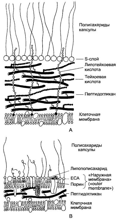
Gram-positive and Gram-negative bacteria differ distinctly in their cell wall architecture. (Gram staining—gentian violet + iodine—can be washed out of Gram-negative bacteria with ethanol, but not out of Gram-positive ones.) In Gram-positive bacteria, the peptidoglycan layer is thick and
composed of multiple murein sheets. In Gram-negative bacteria and cyanobacteria, by contrast, the murein sacculus is relatively thin. However, In addition to the sacculus, these cells possess a characteristic layer known under the Electron microscope, based on its cross-sectional appearance, as the outer membrane. In its molecular organization, it resembles a biomembrane in that it forms a lipid bilayer, with the inner lipid leaflet consisting predominantly of Phospholipids. The outer lipid leaflet, by contrast, is built of lipopolysaccharides—complex polymers featuring fatty acid residues as the lipophilic moiety and characteristic outward-projecting oligo- and polysaccharide chains. Together, they form a hydrophilic protective barrier around prokaryotic cells that prevents the penetration of lipophilic molecules. Hydrophilic particles, however, can pass through. Embedded in The Lipid Bilayer of the outer membrane are trimeric complexes of a transmembrane protein, the porin, which forms hydrophilic pores approximately 1 nm in diameter. (The outer membrane porins of mitochondrial and plastid envelopes perform a comparable function, yet their Amino acid sequences show no close Homology to bacterial porins, which themselves are highly diverse.) The outer membrane is a component of the cell wall rather than a true biomembrane. Unlike genuine Biomembranes, it can also be formed de novo; for instance, it regenerates following the complete loss of the wall. The outer membrane does not interface with the cytoplasm at any point and lacks translocators for specific or Active Transport. The space between the cytoplasmic and outer membranes is termed the periplasmic space.
Under unfavorable conditions, most prokaryotes are capable of forming spores with exceptionally tough and impermeable walls.
Last update: 07/08/2026
Editorial and Educational Adaptation: This material has been compiled based on the primary/original source text. The project team performed an editorial review, corrected technical inaccuracies, structured sections, and adapted the content for an educational format.
What was processed:
- elimination of formatting defects (OCR errors, structural breaks, corrupted characters);
- editorial organization of content;
- standardization of terminology in accordance with academic sources;
- verification of factual statements against the original source text.
All mentions of the author, publication year, and origin of the primary text have been preserved in accordance with the source.