PLANT MORPHOLOGY - T. A. Sautkina - 2012
CHAPTER 3. PLANT TISSUES
3.7. Anatomical, Morphological, and Functional Features of Secretory (Excretory) Structures
Liquid and solid metabolic products secreted to the outside or accumulated inside Cells or intercellular spaces are called excretions or
secretions. Because it is sometimes impossible to accurately determine which substances are excretory and which are secretory, the structures involved in removing substances from metabolic processes are referred to as secretory, or excretory structures.
As a rule, the secreted substances are secondary metabolites: a mixture of terpenoids (Essential Oils, resins, saponins, etc.), Alkaloids, and Flavonoids. Secretory structures vary in Structure and differ in the ways they release substances. The ability of glandular trichomes and hairs, for example, to participate in hormone synthesis and photoreception provides a broader perspective on The Role of these structures in plant life and their potential Applications in human activity.
Secretory structures lack a strictly defined localization and are characterized by diverse origins and structures. They occur in all Organs and are situated among various plant Tissues. Excretory structures may be derivatives of the protoderm, ground meristem, or specialized permanent tissues (epidermis, parenchyma, phloem, xylem). The cytological Features of the cells in secretory structures depend on the Chemical Nature of the secreted substances. The Endoplasmic reticulum and the Golgi apparatus are believed to play The most significant role in carrying out secretion or excretion Functions.
Depending on their Location, secretory structures are divided into exogenous (external) and endogenous (internal) types.
3.7.1. Exogenous Secretory Structures
By origin, exogenous secretory structures are usually associated with the protective tissues of various plant organs. These include glandular hairs, glands, nectaries, osmotrophs (osmophores), digestive glands, and hydathodes.
Glandular hairs consist of a unicellular or multicellular stalk formed by non-glandular cells, and a HEAD (globular or oval) that produces the secretion and may also be unicellular or multicellular. The head of the glandular Hair is covered by a cuticle. The glandular Cells of the head synthesize essential oils, which are released through The Cell wall and accumulate between The Cell wall and the cuticle. Over time, under the pressure of the excreted substances, the cuticle ruptures, and the essential oils are released to the exterior. After the release of essential oils, the glandular hairs die off in some plants, whereas in others, a new cuticle forms over the head cells, and the secretory function is restored. Glandular hairs are characteristic of plants in the families Geraniaceae, Primulaceae, Solanaceae, and Cannabaceae.
A specific type of glandular hair is represented by the digestive glands of carnivorous plants (such as *Drosera*, *Utricularia*, *Nepenthes*, etc.). These glandular hairs secrete mucopolysaccharides or produce a mucous substance (in *Pinguicula*) that attracts insects, as well as Proteolytic Enzymes that aid in their Digestion (Fig. 45).
Class="center">Fig. 45. Exogenous secretory structures: A—glandular hair of zonal geranium (*Pelargonium zonale*) with excretion accumulated beneath the cuticle; B—glandular hair of rosemary (*Rosmarinus officinalis*); C—glandular hair of potato (*Solanum tuberosum*); D—vesicular hairs of spreading orach (*Atriplex patula*); E—peltate gland from a black currant leaf (*Ribes nigrum*)

Glands are multicellular, globular structures—the head—situated on a short stalk or directly on the epidermis. For example, in plants of the Lamiaceae family, the essential oil gland is formed by 8 cells arranged in a rosette. As essential oil accumulates, the common cuticle of these cells swells in a dome-like fashion, forming an essential oil reservoir. In Asteraceae, glands typically consist of two vertical rows of four cells each (*Matricaria chamomilla*, *Artemisia absinthium*). The specific structure of glands is often an important diagnostic feature of plants.
Both glandular hairs and glands can also secrete salt solutions. This phenomenon is characteristic of plants from the families Chenopodiaceae, Verbenaceae, Plumbaginaceae, and Poaceae.
Nectaries are diverse glandular structures that secrete nectar—a sugary liquid containing a solution of sugars mixed with Proteins, alcohols, and aromatic substances (Fig. 46). The secretory cells of nectaries have dense Cytoplasm and high metabolic activity. A vascular bundle may extend to the nectary (e.g., in *Tagetes*). The secreted nectar primarily attracts pollinators, such as insects and birds.
Fig. 46. Structure of floral nectaries in various plants: A—nectar pit covered with a scale (creeping buttercup—*Ranunculus repens*); B—spur formed by a petal (winged larkspur—*Delphinium elatum*); C—nectary formed by a petal (monkshood—*Aconitum napellus*); D—spur formed by a sepal (Touch-me-not balsam—*Impatiens noli-tangere*); E—epigynous disc of Apiaceae; F—hypogynous disc of Lamiaceae; G—glands of Salicaceae (1—nectaries)

In most cases, nectaries are located within flowers—at the Base of the Ovary, stamens, on petals, receptacle, or sepals. Frequently, nectaries are formed from stamens (bog bean, *Spiraea chamaedryfolia*) or corolla petals (*Aconitum napellus*, *Tropaeolum majus*). Such nectaries are called floral (from Latin *flos (floris)* — flower). Extrafloral (from Latin *extra* — outside and *flos* — flower) nectaries are formed on the Water/115.html">Vegetative organs of plants—on the stem, leaf petioles, stipules, flower stalks, and inflorescence axes. One of their functions is to attract protective animals, such as ants, which save the plant from "nectar thieves" and thereby promote successful flower pollination and seed dispersal. Extrafloral nectaries are widely represented in species of the genus *Impatiens* and members of the Bignoniaceae family. The shape and structure of nectaries are highly diverse: they can be tubular, capitate, petaloid, filiform, etc.
Osmophores are specialized glandular formations that secrete aromatic substances and determine the scent (aroma) of plants. Morphologically, they are very diverse, appearing as cilia, villi, glandular spots, etc. The aroma emitted by plants plays a huge role in their life, primarily acting as an attractant for pollinators. It is believed that there are about 500 different floral scents. A strong aroma is characteristic of many tropical plants. In night-blooming plants, scent is the primary means of attracting insect pollinators. Some day-blooming plants also have a strong scent (e.g., mignonette). Sometimes flower scents mimic the smell of animal Skin, decomposing protein, or fruit. Blood-sucking midges, flies, or scavenger insects are attracted by these odors and act as pollinators. Scents also perform the crucial function of repelling herbivores and pest insects.
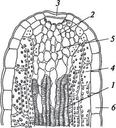
Hydathodes (from Greek *hydor* — water and *hodos* — path), or water Stomata, consist of a system of loosely arranged leaf mesophyll cells—the epithem—supplied by a small vascular bundle of spiral tracheids (Fig. 47). At the apex of the hydathode, there is either a single wide pore, as in primroses and aconites, or a group of narrow pores, as in many succulents, Asteraceae, and Apiaceae. Guttation—the extrusion of water-salt solution droplets—occurs through the water stomata. In this way, the plant rids itself of excess water and salts. Guttation is particularly intensive under conditions where Transpiration (the evaporation of water by leaves) is hindered. Such conditions occur on cool, windless nights with high air and soil humidity. In the early morning hours after such nights, water droplets can be observed on plant leaves. Hydathodes are usually located at the tips of leaf blades or on leaf Teeth. Unlike true stomata, hydathodes function passively. Hydathodes are characteristic of grasses and Araceae (*Monstera*, *Philodendron*, etc.).
Fig. 47. Longitudinal section through a hydathode on a leaf tooth of Chinese primrose (*Primula sinensis*): 1—spiral tracheids; 2—epithem cells; 3—water stoma pore; 4—chlorenchyma cells; 5—intercellular spaces; 6—leaf epidermal cells
3.7.2. Endogenous Secretory Structures
Endogenous secretory structures can develop in various plant tissues. They are represented by idioblast cells, secretory cavities, resin ducts, essential oil canals, and laticifers.
Idioblasts are isolated thin-walled secretory cells or small groups of such cells. They occur in various tissues and may contain diverse secretions, such as mucilages, Tannins, mixtures of terpenoids, and salt crystals. For instance, mucilage cells are quite common in plants of the Malvaceae, Tiliaceae, and Cactaceae families. Idioblasts containing essential oils (terpenoids) are characteristic of Representatives of the Lauraceae, Aristolochiaceae, Piperaceae, Magnoliaceae, and other families. Vegetative and reproductive organs of many plants frequently contain cells with crystals of various SHAPES AND SIZES. Star-shaped clusters of calcium oxalate crystals, known as druses, are widely distributed. Furthermore, thin needle-like crystals collected in bundles and surrounded by a mucilaginous sheath, called raphides, are often found in plants of the Orchidaceae, Balsaminaceae, Vitaceae, and Commelinaceae (Tradescantia) families. Occasionally, large single crystals—styloids—are formed (e.g., in the leaves of the lily of the valley). In the leaves of plants belonging to the Moraceae, Urticaceae, and Cucurbitaceae families, large lithocyst cells occur, within which a botryoidal body made of calcium carbonate, known as a cystolith, is formed (Fig. 48).
Fig. 48. Crystalline inclusions in plant cells: A—cystolith in the leaf epidermis of the common fig (Ficus carica); B—raphides in the leaf epidermis of wandering Jew (Zebrina pendula); C—druses in the palisade tissue of the common fig leaf (Ficus carica); D—druses and single crystals in the cells of the petiole of the royal begonia (Begonia rex); E—single crystals in the scale epidermis of the bulb of the common onion (Allium cepa)

Secretory cavities are spaces of various shapes and origins located within parenchymal or water-conducting tissues. Depending on their mode of formation, they are classified as schizogenous, lysigenous, and schizolysigenous.
Schizogenous cavities (from the Greek schizeo, meaning to split) are formed As a result of The breakdown of intercellular pectin substances and the Separation of cells. The resulting cavity is lined with living epithelial cells, which secrete metabolic products into the schizogenous cavity. Most frequently, schizogenous cavities contain mucilage (plants of the Araliaceae family, some ferns, cycads), and less commonly essential oils (Hypericaceae, Apiaceae), resins, and balsams (conifers) (Figs. 49, 51).
Fig. 49. Formation (A) and structure (B) of a schizogenous resin duct in Norway spruce (Picea abies): 1–3—successive stages of cell separation; 4—resin canal cavity; 5—epithelial cells

Fig. 50. Formation of a lysigenous resin duct in the bark of common juniper (Juniperus communis): A–C—successive stages of cell degradation:
1—resin idioblasts; 2—formed resin duct; 3—resin duct cavity; 4—epithelial cells

Fig. 51. Essential oil cavities in various plants: A—schizogenous cavity in the leaf of common St. John's wort (Hypericum perforatum); B—lysigenous cavity in the leaf of dittany (Dictamnus album); C—lysigenous cavity in the peel of the mandarin fruit (Citrus unshiu): 1—epithelium; 2—essential oil; 3—cavity

Lysigenous cavities (from the Greek lisis, meaning dissolution) are formed through the destruction—lysis—of cells following the accumulation of secretions in intercellular spaces (Figs. 50, 51). Lysigenous cavities are well developed in the peel of citrus fruits (lemon, mandarin, orange) and in the leaves of rue and eucalyptus. Various intermediate forms exist between schizogenous and lysigenous cavities. For instance, in the secondary phloem of conifers belonging to the Cupressaceae family, resinous cavities begin to develop as schizogenous ones, after which the surrounding cells undergo lysis. Such cavities are referred to as schizolysigenous. They can also be observed in sweet bay and the camphor tree. These cavities occur predominantly in plant leaves and are frequently visible to the naked eye as light, transparent dots, as, for example, in common St. John's wort.
Resin ducts and canals (tubules) are closely related to schizogenous cavities, differing in their more or less elongated shape and ability to branch. The arrangement of resin ducts is complex; they run in both vertical and horizontal directions, interconnecting into a unified system. Such structures are formed primarily in stems, fruits, and roots, and less frequently in leaves. A typical resin duct is a long tubular intercellular space surrounded by living epithelial cells. The lining epithelial cells secrete excretes in the form of resins and essential oils into the canal cavity. These epithelial cells are surrounded by sheath cells with strongly thickened cell walls that isolate the secretion from surrounding tissues. Resin ducts are found in many conifers, umbellifers, and composites.
Laticifers are unicellular or multicellular structures whose vacuoles contain milky sap, or latex. Latex is an emulsion, suspension, or aqueous solution in which hydrophobic droplets of various substances and solid particles (resins, Gums, rubber, etc.) are suspended. In addition to resins and rubbers, latex contains sugars, proteinaceous substances, essential oils, alkaloids, and Glycosides. Typical latex is white (in dandelion, spurge, poppy, and ficus), but it can also be orange (in celandine) or transparent (in mulberry and oleander).
Laticifers have been identified in 12,500 species across 900 genera of flowering plants. Two Types of laticifers are distinguished: articulated and non-articulated.
Articulated laticifers are formed by the fusion of many latex-producing cells into a single branched system. Such structures are found in Asteraceae, Papaveraceae, Campanulaceae, Araceae, Musaceae, and other families.
Non-articulated laticifers consist of a single giant branched cell that originates in the embryo, grows, and permeates all plant organs. Such structures are characteristic of plants belonging to the Moraceae, Euphorbiaceae, Apocynaceae, and Asclepiadaceae families.
The exact Physiological Role of laticifers in plants remains not fully understood. Only a secondary protective role is certain for some laticifers, specifically when the latex contains bitter or poisonous compounds that deter herbivores from consuming the plant.
Metabolic products excreted by the plant or accumulated within it cannot be considered mere waste. Currently, the most substantiated hypothesis suggests that secondary metabolites, unlike primary ones, have functional significance not at THE CELLULAR LEVEL, but at the level of the whole organism. For instance, the aromas of essential oils attract pollinators, and their vapors protect plants from overheating. Resins, balsams, and tannins protect plants against decay, while alkaloids defend them against pests. Thus, the functioning of secretory structures is of great adaptive importance and ensures the normal vital activity of plants.
Last update: 07/08/2026
Editorial and Educational Adaptation: This material has been compiled based on the primary/original source text. The project team performed an editorial review, corrected technical inaccuracies, structured sections, and adapted the content for an educational format.
What was processed:
- elimination of formatting defects (OCR errors, structural breaks, corrupted characters);
- editorial organization of content;
- standardization of terminology in accordance with academic sources;
- verification of factual statements against the original source text.
All mentions of the author, publication year, and origin of the primary text have been preserved in accordance with the source.