Phycology - Kostikov I.Yu. - 2009-2013
Chapter 24. Green Algae – Chlorophyta
24.3. The Streptophyte Lineage
The streptophyte Lineage comprises those green Algae that are closely related to land plants. It is believed that the ancestors of the earliest land plants belonged precisely to the streptophyte lineage. Streptophytes include algae in which cytokinesis involves a primitive or developed phragmoplast, and mitosis is open or semi-open; in representatives with flagellated stages, the Cytoskeleton of the monad Cells is asymmetrical. The streptophyte lineage is represented by two classes: Charophyceae and Zygnematophyceae (=Conjugatophyceae).
24.3.1. Class Charophyceae
Charophytes are relatively modest in taxonomic diversity. The Class comprises about 400 extant species and around 1,000 fossil species. Nevertheless, Charophyceae are of paramount phylogenetic importance, as it was the primitive charophytes that gave rise to land plants.
The class encompasses both micro- and macroscopic algae which, like the conjugates, represent the phragmoplast line of evolution. However, unlike Conjugatophyceae, charophytes have retained flagellated stages; similar to the spermatozoids of bryophytes and lycophytes, these stages possess an asymmetrical system of flagellar roots and are covered with submicroscopic scales.
Representatives of the class are found in freshwater and saline Water bodies, soils, and aerophytic habitats.
Features and CHARACTERISTICS OF THE Class
Biochemical Features
The composition of pigments and assimilation products in charophytes is identical to that in conjugate, chlorophycean, and trebouxiophycean algae, meaning that no specific biochemical traits have been identified within the class.
Cytological Features
Cell Coverings. The cells of charophytes are enclosed by a Cellulose-pectin wall. In one of the orders (Charales), the synthesis of cellulose microfibrils is carried out by terminal enzyme complexes, and The Cell walls become encrusted with lime. In other orders, terminal complexes have not been found, and the cell walls lack carbonates. Monad cells—zoospores and spermatozoids—are covered by a Plasmalemma upon The surface of which submicroscopic organic scales are deposited, similar to the scales of monad cells in codiolalean and prasinophycean algae.
Nuclear Apparatus and Mitosis. The nuclei contain one or several nucleoli, which, unlike those in conjugates, disappear at the onset of mitosis. Numerous specific features have been discovered in the nuclear Organization of charalean algae (see below).
Mitosis is open. Centrioles are present and, at least during The formation of monad cells, participate in assembling the mitotic spindle. The spindle persists into telophase and becomes the organizing center for the phragmoplast.
In coccoid, sarcinoid, and filamentous forms, similarly to conjugates, cytokinesis occurs through the inward growth of a Cleavage furrow. The ingrowth of the furrow into the cell is driven by the fusion of Golgi-derived vesicles with the nascent furrow of the plasmalemma. The movement of vesicles toward the furrow is directed by a microtubular system composed of remnant spindle fibers and a complex of peripheral microtubules that forms directly adjacent to the cleavage furrow. In addition to microtubules, the Actin microfibrils are also part of the machinery governing vesicle transport. This entire microtubule-actin complex is regarded as a phragmoplast. A cell plate and pores with plasmodesmata are not formed in coccoid, sarcinoid, and filamentous forms (Fig. 24.59).

Fig. 24.59. Mitosis and cytokinesis in charophyte algae. A - Cell Division in the filamentous alga Klebsormidium; B - cell division in the heterotrichous alga Coleochaete; 1, 5 - prophase; 2, 6 - metaphase; 3, 7 - telophase; 4, 8 - early interphase (a - microbody, b - perinuclear microtubules, c - cell sap vacuole, d - peripheral microtubules of the phragmoplast; e - phragmoplast with cell plate) (A - after Lokhorst, Star, 1985; B - after Marchant, Pickett-Heaps, 1973).
In heterotrichous representatives, cytokinesis is accomplished exclusively through the formation of a cell plate mediated by the phragmoplast. The expansion of the plate in all representatives occurs via the fusion of membranous vesicles—derived from the Golgi apparatus and containing material for the future cell partition—with one another. As a result, a cell plate pierced by pores with plasmodesmata is formed.
Flagellated (Monad) Stages are biflagellate; in highly specialized representatives (Charales), they are represented solely by spermatozoids, whereas in others they include Gametes and p-type zoospores. Monad cells are dorsiventral and invariably covered with a single layer of submicroscopic scales. The flagella are positioned on the ventral side near the cell apex and are directed sideways rather than forward (the so-called unilateral position).
The basal bodies diverge at a narrow angle and are interconnected by a striated bridge. A single microtubular ROOT, consisting of several dozen microtubules arranged in a single row, originates beneath the basal bodies. This root extends beneath the plasmalemma along the ventral side of the cell down to its posterior end. Directly adjacent to the basal bodies, the root microtubules transition into a multilayered Structure (Fig. 24.60). A similar type of flagellar root organization is characteristic of land plant spermatozoids and has no analogs in other classes of green algae.

Fig. 24.60. Flagellar apparatus of spermatozoids in Chara (A) and zoospores in Chaetosphaeridium (B): 1 - basal body, 2 - upper connecting fiber, 3 - multilayered structure, 4 - unilateral microtubular root composed of multiple microtubules (microtubular splint), 5 - submicroscopic scales (A - after Masiuk, 1989; B - from Van den Hoek, Mann, Jahns, 1995 after Moestrup, 1982).
Types of Morphological Organization
Algae within the class exhibit four types of morphological organization: coccoid, sarcinoid, filamentous, and heterotrichous. The first two types are currently known in only two genera, Chaetosphaeridium and Chlorokybus, respectively. Two genera, Klebsormidium and Raphidonema, possess a filamentous structure and form unbranched uniseriate filaments that readily break apart into individual cells in A number of species. The majority of charophytes feature a heterotrichous type of organization. In this case, thalli may appear either as prostrate branched filaments that fuse to form parenchymatous discs bearing colorless hairs on their surface (Coleochaete), or exhibit a complex articulated-whorled structure resembling horsetails (representatives of the order Charales).
Reproduction and Life Cycles
Charophytes reproduce both asexually and sexually. Asexual reproduction in unspecialized forms occurs via cell division, thallus fragmentation, biflagellate zoospores, and occasionally akinetes. In highly specialized algae of the order Charales, Vegetative Reproduction by means of gemmae is widespread.
The sexual process is predominantly oogamous, less frequently heterogamous. The life cycle in all euhagamous charophytes is haplontic, with zygotic reduction; in coccoid and filamentous forms, it occurs without Morphology/12.html">ALTERNATION OF GENERATIONS, while in heterotrichous representatives, it features heteromorphic alternation. Agamospecies are few in number, represented by coccoid, sarcinoidean, and several filamentous forms; in these, the life cycle represents a simple cyclomorphosis.
Systematics of the Class
Based on molecular criteria, which are in good agreement with numerous cytological and morphological features, the class is divided into five orders: Chaetosphaeridales, Chlorokybales, Klebsormidiales, Coleochaetales, and Charales. The main phenotypic characteristics of the orders include the type of morphological structure and body plan (specifically, the presence of a jointed-whorled structure), features of cytokinesis (in particular, whether a cleavage furrow is involved), The ability to reproduce via zoospores, the presence of a sexual process, and others (Table 24.7).
Chaetosphaeridales. This order includes only a single genus, Chaetosphaeridium, with four species. According to molecular phylogenetic data, this genus is the most primitive group of charophytes, which almost simultaneously gave rise to all other orders of the class, as well as to the conjugates and the lineage that directly led to the evolution of higher plants.
Table 24.7. Some main taxonomic Features of the orders of charophyte algae
Feature → Order ↓ |
Structural type |
Jointed-whorled structure |
Zoospores |
Sexual process |
Chaetosphaeridales |
coccoid |
- |
+ |
oogamy |
Chlorokybales |
sarcinoidean |
- |
+ |
- |
Klebsormidiales |
filamentous |
- |
+ |
hetero-, oogamy |
Coleochaetales |
heterotrichous |
- |
+ |
oogamy |
Charales |
heterotrichous |
+ |
- |
oogamy |
Abbreviations: Jointed-whorled structure — presence of a jointed-whorled thallus structure (this feature applies only to heterotrichous forms).
Species of the genus Chaetosphaeridium are coccoid freshwater periphytic algae. Cells are solitary or united into colonies by common mucus or mucous strands. Each cell, in addition to The Cell wall, is covered by a mucous capsule and bears a long cellulosic seta, which is a modified part of the cell wall. The cell contains one or two parietal plate-like METABOLISM/14.html">Chloroplasts with pyrenoids and a single nucleus. The protoplast has limited motility; together with the chloroplasts, it slowly rotates around the cell axis (Fig. 24.61).
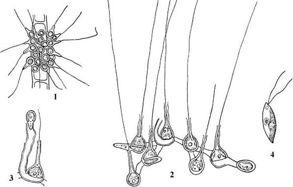
Fig. 24.61. Chaetosphaeridium: 1 — general view of the colony, 2 — formation and release of zoospores, 3 — connection of cells within the colony, 4 — zoospore (1–3 — after Moshkova, 1979; 4 — orig.).
Reproduction occurs via cell division and zoospores. A sexual process has been discovered in one species. During binary fission, the cell protoplast divides in a plane parallel to the substrate; subsequently, the lower cell turns on its side and continues to grow, forming a small cylindrical strand that ruptures the mucous envelope of the mother cell. Next, each daughter cell develops its own cell wall, seta, and new mucous envelope, after which the mother cell wall undergoes gelatinization. Unfortunately, electron microscopic data on the features of karyokinesis and cytokinesis are currently lacking.
Zoospores are biflagellate, dorsoventral, lack a stigma, and are produced in groups of four. Each zoospore is covered by a plasmalemma bearing submicroscopic scales on its surface. The Root System is asymmetrical, comprising a single microtubular root composed of approximately 60 microtubules and a multilayered structure.
An oogamous sexual process has been found in C. globosum. The life cycle in this alga is haplontic, with zygotic reduction and without alternation of generations.
Chlorokybales. This order is monotypic, including only a single species, Chlorokybus atmophyticus, which inhabits soils and damp aerophytic substrates. The thalli of the alga are sarcinoidean, appearing as cubic packets enveloped in a common colonial mucilage. Each cell contains a parietal lobed chloroplast with a naked pyrenoid. Between the cell wall and the chloroplast, There is a special spherical structure known as the pseudopyrenoid. Cytokinesis involves a cleavage furrow, although additional bundles of microtubules resembling a phragmoplast are observed around the spindle in telophase (Fig. 24.62).
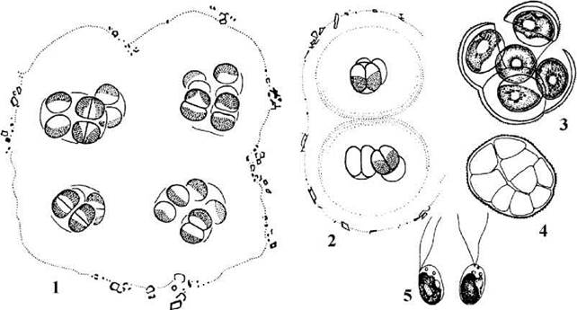
Fig. 24.62. Chlorokybus: 1, 2 — general view of colonies, 3 — aplanosporangium, 4 — zoosporangium, 5 — zoospores (after Geitler, 1955; Rith, 1972).
Reproduction occurs through packet fragmentation, aplanospores, and biflagellate zoospores formed singly from the protoplast of any cell. The ULTRASTRUCTURE OF THE zoospores is identical to that of Chaetosphaeridium.
Klebsormidiales. This order unites algae with a filamentous structural type. In klebsormidialeans, cytokinesis involves a cleavage furrow, and the transverse cell septum lacks plasmodesmata. The ultrastructure of the zoospores is generally similar to that of Chaetosphaeridium. As in Chlorokybus, a single mother cell can give rise to only one monad cell — a zoospore or a gamete.
The order comprises about 40 species belonging to two genera, Klebsormidium and Raphidonema (Fig. 24.63). There is evidence that certain species of the genus Stichococcus from the class Trebouxiophyceae also belong to the Klebsormidiales.

Fig. 24.63. Klebsormidialean algae. A — Klebsormidium (1 — gametosporophyte; 2 — rudimentary branching; 3 — formation of a zoospore; 4 — release of a zoospore; 5 — akinetes; 6, 7 — micro- and macrogametes; 8 — initial stage of zygote germination; 9 — aplanospore and its germination). B — Raphidonema (A: 1–4, B — orig., 5–9 — after Moshkova, 1979).
The thallus of Klebsormidium appears as an unbranched uniseriate filament, which closely resembles Ulothrix in appearance. The Cells of the filament are morphologically and functionally identical, each containing a single parietal chloroplast with a pyrenoid. Klebsormidium reproduces via filament fragmentation, akinetes, zoospores, and sexual reproduction. The sexual process is heterogamous. Furthermore, gametes are capable of parthenogenetically germinating into new gametosporophytes, thereby functioning as zoospores. The life cycle is haplontic, with zygotic reduction and without alternation of generations.
Species of Klebsormidium are extremely widespread in soils (especially steppe and meadow soils) and in aerophytic habitats, and are occasionally found in freshwater bodies.
Raphidonema predominantly comprises cryophilic species inhabiting snow and glaciers. The filaments in these algae are short, typically consisting of only two cells, and generally fragment easily. The cells themselves are pointed at the ends and contain a single parietal chloroplast with or without a pyrenoid. Reproduction occurs through filament fragmentation. In one species, R. longiseta, an oogamous sexual process is also known, with the spermatozoa exhibiting the dorsiventral structure and asymmetric cytoskeleton typical of klebsormidiacean algae. The life cycle is haplontic, with zygotic reduction and no alternation of generations.
Coleochaetales. This order includes a single genus of freshwater heterotrichous algae, Coleochaete (about 10 species). Their thalli appear as branched creeping filaments or single-layered parenchymatous plates, typically bearing hairs (setae) of a specific structure: each Hair features a collar at its base. Cytokinesis occurs via cell plate formation involving a well-developed phragmoplast. Unlike klebsormidiacean algae, the transverse septa have pores with plasmodesmata, and the cleavage furrow does not take part in cell division. Similar to chaetosphaeridialean algae, the cell protoplasts are capable of limited oscillatory movement. The Cytoplasm contains a parietal chloroplast with one or two pyrenoids and a single nucleus.
Coleochaetalean algae reproduce through thallus fragmentation, naked biflagellate zoospores similar to those of Chaetosphaeridium, and sexually.
The sexual process is oogamy. The egg cell develops within an oogonium, which in some species possesses a long neck called a trichogyne. Single biflagellate spermatozoa are formed within unicellular antheridia. Interestingly, like those of Klebsormidium, these spermatozoa are capable of parthenogenetic germination.
Following Fertilization, the zygote enters a resting stage, during which lateral branches begin to grow from the oogonial cell and adjacent cells, closely pressing against the surface of the oogonium. Because of this, the oogonium with the fertilized egg somewhat resembles the archegonium of primitive higher plants. At the end of the resting period, Meiosis occurs within the zygote, and it germinates into a haploid sporophyte. The sporophyte appears as a disc composed of 8–16 wedge-shaped cells. Each such cell gives rise to a single biflagellate zoospore that grows into a new haploid gametosporophyte. Thus, the life cycle in Coleochaete is haplontic, with zygotic reduction and heteromorphic alternation of generations.

Fig. 24.64. Coleochaete: 1, 2 — general view of the gametosporophyte in species with lamellate (C. scutata) and branched filamentous (C. pulvinata) thallus forms; 3 — antheridia and release of a spermatozoon; 4 — mature oogonium covered by a cortex; 5 — eight-celled sporophyte developing from a zygote; 6 — germination of the sporophyte into zoospores; Initial Stages of gametosporophyte development (after Moshkova, 1979; Pringsheim, 1860).
Following the discovery of structural similarities in the flagellated stages of charophyte algae and sporous higher plants, the order Coleochaetales was regarded as the lineage that gave rise to bryophytes and vascular plants. The presence of a heteromorphic alternation of generations, heterotrichous structure with the ability to form parenchymatous discs, oogonia with a trichogyne, and archegonium-like structures of oogonia containing fertilized eggs strongly supported this hypothesis. However, molecular phylogenetic reconstructions have shown that the lineage leading to higher plants diverged not from coleochaetalean algae themselves, but from their ancestor, which occupied an intermediate position between Chaetosphaeridium, on the one hand, and Coleochaetales, Klebsormidiales, and Chlorokybales, on the other.
Charales. This group unites algae whose sporophyte thalli resemble horsetails: they are macroscopic, complexly articulated-whorled in structure, and differentiated into nodes and internodes (Fig. 24.65). Representatives of the order exhibit a whole range of specific cytological and morphological features, as well as a life cycle with a heteromorphic alternation of generations, wherein the gametophytes are embryonized and develop on the sporophyte.

Fig. 24.65. Thallus structure and cellular features of charalean sporophytes. A — general view of the Chara thallus (1 — node, 2 — internode, 3 — lateral "branch" of limited growth); B — apex of a limited-growth branch of Nitella (4 — line separating opposite cytoplasmic streams); C — peripheral part of an internodal cell (5 — cell wall, 6 — stable cytoplasmic zone, 7 — labile cytoplasmic zone, 8 — vacuole with cell sap, 9 — plasmalemma, 10 — chloroplast, 11 — actin microfilament bundles "guiding" cytoplasmic streaming, 12 — mitochondrion, 13 — Endoplasmic reticulum, 14 — nucleus, 15 — Myosin microfilaments); D — node Cell Nucleus during mitosis; E — internodal cell nucleus during amitosis; F — apical cells of the sporophyte (16 — apical cell, 17 — biconcave cell giving rise to nodal cells, 18 — biconvex cell differentiating into an internodal cell); G — diagram of thallus apex structure (19 — nodal cell, 20 — internodal cell, 21 — stem cortex cells, 22 — antheridium primordium, 23 — oogonium primordium); H — fragment of an internode covered with a spiny stem cortex; I — cross section through an internode with a stem cortex; J — nodes with a whorl of "stipules" (A–B, D–J after Gollerbakh, 1977; C after Stebbings, Hyams, 1979).
Charalean algae inhabit fresh and brackish continental water bodies as well as marine estuaries. The order comprises about 150 extant species and over 1,000 fossil species.
Cytological features. The cells are covered by walls in which cellulose microfibrils are synthesized by rosette terminal enzyme complexes (a similar type of wall formation is also characteristic of conjugation algae and higher plants), and are usually encrusted with lime.
The cell center is occupied by a large vacuole containing cell sap. The cytoplasm with Organelles forms a thin peripheral layer, differentiated into two zones: stable and labile. The stable zone adheres to the plasmalemma, where numerous small disk-shaped chloroplasts lacking pyrenoids are arranged in a single layer in regular longitudinal rows. Beneath the stable zone lies the labile zone, which concentrates numerous nuclei, Mitochondria, elements of The endoplasmic reticulum, and myosin microfilaments attached to them; this entire complex is rapidly driven by cytoplasmic streaming along the cell axis. The rate of cytoplasmic streaming is 50–100 µm/s. The stable and labile zones are separated by a layer of parallel actin bundles forming guiding "tracks" along which myosin microfilaments slide together with the organelles attached to them. Opposing cytoplasmic streams do not mix, and the boundary between them appears as a colorless stripe.
Internodal cells are multinucleate. Their nuclei have a characteristic lobed shape and, in addition to standard nuclear structures (Chromatin, nucleoli, preribosomes), contain bundles of microtubules and divide amitotically. Nodal cells, by contrast, are uninucleate and spherical, and their division occurs via mitosis. All sporophyte cells lack centrioles. In male gametophyte cells, conversely, centrioles are present and participate in forming the mitotic spindle during spermatozoon formation. Mitosis is open. Cytokinesis involves a phragmoplast, with primary pores containing plasmodesmata forming in the transverse walls between daughter cells.
Charalean sporophytes look like branched tufts consisting of upright support filaments of unlimited growth (so-called "stems", lateral whorled assimilatory filaments of limited growth, i.e., "leaves"), and rhizoidal filaments. Each filament ("stem", "leaf", rhizoid) is differentiated into regularly alternating nodal and internodal cells.
Nodes and internodes are formed through the division of an apical cell. It divides in a plane transverse to the filament axis, alternately cutting off cells of two types: biconvex and biconcave. The biconvex cell is incapable of further division—it only grows and stretches, gradually differentiating into a giant multinucleate internodal cell. The biconcave cell is capable of further divisions, which occur in radial directions in a plane longitudinal to the cell axis. The daughter cells form a node. Each nodal cell, in turn, is capable of division occurring in the same manner as in the apical cell, thereby producing "leaves" and lateral "stem" branches.
In some representatives (e.g., in Chara), stem cortex and stipules develop from stem nodal cells, while sporangia giving rise to male or female gametophytes develop from leaf nodes. Brood buds (bulbils), which serve as vegetative reproductive Organs, are formed on rhizoidal nodes.
Gametophytes develop from sporangial cells formed in the nodes of lateral branches of limited growth. The female gametophyte consists of a large egg cell and one to three small accessory cells. The accessory cells connect the gametophyte to a specialized internodal cell of the sporophyte called the stalk cell. Five oogonial cortex cells depart from the node located beneath the stalk cell. They spirally and tightly wrap around the gametophyte and cut off one or two closing cells at their apices, which together form a coronula. Accordingly, in the first case, the coronula is single-tiered and five-celled; In the second, it is two-tiered and ten-celled. Thus, a two- or four-celled female gametophyte is protected by ten or fifteen specialized sporophyte cells. The system consisting of the gametophyte, its cortical cells, and the coronula forms an oogonium (Fig. 24.66).

Fig. 24.66. Structure and Development of the gametophyte in charalean algae. A — fragment of a Chara sporophyte thallus with female and male gametophytes; B–D — successive stages of antheridium development; E — octants; F — spermatogenous filaments; G — fragment of a spermatogenous filament and release of spermatozoa; H — spermatozoon; I–M — successive stages of Nitella oogonium development (1 — egg cell, 2 — oogonial cortex, 3 — coronula, 4 — male gametophytes forming an antheridium, 5 — octant, 6 — shield, 7 — manubrium, 8 — primary HEAD, 9 — secondary head, 10, 11 — inner (10) and outer (11) basal cells, 12 — leaf nodal cell, 13 — female gametophyte initial cell, 14 — female gametophyte accessory cells) (after Gollerbakh, 1977).
Male gametophytes, or octants, are multicellular and unite in groups of eight to form spherical antheridia. An octant consists of a shield, heads, and spermatogenous filaments. The outer cells of the octants (shields) connect with each other laterally to form a closed sphere. A manubrium cell with one primary and six secondary head cells extends from the shield into the sphere cavity. The secondary heads bear four 200-celled spermatogenous filaments each, with every cell giving rise to a single spermatozoon. Spermatozoa are spirally coiled, bear two flagella, feature an asymmetric root system, and are covered with submicroscopic scales.
The sexual process is oogamous. Mature shields of the antheridium separate from one another, the walls of the spermatogenous filament cells mucilaginize, and spermatozoa enter the water. They then swim towards the oogonium, where the coronula cells part to form an opening through which a spermatozoon enters the egg cell. Following copulation, a ribbed zygote (oospore) is formed, which enters a resting stage. Upon the end of the resting period, meiosis occurs within the zygote, with three of the four haploid nuclei degenerating. Mitosis follows: one of the resulting cells becomes the apical cell of the future stem, and the other becomes the apical cell of the rhizoids. Through division and subsequent Cell Differentiation, the creeping (rhizoidal) and ascending PARTS OF THE new thallus are formed (Fig. 24.67). Thus, the Life Cycle of charalean algae is haplontic, with zygotic reduction, heteromorphic alternation of generations, and Sexual Dimorphism.

Fig. 24.67. Charalean algae: 1–3 — germination of the Chara oospore; 4–6 — Nitella; 7–9 — Tolypella (1, 2 — initial stages of oospore development; 3 — development of the sporophyte rhizoidal and ascending system; 4, 7 — sporophytes; 5, 8 — thallus fragments with gametophytes; 6, 9 — oospores) (1–3 after Gollerbakh, 1977; 4–9 after Topachevsky, Masyuk, 1984).
The most common genera of charophyte algae are Chara, Nitella, and Tolypella. In Chara, the internodes are covered with a cortical layer, and the oogonium crown consists of five cells. In Nitella and Tolypella, the cortical layer is absent, and the crown comprises ten cells arranged in two tiers of five cells each. In water bodies, these algae typically form dense beds and play a significant role in aquatic productivity. Oospores (dormant zygotes) serve as a food source for waterfowl, especially during their autumn Migrations.
Fossil charophytes. Because cell walls, and particularly the oogonial cortex, accumulate large amounts of calcium carbonate, charophytes are exceptionally well-preserved in the fossil record. The oldest known remains date back to the Silurian (approximately 420 million years ago). During the Devonian period, charophytes flourished and became one of the dominant groups in shallow marine environments, with fossil remains of over 1,000 species discovered in deposits from this era. In the Carboniferous period, charophytes began to decline rapidly. During the Mesozoic era (around 200–100 million years ago), some Charales adapted to freshwater habitats, and the first modern species of Charales emerged roughly 180 million years ago.
Based on the fossil record, the Evolution of the oogonium within the order is clearly traceable: transitioning from an oogonium with a pore, to one with a lid-like crown, and finally to an oogonium possessing both a crown and a pore simultaneously. In Paleozoic charophytes, the cortical cells of the oogonium were spirally twisted from left to right. In the Mesozoic, these cortical threads reversed, and in modern forms, they wrap around the egg cell from right to left.
Fossil charophytes are used in geology to determine the age of sedimentary rocks, while modern Charales serve as convenient model organisms for physiological and biophysical research. Specifically, internodal cells are widely used to study cytoplasmic streaming and electrophysiological phenomena, such as the generation of surface cell potentials and their impact on various cellular physiological processes.
Charophyte Algae and the Taxonomic Placement of Charales within the Plant Kingdom
Until the 1980s, the class Charophyceae included only the order Charales, with the defining features of the class considered to be the complex, jointed-whorled STRUCTURE OF THE thallus and the specific Morphology of the sex organs. Some leading phycologists placed such high importance on these traits that they proposed treating charophytes as an independent division, Charophyta (this viewpoint is reflected, for example, in the multi-volume work Plant Life, the reference book Algae, and regional identification series such as Freshwater Algae of the USSR and The Freshwater Algae Flora of the Ukrainian SSR).
Adherents of B. Fott’s Classification system regarded charophytes as an independent class within the division of green algae (as seen, for example, in the concise guide Freshwater Algae of the Ukrainian SSR and the textbook Algenkunde). The results of comparative cytological studies ultimately resolved the debate over whether charophytes represent a distinct division or merely a class within Chlorophyta, concluding that charophytes constitute a single order of the class Charophyceae within the division Chlorophyta.
Molecular biology data initially corroborated this Conclusion, as Charales were shown to be closely related to Chlorokybales, Klebsormidiales, and Coleochaetales. However, in the mid-1990s, sequence data from the nuclear 18S rRNA Gene of Chaetosphaeridium and various zygnematophycean algae revealed two unexpected findings: first, that the Zygnematophyceae, land plants, and the Chlorokybales–Klebsormidiales–Coleochaetales lineage descend from Chaetosphaeridales ancestors and represent three parallel evolutionary lines; and second, that Chaetosphaeridales and Charales are sister taxa sharing a common ancestor. Therefore, to preserve monophyly, one must either combine zygnematophytes and charophytes into a single class or subdivide the phragmoplastophyte green algae into a greater number of classes—specifically separating Chaetosphaeridales, Charales, and the group comprising Chlorokybales, Klebsormidiales, and Coleochaetales into distinct classes. Under this latter approach, the zygnematophytes can be retained as a class.
Distribution and ecological significance of green algae. Green algae have colonized virtually all planetary habitats—oceans, continental water bodies, soils, and aerophytic environments—and can even be found in hot springs as well as on snow and ice. Different classes of green algae predominate across these various habitats.
Typical inhabitants of marine plankton include members of the class Prasinophyceae. In marine benthic and periphytic habitats, green algae are dominated by species of Ulvophyceae and Siphonophyceae. Algae belonging to the classes Chlorophyceae, Conjugatophyceae, and Charophyceae inhabit primarily freshwater ecotones. In aerophytic communities, green algae are represented predominantly by the class Trebouxiophyceae. A significant number of species from Chlorophyceae, Trebouxiophyceae, and Charophyceae also dwell in soils.
The ecological importance of Chlorophyta stems primarily from the fact that green algae, like all plants, form the foundation of trophic chains. The biomass of green algae in aquatic ecosystems can reach remarkable levels: for instance, during water blooms in hyperhaline lakes, the biomass of Dunaliella salina occasionally reaches several kilograms per m3, whereas the biomass of Chara in benthic beds or Cladophora mats can reach up to 3–5 kg/m2. In temperate soils, the average biomass of green algae ranges from 10 to 30 kg/ha, rising to 150–200 kg/ha during soil algal blooms.
The annual production of planktonic green algae in freshwater mesotrophic water bodies (per hectare) can reach several centners, and in southern marine benthic communities, it can even amount to several tons.
Green algae are a vital component influencing water quality formation and self-purification processes. Representatives capable of producing substances with antibiotic, insecticidal, and herbicidal activities have been discovered across all classes of green algae.
Unicellular species of Chlorophyta frequently cause water blooms. However, unlike blooms driven by the mass proliferation of Cyanophyta or Dinophyta, green algal blooms are generally non-toxic and can sometimes be beneficial (for example, in fish-rearing ponds). Certain representatives (such as Chloromonas, Chlamydomonas, Haematococcus, and Raphidonema) cause green or red snow blooms, significantly impacting glacial dynamics in mountainous regions. Volvocales, Chlorococcales, and Klebsormidiales algae can trigger soil blooms, while the genera Desmococcus, Stichococcus, and Trentepohlia are responsible for the greening of tree bark and various terrestrial structures.
A considerable number of species within the division live in Symbiosis with various animals, Fungi, and higher plants. For instance, among small prasinophyte algae, there are endosymbionts of radiolarians and the heterotrophic dinoflagellate Noctiluca. Tetraselmis convolutae inhabits the cells of the turbellarian Convoluta roscoffensis. Several species of the genus Chlorella act as photoautotrophic symbionts in Ciliates and Sponges. A large number of green algae, predominantly from the class Trebouxiophyceae, serve as the phycobionts in Lichens (e.g., Trebouxia, Trentepohlia).
Several parasitic representatives are also known among Chlorophyta. Notably, Prototheca parasitizes vertebrates (including humans), causing protothecosis. Higher Plants and other algae are most commonly parasitized by ulvophytes. For example, Cephaleuros causes leaf spot diseases on shrubs and trees in tropical and subtropical regions; certain species of Chlorochytrium parasitize the intercellular spaces of duckweed; and Chlorocystis and the Codiolum stage act as parasites on marine green and red macrophytic algae.
Green algae, together with cyanobacteria, are pioneer colonizers of newly formed substrates (such as volcanic islands) and represent the initial stage in the weathering of rocks, upon which they establish themselves even before lichens.
Human economic importance. Green algae occupy a prominent place in human economic activity. They serve as objects of phycotechnological production and aquaculture, bioindicators of environmental health, and geological dating markers.
The most widely utilized phycotechnological species include Dunaliella salina, Chlorella vulgaris, and species of Scenedesmus and Chlamydomonas. The hyperhaline alga Dunaliella salina is cultivated as a source of β-carotene, the content of which can reach approximately 2% of dry cell weight under optimal conditions. Additionally, Dunaliella yields glycerol, Hydrocarbons, food colorings, and is used to manufacture radioprotective and antioxidant agents.
Chlorella vulgaris was the first alga to be cultivated on an intensive industrial scale. Chlorella is used as a vitamin Supplement and dietary food product in Human Nutrition, as well as a biostimulant in animal husbandry. It has also been extensively studied in space life-support research for closed ecological systems. Overall, however, this alga proved to be a suboptimal biotechnological subject because its cells possess a very robust and chemically resistant cell wall containing a sporopollenin layer. Scenedesmus has emerged as a more successful alternative suitable for the same Applications as Chlorella.
Soil-isolated species of the genus Chlamydomonas have found application in agriculture as soil-stabilizing and water-retaining agents. This is because technologically selected strains of chlamydomonas produce substantial amounts of mucilage in the soil (during their palmelloid state), which stores moisture and binds soil particles together. Chlamydomonas mexicana has become the most prominent Organism in this field.
Edible ulvophyte algae such as Ulva, Enteromorpha, and Monostroma—commonly known as green sea pearls or sea lettuce—are cultivated in marine aquaculture on coastal farms in warm seas, in addition to being harvested directly from the wild.
A large group of green algae serves as bioindicators of environmental conditions. Certain species have been incorporated into saprobic organism atlases and are used to calculate saprobic indices (with Pantle and Buck’s method being the most widely accepted). The aerophytic alga Desmococcus vulgaris has become a standard test organism for air pollution monitoring, and the Desmococcus test is included in international environmental monitoring protocols.
Overall, the applications of green algae are highly diverse, and by the late 20th century, the number of patent documents concerning the cultivation and utilization of Chlorophyta exceeded three thousand.
Last update: 07/08/2026
Editorial and Educational Adaptation: This material has been compiled based on the primary/original source text. The project team performed an editorial review, corrected technical inaccuracies, structured sections, and adapted the content for an educational format.
What was processed:
- elimination of formatting defects (OCR errors, structural breaks, corrupted characters);
- editorial organization of content;
- standardization of terminology in accordance with academic sources;
- verification of factual statements against the original source text.
All mentions of the author, publication year, and origin of the primary text have been preserved in accordance with the source.