BIOLOGY Volume 1 - A Guide to General Biology - 2004
2. THE DIVERSITY OF LIFE ON EARTH
2.8. Kingdom Animalia (Animals)
2.8.3. Phylum Platyhelminthes (Flatworms)
The Classification and key CHARACTERISTICS OF THE phylum Platyhelminthes are presented in Table 2.14.
Class="center">Table 2.14. Classification and Main characteristics of the phylum Platyhelminthes (flatworms)
Phylum Platyhelminthes |
||
Characteristic features Triploblastic animals Bilateral body Symmetry Unsegmented (unlike Roundworms and unlike segmented Annelids) Acoelomate Flattened body shape Mouth present; anus absent |
||
|
(turbellarians or flatworms) |
(flukes) |
(tapeworms) |
Free-living aquatic animals |
Endoparasites (living inside the host) or ectoparasites (living on the outer surface of the host) |
Endoparasites (living inside the host) |
Body is delicate and soft |
Body is leaf-shaped |
Body is elongated and divided into proglottids, which are capable of breaking off |
Suckers are rare |
Usually, In addition to the oral sucker on the 'HEAD' used for attaching to the host, there is another sucker on the lower (ventral) side |
Suckers and hooks on the 'head' (scolex) for attachment to the host |
Body surface covered with cilia required for locomotion; cuticle absent |
Thick cuticle with spines (for protection); adult form lacks cilia (no need for locomotion as they lead a parasitic lifestyle) |
Thick cuticle (for protection); adult form lacks cilia |
Gut present |
Gut present |
Gut absent (they do not need to digest food themselves, as they absorb nutrients digested by the host across their entire body surface) |
Sensory organs present only in free-living larval stages |
Sensory organs present only in free-living larval stages |
|
EXAMPLE: Planaria |
EXAMPLE: Fasciola (Liver fluke) Schistosoma — the CAUSATIVE AGENT OF Schistosomiasis (bilharziasis) in many tropical countries |
EXAMPLE: Taenia (tapeworm) |
Triploblastic animals
The triploblastic Structure of flatworms is due to The Development of a third germ layer in the embryo—the mesoderm, which is located between the ectoderm and endoderm (Fig. 2.55, A). The presence of the mesoderm is significant in several ways.
1. A triploblastic structure allows for an increase in body size, placing the digestive tract at a greater distance from the animal's body wall.
2. The mesoderm gives rise to various organs whose coordinated function leads to a higher level of Organization—the development of Organ Systems. Examples of such systems include the Central Nervous System, as well as the digestive, excretory, and reproductive systems.
3. In triploblastic animals, Muscle Function becomes significantly more sophisticated. This necessity arises from the increase in the animal's size, as Cilia and flagella can no longer meet the requirements for locomotion.
However, as the animal grows larger, the Transport of substances between the endodermal and ectodermal layers becomes a challenge. In some animals, the mesoderm completely fills the space between the endoderm and ectoderm (acoelomate animals; Fig. 2.55, A). In this case, transport problems are solved by the flattening of the body, which ensures a large surface-area-to-volume ratio. As a result, simple diffusion is sufficient to maintain the necessary level of Metabolic exchange between the environment and the animal's Tissues. In other animals, where a body cavity—the coelom—develops within the mesoderm (coelomate animals), specialized transport systems have evolved to carry substances from one part of the body to another.
Flatworms are triploblastic. They represent the earliest group of animals in which organs and organ systems derived from the mesoderm first appeared. Flatworms are acoelomate, and consequently, their bodies are flattened—hence their name. The bulk of the mesoderm remains undifferentiated, forming a packing tissue known as mesenchyme, which provides structural support and protection for the Internal Organs.
The phylum Platyhelminthes is divided into three classes; members of two classes lead an exclusively parasitic lifestyle, whereas the third, most typical class comprises free-living forms. Flatworms have a well-differentiated head at the anterior end and a clearly defined posterior end. In addition, their dorsal (upper) and ventral (lower) surfaces are easily distinguishable. Many structures (such as eyes) are arranged symmetrically on the right and left sides of the body. This type of symmetry, where the right side is essentially a mirror image of the left and There is a distinct anterior end, is called bilateral symmetry.
Flatworms lack a specialized transport system because, given their overall body plan, all parts of their body are located close to sources of food and oxygen. The body of all members of this phylum is flat and thin, resulting in a high surface-area-to-volume ratio, which facilitates gas exchange. Many forms have a highly branched gut, which AIDS in the uptake of nutrients. Furthermore, they possess a system of branching excretory tubules that collect Metabolic waste products destined for elimination.
Class Turbellaria
The planarian (Planaria) is a free-living flatworm inhabiting freshwater streams and ponds. By day, the planarian hides under stones, and by night, it hunts. It is black in color and reaches up to 15 mm in length. The planarian has an elongated, highly flattened body with a relatively broad anterior 'head' and a tapered posterior end. A pair of eyes is located on the DORSAL SIDE OF the 'head'. Planarians exhibit bilateral symmetry, which is associated with their active lifestyle (Fig. 2.49).
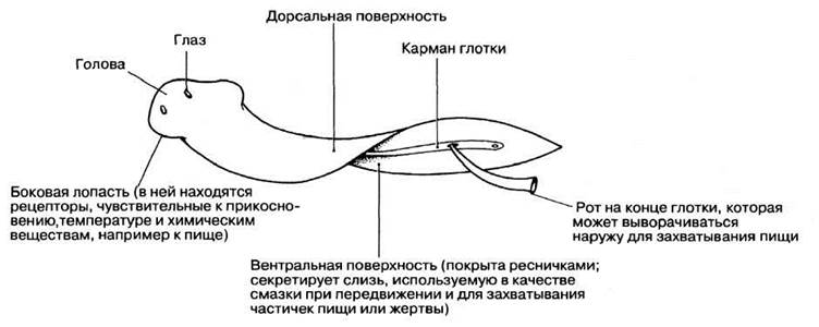
Fig. 2.49. External structure of a planarian.
On the ventral surface, closer to the posterior end, lies the mouth—the single opening through which the gut communicates with the external environment. The diet of planarians consists of small worms, crustaceans, and decaying remains of larger organisms.
Class Trematoda
The liver fluke (Fasciola hepatica) (Fig. 2.50) belongs to the class Trematoda, which constitutes one of the major parasitic groups in the animal kingdom. The liver fluke is an endoparasite, meaning it lives inside the host's body. Its habitat is the Bile ducts of the sheep, which serves as its primary or definitive host. Cattle and occasionally humans may also act as the primary host.
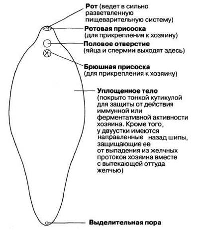
Fig. 2.50. External STRUCTURE OF THE liver fluke (Fasciola hepatica). The fluke inhabits the host's bile ducts.
Numerous differences exist between the liver fluke and the free-living planarian. These differences are evidently driven by the evolution of adaptations required to survive as an endoparasite. The parasitic lifestyle is associated with a complex life cycle comprising three larval stages (miracidium, redia, and cercaria), which helps maximize offspring numbers throughout The life cycle. Producing massive numbers of offspring compensates for the high mortality rate inevitable during host switching. A portion of the liver fluke's life cycle takes place within the body of a secondary (intermediate) host—the freshwater snail Limnaea (pond snail)—in whose tissues certain larval Stages of the parasite grow and reproduce.
At all stages of its life cycle, the fluke exhibits morphological, physiological, and reproductive adaptations to a parasitic way of life. Some of these are listed below and in Fig. 2.50. The Life Cycle of the liver fluke is shown in Fig. 2.51.
Fig. 2.51. Life cycle of the liver fluke.
ADULT FLUKE. The fluke's body is flattened and thin, adapted for living in the bile ducts. The body wall protects the fluke from the host's Enzymes, while secretions from glandular Cells located within the wall protect it against the action of the host's antitoxins.
The hermaphroditic Reproductive System (the presence of both male and FEMALE REPRODUCTIVE ORGANS in the same Organism) allows for both self-Fertilization and cross-fertilization. The liver fluke is also capable of surviving under conditions of oxygen deprivation.
MIRACIDIUM. The miracidium is the first larval stage in the developmental cycle of the fluke (Fig. 2.51). The main function of the miracidium is to locate an intermediate host, which requires Sensory Organs and powers of locomotion. In addition, the miracidium gives rise to new larvae (sporocysts; see below). The epidermis of the miracidium is covered with cilia, enabling the larva to swim in Water or in droplets of moisture clinging to vegetation.
Driven by specific chemical cues, miracidia swim toward their intermediate host, the pond snail. (The ability to move in response to a chemical stimulus is called chemotaxis.) Using its apical papilla, the miracidium attaches to the FOOT of the snail, while proteolytic (i.e., protein-digesting) enzymes secreted by the apical gland onto The surface of the foot facilitate penetration into the host tissues. Further entry is aided by muscle cells that assist the miracidium in burrowing into the snail's body. Thus, the miracidium migrates into the digestive gland of the snail. Within the miracidium are specialized Germ Cells from which the subsequent larval forms develop.
SPOROCYST. The Role of this stage is to multiply the number of larvae to compensate for losses stemming from the fact that many larvae fail to find a host. A sporocyst is a non-motile, enclosed sac containing germ cells that develop into rediae—the next larval stage.
REDIA. This is a reproductive and feeding stage. The larva at this stage possesses a muscular Pharynx used for sucking fluids from the host's tissues. Rediae move about with the help of muscle cells. Germ cells give rise to new rediae or cercariae. A new generation of rediae or cercariae exits through a specialized birth pore.
CERCARIA. This larva closely resembles the adult fluke in many respects. Specifically, it features oral and ventral suckers for attaching to a suitable substrate, such as grass. Additionally, it possesses a tail that enables it to swim in water or in moisture droplets on plants. These larvae possess cyst-secreting glands (Fig. 2.51). No further development occurs until the encysted cercaria is ingested by a sheep. Encysted cercariae are highly resistant to low temperatures, but sensitive to desiccation.
Limnaea is an amphibious pond snail inhabiting ponds, muddy river banks, and flooded meadows. It is able to endure adverse environmental conditions; consequently, the larval forms of the fluke developing inside its body (sporocysts and rediae) remain shielded from such unfavorable conditions. Indeed, at low temperatures, rediae produce daughter rediae rather than cercariae. The rediae stay within the body of the pond snail and can overwinter there. Cercariae begin to develop only in spring with the onset of warm weather. The pond snail also reproduces very rapidly. It has been estimated that in 12 weeks a single snail can produce up to 160,000 offspring. If all these offspring are infected with developing stages of the fluke, the probability of cercariae being released into the environment to infect new definitive hosts increases significantly. The amphibious lifestyle of Limnaea ensures that cercariae are deposited in a moist environment where they can disperse.
The release of young flukes from metacercariae takes place in the intestine of a sheep or cow. This process is triggered in The Stomach by a high concentration of carbon dioxide and elevated temperatures (up to 39 °C). Under these conditions, the parasite secretes Proteolytic Enzymes that digest the cyst wall, creating an opening in it. The Emergence of young flukes from the cyst is triggered by the presence of bile in the digestive juices of the Small Intestine.
After penetrating the intestinal wall, the newly emerged young flukes migrate to the liver. For some time they feed on liver tissue, but eventually, roughly 6 weeks post-infection, they permanently take up residence in the bile ducts.
The consequences of fluke infection for the primary host vary widely. In severe cases, fascioliasis can cause the death of the host. The migration of cercariae through the liver disrupts the organ's metabolic processes. This results in the destruction of liver cells and, consequently, the blockage of bile ducts; extensive erosion of the liver (cirrhosis) can lead to the development of ascites. Insufficient bile or a complete lack thereof in the intestine impairs Digestion, and the metabolic waste products of the fluke may exert toxic effects on the host.
Various Methods are employed to control flukes. Pasture drainage and the grazing of geese and ducks that feed on snails (biological control) are aimed at eradicating the intermediate hosts—pond snails (Limnaea). Artificial filling of ponds and The Use of drinking-water lifting systems also contribute to this goal. Soil liming can likewise be an effective measure, since the parasite's eggs cannot develop in an alkaline environment (pH 7.5). To rid infected sheep of the liver fluke, animals are administered carbon tetrachloride, which kills the flukes directly inside the liver.
Last update: 06/08/2026
Editorial and Educational Adaptation: This material has been compiled based on the primary/original source text. The project team performed an editorial review, corrected technical inaccuracies, structured sections, and adapted the content for an educational format.
What was processed:
- elimination of formatting defects (OCR errors, structural breaks, corrupted characters);
- editorial organization of content;
- standardization of terminology in accordance with academic sources;
- verification of factual statements against the original source text.
All mentions of the author, publication year, and origin of the primary text have been preserved in accordance with the source.