Practical Guide to Zoology: A Textbook - T. A. Dauda 2014
Multicellular Animals
Roundworms
Class Nematoda, or Roundworms
Nematodes represent the largest Class of roundworms, inhabiting marine environments, freshwater bodies, and damp soil. A vast number of species within this class are parasites affecting animals, humans, and plants alike.
The Scope of this practical guide does not permit a detailed examination of the entire diversity within this extensive group of roundworms. Therefore, we will focus on a thorough analysis of one of the major model organisms—the ascarid worm, along with Trichinella and the pinworm.
Equine ascarid (Parascaris equorum).
The equine ascarid frequently parasitizes the Small Intestine of horses. Anatomically and biologically, the porcine and human ascarids (Ascaris suum and A. lumbricoides) are very closely related to this species. Relatively rarely, they may be found in other Organs, such as the Liver, Lungs, Heart, and Urogenital System. The disease caused by this parasite is known as ascariasis. The Development of ascarids takes place within a single host (human, pig, or horse), though it requires the eggs to be released into the external environment.
Materials and equipment. Laboratory work should utilize the equine ascarid, which does not act as a human parasite in either the larval or adult stage (unlike the larvae of the porcine ascarid, which can survive in The Human Body). Material must be sourced from slaughterhouses, primarily from older or injured horses. Porcine ascarids, which are anatomically similar to equine ascarids, can also be obtained there. Specimens are fixed using 4% formalin or 50% alcohol.
The required equipment includes: a Microscope; a hand lens; dissection tools (scissors, scalpel, safety razor blade, forceps, dissecting needles, pins, dissecting trays); beakers or jars with Water; formalin-fixed ascarids (male and female); wet mounts of equine, porcine, and human ascarids; a wet mount of a dissected female ascarid; a microscopic cross-section slide; and a life cycle diagram.
Assignment. Examine and sketch the external Morphology of the equine ascarid. Compare it with the human and porcine ascarids. Dissect the ascarid to observe and sketch the general arrangement of Internal Organs. Isolate and examine the digestive and reproductive systems (in both male and female specimens). Examine the Skin-muscular sac. Locate the excretory canals, phagocytic Cells, and nerve trunks. Study the cross-section of the ascarid. Review the developmental cycle and the preventive measures against ascariasis.
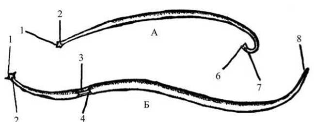
Fig. 51 External appearance of the ascarid:
A — male; B — female; 1 — oral aperture and Lips; 2 — excretory system pore; 3 — female genital pore; 4 — constriction on the female body (clitellum-like girdle); 5 — female anus; 6 — curled posterior end of the male body; 7 — cloacal aperture.
Examination of the fixed specimen and wet mounts. Observe the equine ascarid in a dissecting tray with a wax-lined bottom. This is a large worm characterized by a fusiform body externally covered by a dense, smooth cuticle. In living specimens, ascarids appear white, grayish, or pink, though fixation may alter their coloration.
Learn to distinguish males from females. Females reach a length of 17–40 cm, while males measure 15–25 cm. Aside from their smaller size, males differ from females by having the posterior end of the body curved into a hook shape (Fig. 51). Using a hand lens, locate the posterior and anterior ends of both male and female specimens. When examining the body surface of the ascarid, one can discern the tangled, tubular reproductive organs shining through, alongside four longitudinal stripes: one ventral, one dorsal, and two lateral. These are formed by thickenings (cords) of the hypodermis projecting into the body cavity. The lateral stripes are significantly more visible than the ventral and dorsal ones.
Examine the anterior end to locate the Mouth, surrounded by three lips. Find the anal opening in the female, situated on the ventral side slightly anterior to the posterior tip of the body. Examining the curved posterior end of the male in profile using a magnifier reveals small cuticular spicules protruding from the cloaca. These serve as copulatory organs, which the male uses to grasp the female's genital pore during mating. The slit-like genital pore of the female is located on the ventral side in the anterior third of the body (visible under a lens) within a ring-like depression. The male lacks a dedicated genital opening; reproductive products are emptied into the posterior section of the intestine, meaning the anal opening also Functions as a cloaca. The excretory pore, in both males and females, is likewise situated on the ventral side of the body, 3–4 mm posterior to the mouth (though not always visible even under magnification). Examine the wet mounts of equine, porcine, and human ascarids and compare them.
Dissection of the ascarid and study of its internal anatomy. Examine the organs concurrently on the dissected ascarid and via the wet mount. During dissection, care must be taken to prevent the coelomic fluid from spraying into the eyes, as this may trigger an inflammatory response.
Take formalin-fixed ascarids (male or female). Determine the sex of the specimen being dissected, and identify the lateral, ventral, and dorsal sides. Pin the worm down to the tray floor, ventral side down. Insert one pin into the anterior end (slightly to the side) and another into the posterior end. Holding the ascarid with forceps, carefully make a shallow longitudinal incision along the body wall, several centimeters long, using a scalpel or safety razor blade (along the middle region of the worm's body). Fold the edges of the incision outward and pin them to the bottom of the dish.
Using scissors, extend the incision toward the anterior and posterior ends of the body, pinning the cut edges to the wax-lined bottom of the tray. The pins should be inserted as obliquely as possible to keep the dissected specimen fully exposed. Pour water from a beaker or jar into the tray until the ascarid is submerged, and observe the general layout of the internal organs.
Cutting through the body wall opens the body cavity, which is filled with coelomic fluid. Within the cavity, the intestine is visible, entwined by the tubular Organs of the Reproductive System. Using dissecting needles and forceps, isolate the reproductive system, untangle and straighten out the loops of the reproductive organs (tears and damage are inevitable when working with fixed material), and examine them.
The female possesses a paired reproductive apparatus (Fig. 52). While examining the dissected female, locate the genital pore, which leads into a short Vagina that bifurcates into thicker tubes known as the uteri. Each Uterus transitions into a slender tube—the oviduct—which gradually merges without a sharp boundary into the thread-like Ovary.
Locate the reproductive organs in the dissected male: they are unpaired, consisting of a single thread-like Testis that gradually transitions into a thicker tube, the vas deferens, which empties into the Seminal Vesicle. Spermatozoa produced in the testis travel via the vas deferens to the seminal vesicle, where they accumulate. A muscular ejaculatory duct extends from the seminal vesicle and empties into the hindgut, which terminates in the cloaca.
Fertilization in the ascarid is internal. During copulation, the spicules protrude through the cloacal aperture and are inserted into the female's genital pore, widening it. The male injects spermatozoa into the vagina, and they migrate to the oviduct, where fertilization of the egg cells takes place. The fertilized eggs then enter the uterus and become encased in a protective shell.

Fig. 52 Internal Structure of the ascarid:
A — dissected female; B — male reproductive apparatus; C — posterior body tip in female and male; 1 — mouth and lips; 2 — Pharynx; 3 — intestine; 4 — circumesophageal nerve ring; 5 — ventral hypodermal cord with nerve trunk; 6 — right muscular fields; 7 — lateral hypodermal cord; 8 — ovary; 9 — oviduct; 10 — uterus; 11 — vagina; 12 — testis; 13 — vas deferens; 14 — ejaculatory duct; 15 — spicules; 16 — female anus, male cloacal aperture.
Once the reproductive organs have been removed, examine the digestive, excretory, and nervous systems, as well as the body wall.
Locate the mouth—it leads into a short, muscular pharynx, followed by the midgut, which appears as a grayish, thin-walled tube marked by longitudinal folds. Trace how it transitions without a distinct boundary into a brief hindgut, which terminates in the female's anal opening (in formalin-fixed ascarids, the intestine frequently fragments).
After removing the intestine, examine the body wall. Shiny and smooth on the outside due to the cuticle, it feels rough on the inside facing the body cavity. This roughness is caused by Muscle cells tightly fused with the skin (the musculocutaneous sac). Each muscle Cell is spindle-shaped, featuring a large process directed into the body cavity. Between the cuticle and the muscle cells lies a non-cellular epithelial layer, the hypodermis. It contains no separate cells, representing instead a protoplasmic mass with numerous nuclei. The longitudinal stripes—the hypodermal cords observed during external inspection—can be felt with a dissecting needle on the inner side of the musculocutaneous sac. Each cord is separated from the neighboring one by a layer of muscle cells. Within the lateral hypodermal cords, two excretory canals run along the body of the Ascaris. At the posterior end of the body, they end blindly, while at the anterior end, they merge and open via a common excretory pore.
During dissection, the lateral hypodermal cords are best visible. Locate four large, yellowish-brown, stellate cells—phagocytic cells—on the lateral cords within the anterior third of the body. They engulf insoluble Metabolic waste products and Bacteria that have penetrated the body cavity from the coelomic fluid. Their phagocytic ability can be proven by a simple experiment: if a living Ascaris is injected into the body cavity with carmine powder ground in saline solution, carmine granules concentrate within the protoplasm of the phagocytic cells within a few hours. Subsequently, the substances accumulated in them are apparently eliminated through the excretory canals.
Examine The Nervous system: it consists of two longitudinal nerve trunks running inside the ventral and dorsal hypodermal cords; they can be detected by gently scratching the corresponding hypodermal thickenings with a dissecting needle. Using a magnifying Glass, locate the nerve ring surrounding the pharynx (the circumpharyngeal nerve ring) that connects both trunks. In addition to the main trunks, there are several smaller ones that are not visible upon dissection or in cross-section. Ascaris lacks respiratory and circulatory organs.
Thoroughly wash your hands after dissection.
Study of an Ascaris cross-section slide. Examine the cross-section of a female Ascaris under low magnification, and certain details under high magnification of the microscope (see Fig. 53). Compare the Ascaris cross-section with the previously studied cross-section of the liver fluke, a representative of Flatworms. Examine the musculocutaneous sac of Ascaris under high magnification, including the cuticle, hypodermis, and the layer of muscle cells. Note that the muscle cells and their protoplasmic processes directly line the body cavity of Ascaris. The absence of an epithelial lining is characteristic of a pseudocoelom (primary body cavity).
Blister-like processes of the muscle cells are directed toward the nerve trunks. Locate the lateral hypodermal cords and the excretory canals within them. Orient the slide using the lateral hypodermal cords. Examine the dorsal and ventral hypodermal cords containing the nerve trunks. Observe the organs located in the body cavity under low magnification: the intestine, both uteri filled with eggs, the oviducts, and the Ovaries.
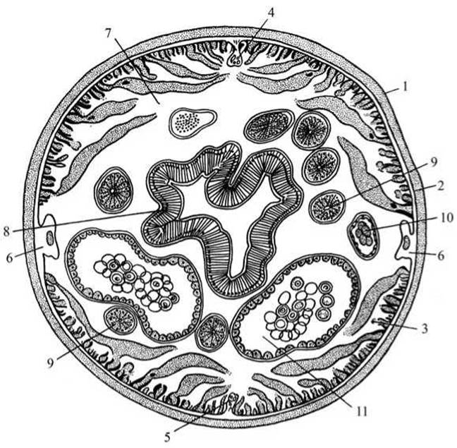
Fig. 53 Cross-section of a female Ascaris body:
1 — cuticle; 2 — hypodermis; 3 — muscle cells; 4 — dorsal nerve cord; 5 — ventral nerve cord; 6 — lateral hypodermal cords with the excretory system canal; 7 — primary body cavity (pseudocoelom); 8 — intestine; 9 — ovary; 10 — oviduct; 11 — uterus.
The shape of the intestine in cross-section varies depending on where the cut was made. The intestinal wall consists of a single layer of tall cylindrical epithelial cells. Numerous cross-sections of FEMALE REPRODUCTIVE SYSTEM tubes of varying diameters are visible around the intestine. Two large ovals or circles filled with eggs are sections of the Ascaris uteri. Sections of smaller diameter tubes represent oviducts, appearing as ovals or small circles. Ovarian sections are distinguished by smaller dimensions and contain an internal central axis called the rachis, along with radial striation.
Make a drawing of the female Ascaris cross-section.

Fig. 54 Life Cycle of the human roundworm (Ascaris lumbricoides)
Study the Ascaris developmental cycle using the diagram. Fertilized Ascaris eggs are eliminated into the external environment along with the host's excrement. The eggs cannot develop in the host's intestine because their development requires free atmospheric oxygen. Under favorable environmental conditions (oxygen, soil Temperature, and humidity), larvae begin to develop within the eggs, which possess unusually dense and durable membranes.
For further development, the egg containing the infective larva must enter the host's Organism (human for the human roundworm, horse for the equine roundworm, and pig for the porcine roundworm). Inside the host's intestine, the larva hatches from the egg, penetrates the wall of the small intestine, enters Blood Vessels, and undergoes a complex migration via the bloodstream (Fig. 54). The larva passes through the liver, enters the right atrium and right ventricle of The Heart via the systemic Circulation, and then travels through the Pulmonary Circulation into the lung capillaries. Through active movements, the larva pierces the walls of the pulmonary capillaries and enters the lung alveoli.
The presence of larvae in the lungs frequently causes pulmonary disorders. The larva grows, develops, and gradually moves upward through the respiratory tract—via the Bronchi and Trachea—into the pharynx, and from there, via saliva, into the Esophagus, Stomach, and small intestine. Upon entering the intestines for the second time, the larva reaches sexual maturity. The lifespan of an adult Ascaris in the human intestine is approximately one year.
Personal prophylaxis: observance of general hygienic rules—hand washing, protecting food from flies that carry Ascaris eggs, maintaining cleanliness, and consuming washed vegetables and fruits.
Public prophylaxis: protection of soil and water bodies from contamination by Human and Animal excrement, prohibition of using raw human and animal feces to fertilize garden soil without prior Treatment, and dehelminthization of humans and animals suffering from ascariasis.
Trichina worm (Trichinella spiralis).
The Significance of veterinary and sanitary inspection and the proper Organization of animal husbandry are clearly demonstrated when studying trichinae, or Trichinella (Fig. 55).
In its sexually mature state, Trichinella parasitizes the intestine of various carnivorous or omnivorous animals, both domestic and wild. In the larval stage, Trichinella encysts in the Muscles of the same animals.
Materials and equipment. Larval material is best obtained from a slaughterhouse from trichinellotic pigs, in which the muscles of the Diaphragm, intercostal muscles, Larynx, and neck are most heavily infested. Encysted larvae are studied either after clearing in glycerin or on carmine-stained preparations.

Fig. 55 Trichinella (Trichinella spiralis):
A — sexually mature female Trichinella; B — sexually mature male Trichinella; C — non-encysted muscle trichinae; D — encysted muscle trichinae: 1 — nerve ring; 2 — esophageal cells; 3 — female genital pore; 4 — uterus; 5 — ovary; 6 — midgut; 7 — testis;
8 — anus; 9 — cloacal aperture; 10 — host muscle fibers; 11 — muscle trichina; 12 — capsule surrounding the trichina.
To study sexually mature trichinae, one or two laboratory rats should be specifically fed trichinous meat. Within 30–40 hours after infection, young trichinae in the small intestine reach sexual maturity and copulate; 6–7 days post-infection, the females penetrate the intestinal wall, where they give birth to live larvae. Rats should be dissected before the females penetrate the intestinal wall, i.e., 4–5 days after infection!
Trichinae should be searched for in the Contents of the small intestines using a magnifying glass. Adult trichinae are studied *in vivo*, on material fixed in 70% alcohol, or on whole mounts.
Required equipment and materials: a microscope; whole-mount microscopic preparations of male and female trichinae; prepared microscopic slides of non-encapsulated and encapsulated trichinae in Muscle tissue; and a developmental cycle diagram.
Task. Examine the male and female trichina. Examine and sketch a preparation of trichinous meat containing non-encapsulated and encapsulated larvae. Study the developmental cycle of the trichina and control measures against trichinosis.
Examination of the whole-mount microscopic preparation.
Examine, without sketching, the whole-mount preparation of male and female trichinae under low and then high magnification of the microscope. These are small roundworms: the female is 3–4 mm long, and the male is 1.4–1.6 mm long. Like all nematodes, the body of trichinae is cylindrical, tapering toward the anterior end and slightly thickened at the posterior end (see Fig. 55). The male lacks spicules; their function is performed by two cutaneous processes located at the posterior end of the body. The Digestive System begins with the oral opening (lacking lips) leading into the esophagus, which is located in the slender anterior end of the body. This is followed by the Midgut and Hindgut, which opens via the anus (the cloaca in the male) at the posterior end of the body.
The circulatory and respiratory systems are absent, as in all roundworms. Trichinae are dioecious. The reproductive system, similar to that of ascarids, is tubular in structure, yet in both male and female it consists of a single unpaired tube. The reproductive system fills the entire body of adult trichinae and often hinders the examination of internal anatomy.
Locate the intestine, uterus, and female genital pore in the female, and the intestine and testis in the male.
Study of the trichina developmental cycle based on the diagram (Fig. 56).

Fig. 56. Diagram of The life cycle of Trichinella
Adult trichinae inhabit the small intestine of the host—this is the intestinal phase of trichinosis. Fertilization takes place within the host's intestine. Fertilized females penetrate the intestinal mucosa and enter the intestinal Lymphatic vessels. Here, each female produces up to 1,500 or more tiny larvae over a period of 1.5–2 months. The females then return to the intestinal lumen and perish. The larvae are disseminated throughout the host's body via the lymphatic and blood streams. Most frequently, trichina larvae encyst in the diaphragmatic muscles, intercostal muscles, and muscles of the Tongue, larynx, neck, and eyes. Here they actively penetrate inside the muscle fibers, feed by destroying the fiber, and grow—this represents the muscular phase of trichinosis development.
Two weeks later, the larva becomes quiescent, coils spirally, and a connective-tissue capsule forms around it. After approximately 6 months, the walls of the capsule begin to impregnate with layers of lime (becoming encapsulated), and the capsule turns milky-white with a characteristic lemon-like shape. Consequently, in this case, the same organism serves as both the definitive and intermediate host. The parasite is not released into the external environment. Encapsulated trichinae are hardy, capable of surviving for many years while remaining infective. For further dissemination of the parasite, animals must consume meat infected with encapsulated trichinae. Humans become infected by eating pork containing trichinae. Pigs become infected by eating dead rats or scraps of infected pork, whereas rats become infected by eating infected pork or each other.
Prophylaxis: veterinary and sanitary inspection of pork and pork products, proper hygienic maintenance of pigs, ensuring that pigs do not consume dead rats, and rodent control in pigsties.
Examination of microscopic preparations of trichinous meat. Under low microscope magnification, alternately darkening and illuminating the visual field, locate non-encapsulated trichinae within a piece of trichinous meat (among the muscle fibers). By moving the slide, observe the small worms—trichinae—lying within the muscle fibers in various positions; for the most part, they are spirally coiled.
Examine the muscle fibers containing trichinae under high magnification using the same preparation and sketch them.
Examination of the microscopic preparation of encapsulated larvae in muscle tissue. Under low and high microscope magnification, examine the striated muscle fibers and locate the capsules containing trichinae. Inside each capsule lies a spirally coiled small worm (about 5 mm in length)—the trichina larva. Sometimes two larvae are found within a single capsule. In capsules whose walls are heavily impregnated with calcium salts, the larvae are obscured, and the capsules assume a characteristic lemon-like shape. Sketch the preparation, highlighting the striated muscle fibers and the capsules containing trichina larvae.
Human pinworm (Enterobius vermicularis).
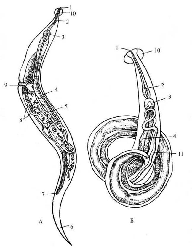
Fig. 57. Human pinworm (Enterobius vermicularis):
A — female; B — male: 1 — mouth; 2 — esophagus; 3 — bulb; 4 — midgut; 5 — ovary; 6 — caudal end; 7 — anus; 8 — uterus; 9 — female genital pore; 10 — vesicle; 11 — testis.
Pinworms (Fig. 57) are parasites inhabiting the small intestine, cecum, and Large Intestine of humans, predominantly children (Enterobius vermicularis), and horses (Oxyuris equi). Pinworm infestation causes enterobiasis in humans and oxyuriasis in horses.
Required equipment and materials: a microscope; a hand lens; forceps; dissecting needles; a watch glass; pinworms (males and females) fixed in formalin; and whole-mount microscopic slides of male and female specimens.
Task. Examine and sketch the human pinworm.
Examination of fixed specimens. Examine fixed pinworms—both male and female—placed in a watch glass using a hand lens. Learn to distinguish males from females based on body shape and size. Pinworms are small, white nematodes. Females are considerably larger: an adult female human pinworm reaches up to 10–12 mm in length, whereas the male is 2–5 mm (see Fig. 57).
The posterior end of the female's body is pointed, while in the male it is curved ventrally in a spiral. Note that, as in all roundworms, the body of the pinworm is fusiform and circular in cross-section.
Examination of a whole-mount stained preparation of a female pinworm. Observe the internal anatomy of a young female pinworm under low magnification of the microscope (in mature females, the highly developed reproductive system obscures the other internal organs, making them difficult to study).
Sketch the body outline and fill in the details of the internal structure.
By diming the microscope's field of view, locate a vesicle—a bladder-like cuticular Swelling at the anterior end—which the pinworm uses to anchor itself in the host's intestinal mucosa.
Examine the digestive system by moving the slide from the anterior to the posterior end of the body. Locate the mouth opening surrounded by three lips. Pinworms lack a pharynx. The esophagus is visible, consisting of a slender tube and a spherical expansion—the bulb—equipped with masticatory plates. Posterior to the bulb lies the intestine, in which the midgut and hindgut are difficult to differentiate. The intestine terminates in an anus situated at some distance from the posterior end of the body.
Like all roundworms, the circulatory and respiratory systems are absent. The nervous and excretory systems of pinworms are of the same type as those of ascarids. On some preparations, the lateral excretory canals can be distinguished. The reproductive system of pinworms is tubular, bearing structural similarities to that of ascarids.
Examine and sketch the tubular ovaries, oviducts, and the uteri packed with eggs. By dimming the field of view, locate the vagina and the genital pore on the lateral side in the anterior third of the body, and draw them.
Examination of a whole-mount stained preparation of a male pinworm. Study the internal Anatomy of the male under low magnification. The digestive System of the male differs from that of the female in that the hindgut merges with the ejaculatory duct to form a cloaca. Locate the mouth opening, vesicle, esophagus, bulb, intestine, and cloacal aperture. Identify the unpaired tubular reproductive organ of the male—the testis, vas deferens, ejaculatory duct, and spicule. Unlike the ascarid, the male pinworm possesses a single spicule.
Sketch the body shape and internal structure.
Last update: 13/08/2026
Editorial and Educational Adaptation: This material has been compiled based on the primary/original source text. The project team performed an editorial review, corrected technical inaccuracies, structured sections, and adapted the content for an educational format.
What was processed:
- elimination of formatting defects (OCR errors, structural breaks, corrupted characters);
- editorial organization of content;
- standardization of terminology in accordance with academic sources;
- verification of factual statements against the original source text.
All mentions of the author, publication year, and origin of the primary text have been preserved in accordance with the source.