Biochemistry: The Chemical Reactions of Living Cells, Volume 1 - D. Metzler 1980
How Molecules Interact with One Another
Muscles
Structural Organization of Striated Muscle
Among the vast array of complex structures assembled from protein subunits, none has commanded more attention than contractile Muscle fibers. The Human Body features several Types of Muscles. Striated skeletal muscles operate under voluntary control. Closely resembling them in Structure is The cardiac muscle, which exhibits a characteristic striated pattern and is involuntarily controlled. The third type comprises involuntary smooth muscles. Other species possess specialized muscle types; for instance, the asynchronous flight muscles of certain insects enable them to beat their wings at frequencies of ~100–1000 strokes per second. In these muscles, nerve impulses serve solely to «trigger» and halt wing movement, whereas the contraction–relaxation cycle proceeds automatically.
Striated muscles consist of bundles of long filaments (fibers) with a diameter of 10–100 µm, typically formed by the fusion of numerous embryonic Cells. In mammals, such fibers generally measure 2–3 cm in length, though they can occasionally reach up to 50 cm. Each fiber can be viewed as a single Cell containing anywhere from 100 to 200 nuclei. These cells contain conventional cellular Organelles, which nevertheless bear specialized names: for instance, The Plasma Membrane (Plasmalemma) of muscle cells (fibers) is termed the sarcolemma, their Cytoplasm is called the sarcoplasm, and their Mitochondria are referred to as sarcosomes.
The most distinctive feature of muscle cells is the presence of contractile myofibrils, which are specially organized bundles of protein molecules.
Under a Light Microscope, myofibrils reveal transverse striations spaced approximately 2.5 µm apart (Figs. 4-21 and 4-22). The region between two dense Z-discs, termed the sarcomere, serves as the primary contractile unit of the muscle cell. At the center of the sarcomere lies a dense anisotropic band (exhibiting strong birefringence) designated as the A-band. The Z-discs extend into less intense I-bands. Their name derives from the English word isotropic and is somewhat misleading because, although I-bands do not exhibit birefringence, they are not truly isotropic. Faintly stained M-lines, observable solely by Electron Microscopy, are located at the center of both the A-bands and the sarcomeres.
The detailed STRUCTURE OF THE sarcomere remained elusive until 1953, when H. Huxley, examining thin sections of muscle under an Electron microscope, discovered that protein filaments are arranged in a strictly ordered manner [83]. It turned out that thick filaments, 12–16 nm in diameter and ~1.5 µm in length, are packed in a hexagonal lattice with a spacing of 40–50 nm and span the entire A-band (Fig. 4-21, B). Interspersed between these thick filaments are thin filaments 8 nm in diameter, extending from the Z-disc for a distance of ~1.0 µm. Examination of muscle in a contracted state demonstrated that the I-bands nearly vanish, while the region of overlap between thick and thin filaments increases; this indicates that during contraction, the thin and thick filaments slide past one another. In Skeletal Muscle, the sarcomere shortens to ~1.7–1.8 µm; in insect flight muscle, this shortening is less pronounced, yet the contraction process repeats rapidly and continuously at a very high frequency.
Myofibrillar Proteins account for 50–60% of the total protein content in muscle cells. At low Ionic strength, these proteins are insoluble, but upon raising the ionic strength to ~0.3, they become soluble and can be extracted. Myosin, the principal muscle protein, forms the backbone of the thick filaments. Another protein, Actin, constitutes the primary structural component of the thin filaments (Fig. 7). Associated with the actin filaments are the muscle’s «regulatory proteins» — Tropomyosin and troponin [84] — while α-actin is found within the Z-disc. More recently, a protein designated as the M-protein has been identified as a component of the M-lines [85].
Myosin molecules take the form of very long, slender filaments measuring ~160 × 2 nm (Fig. 4-23). The greater part of the molecule consists of two presumably identical polypeptide chains folded into α-helices [86]. These chains are twisted around each other (Chap. 2, Sec. B.3.g). The C-terminal region of the molecule has a rod-like appearance; at the N-terminus, four subunits with a Molecular Weight of ~16,000–21,000 are associated with both heavy chains, forming a pair of «heads». Brief Trypsin Treatment cleaves the myosin molecule into two fragments. The tail region yields light meromyosin (LMM), a 90 nm long fragment, while the remainder, encompassing the «heads», forms heavy meromyosin (HMM). Heavy meromyosin can be further cleaved by prolonged trypsin treatment to yield a single 52-fragment 40 nm in length with a molecular weight of ~62,000 and two identical S1-fragments with a molecular weight of ~110,000, which represent the «heads» (Fig. 4-23).
Class="center">
FIG. 4-21. A. Schematic representation of The structure of a typical skeletal muscle sarcomere. The longitudinal section shown corresponds to the electron micrograph in Fig. 4-22. B. Diagram illustrating the arrangement of thick and thin filaments in striated muscle (cross-section). C. Left: Electron micrograph of a cross-section of glycerol-treated rabbit muscle. At the center of the circle, one can see that six thin filaments are positioned at the vertices of a hexagon surrounding a central thick filament. The other six thick filaments lie at the vertices of a larger hexagon. Right: Cross-section of a smooth muscle fiber. Thick and thin filaments are arranged in a non-ordered fashion. Filaments of intermediate thickness are visible, forming clusters known as «dense bodies» (1), the presence of which is a characteristic feature of smooth muscles.
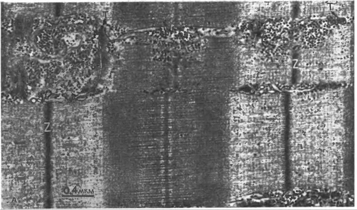
FIG. 4-22. A. Electron micrograph of a Cytology/practical/54.html">Longitudinal section of mammalian skeletal muscle (porcine biceps muscle). The tissue was double-fixed — first with formaldehyde and glutaraldehyde, and subsequently with osmium tetroxide — then stained with uranyl acetate and lead citrate. The photograph shows a white muscle fiber containing several mitochondria and narrow Z-discs. The M-line, A- and I-bands, as well as thick and thin filaments, are clearly visible. Note the periodicity (~40 nm) along the thin filaments; this distance corresponds to the length of the tropomyosin molecule, and the transverse striations are presumably due to the binding of tropomyosin and troponin. The numerous dense particles in the upper portion of the photograph are Glycogen granules, and the horizontal membrane-like structures are the transverse tubules of the sarcoplasmic (endoplasmic) reticulum. They are situated near the tubules extending from The surface of the muscle fiber. In the upper left corner of the photograph, on both sides of the Z-disc, longitudinal sections of T-tubules are visible, whereas cross-sections appear in the upper right corner. It can be seen that a T-tubule lies flanked by two terminal cisternae of the sarcoplasmic reticulum.

FIG. 4-22 B. Longitudinal section of smooth muscle (chicken gizzard); the specimen was fixed as described in the legend to Fig. 4-22, A. Thick filaments (1) are visible, which are substantially thicker than the thick filaments of striated muscle and exhibit a less orderly arrangement. They are surrounded by numerous thin filaments, frequently associated with «dense bodies» (2). A mitochondrion (3) is visible in the center of the photograph, and the boundary interface between two adjacent cells runs along the bottom. Note the pinocytotic vesicles, which are abundant in Plasma Membranes and highly active in smooth muscles. (Kindly provided by Marvin Stromer, Iowa State University.)
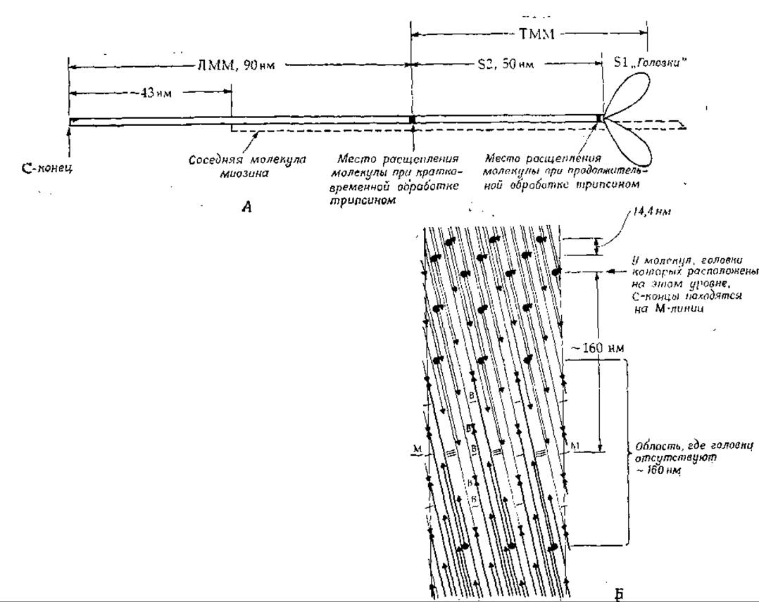
FIG. 4-23. A. Diagram of a myosin molecule. At a distance of 90 nm from the C-terminus lies the site where the molecule is cleaved upon brief trypsin treatment, yielding two fragments: light and heavy meromyosin (LMM and TMM). The total length of the myosin molecule is ~160 nm, with a molecular weight of 470,000; it consists of two heavy chains (molecular weight 200,000) and two pairs of HEAD light chains (molecular weight 16,000–21,000), measuring ~15 × 4 × 3 nm. B. Diagram proposed by Squire [87] illustrating the structure of vertebrate skeletal muscle thick filaments. The head-free (bare) zone near the M-line is shown. Dark circles denote the heads at the ends of the myosin molecules (rods), and dark triangles indicate the opposite ends of the myosin rods. Interactions between antiparallel molecules over spans of 43 and 130 nm are indicated by single and triple transverse lines, respectively. Opposing arrows (triangles) mark the tail-to-tail junction points of the myosin molecules (rods). The molecules extend from the center of the structure, where their C-termini are located, to the surface of the filaments, where their heads reside. At the levels designated by the letter B, an M-bridge attaches to the myosin filament. The M–M level marks the center of the M-line and the entire filament.
Dissolved myosin molecules can aggregate to form rods analogous to thick muscle filaments. Given that the diameter of such filaments is ~14 nm, A large number of myosin molecules (each 2 nm in diameter) must be packed within them. Electron microscopic studies have revealed that heads protrude from the thick filaments at intervals of ~43 nm. However, in the region of the M-line, these heads are absent, suggesting that aggregation of myosin monomers occurs here in a tail-to-tail fashion. It is postulated that approximately 300 myosin molecules (up to 30 rods in the filament cross-section) are densely packed within skeletal muscle myosin filaments [87], leaving a small central cavity (Fig. 4-23). In reality, the structure may be slightly different — there may be 3 (rather than 4) heads per 14.3 nm helical repeat [88]. Myosin filaments also contain small amounts of another protein, C-protein [88]. In insect flight muscles, the packing mode of myosin rods is different.
Actin possesses unique properties exclusive to this protein. Native fibrous F-actin (Fig. 7) is built from monomeric subunits with a molecular weight of ~43,000, each consisting of 374 amino acid residues. Interestingly, actin molecules contain an $N^\tau$-methylhistidine residue at position 73. In low ionic strength media in the presence of ATP, actin filaments can depolymerize into monomeric G-actin. Each G-actin molecule typically contains one bound ATP molecule and a calcium ion. The addition of Mg2+ to a concentration of 1 mM or KCl (0.1 M) triggers the spontaneous formation of filaments resembling thin muscle filaments, each containing 340–380 actin monomers. During this process, ATP is hydrolyzed, while ADP remains bound to the F-actin filaments. Strikingly, this process bears a remarkable resemblance to nucleotide binding by microtubule subunits (Supplement 4-A) and the events occurring during phage tail contraction (Supplement 4-D).
The fact that the actin-myosin complex is responsible for Muscle contraction was known long before the Fine Structure of myofibrils was elucidated. As early as 1929, it was established that ATP serves as the energy source for muscular contraction; however, a decade elapsed before Engelhardt and Lyubimova demonstrated that myosin preparations isolated from muscle catalyze ATP Hydrolysis [88a], thereby proving that the enzymatic mechanisms responsible for utilizing the Free energy released during ATP hydrolysis are intimately linked to the major proteins comprising muscle fibers. Later, A. Szent-Györgyi [88b, c] showed that Mg2+-stimulated ATP hydrolysis (ATPase activity) requires a complex of two proteins — actin1 and myosin (Actomyosin). He also discovered The phenomenon of superprecipitation, which consists of the rapid contraction of actomyosin in a test tube upon the addition of ATP. Although the question of whether superprecipitation is directly related to muscle contraction remains open, the discovery of this phenomenon played a pivotal role at the time by drawing researchers' attention to the cooperative interaction between Actin and myosin during Muscle Function.
1 Actin was discovered by F. Straub [88d].

FIG. 4-24. Hypothetical structure of the actomyosin–tropomyosin complex (viewed from top to bottom, along the axis of the actin filament) [92, 94]. The interaction of actin with the myosin S1 head and ATP is blocked by tropomyosin (solid circles). It is hypothesized that in the presence of Ca2+, tropomyosin shifts to the positions indicated by dashed circles, thereby relieving the steric block [93, 94].
Chemical studies of myosin have revealed that actin-activated ATPase activity resides within its «heads». Electron micrographs frequently show myosin heads linked to neighboring actin filaments, forming cross-bridges. When a skeletal muscle is in a relaxed state (unstimulated by a Nerve Impulse), the cross-bridges are disengaged (allowing the muscle to stretch freely). Conversely, when the muscle is activated and under tension, cross-bridges are readily visible in micrographs. Upon depletion of ATP reserves (i.e., rigor mortis), the muscle enters a state of stiffness. In this condition, nearly all cross-bridges are attached to the thin actin filaments, resulting in absolute muscular rigidity. These findings implied that during contraction, the heads of myosin filaments attach to the thin actin filaments. Subsequently, ATP hydrolysis is coupled, through as-yet-unknown mechanisms, to tension generation. The heads then detach and reattach at new sites. Repetition of this cycle drives the sliding of filaments past one another. The chemical mechanisms underlying these events remain poorly understood [89, 90]; whatever is known about them is detailed in Supplement 10-E.
The mechanisms regulating voluntary muscle contraction are no less remarkable than the contraction process itself. The endoplasmic (sarcoplasmic) reticulum in muscle cells exhibits a high degree of Organization [91, 92]. Connecting tubules run alongside the filaments, threading through bundles of contractile elements, and make close contact at precise intervals with folds of the outer cell membrane (the T-system of membranes, Fig. 4-22, A). A nerve impulse travels along the plasmalemma of the muscle fiber and reaches the T-tubules. At these close-contact sites, the signal is somehow transmitted to the connecting tubules of the sarcoplasmic reticulum—which harbor high concentrations of Calcium Ions—triggering the influx of these ions into the cytoplasm and myofibrils. Here, calcium binds to the C-subunit of troponin, an oligomeric protein that, together with tropomyosin (Chap. 2, Sec. B.3.g), forms a regulatory complex. It is this complex that associates with the actin filaments (Fig. 4-24).
If regulatory proteins are completely removed from actin filaments, contraction will proceed unabated until ATP reserves are exhausted. Conversely, in the presence of regulatory proteins and the absence of calcium, both contraction and ATP hydrolysis are inhibited. The working hypothesis explaining the function of this system [93, 94] posits that the elongated tropomyosin rods reside within the grooves between actin and the myosin heads [92]. Fig. 4-24 schematically illustrates the structure of the actomyosin–tropomyosin complex (top view). The myosin molecule's head (S1) is attached to one of the actin subunits. In resting muscle, tropomyosin lies close to the site on actin where the S1 domain binds. Consequently, the tropomyosin rod blocks the attachment of S1 myosin cross-bridges to actin and prevents actin-activated ATP hydrolysis. The tropomyosin molecule, measuring ~41 nm in length, contacts seven actin subunits simultaneously [95]. Thus, the tropomyosin–troponin complex synchronously controls the function of seven actin subunits.
The troponin molecule consists of three polypeptide chains with molecular weights ranging from 18,000 to 37,000 daltons. One polypeptide (T) firmly anchors troponin to tropomyosin at a site located approximately one-third of the distance from the C-terminus to the N-terminus, on the C-terminal side. A second polypeptide (I) component of troponin interacts with actin in the absence of Ca2+ ions and acts in concert with the other two Polypeptides to hold tropomyosin in a position where it inhibits ATP hydrolysis. When the third polypeptide (the C-subunit) binds calcium ions, this inhibition is lifted, and contraction can ensue. Nevertheless, the overall operational picture of this «machine» remains elusive. According to X-Ray Diffraction and electron microscopy data [93, 94], upon calcium binding to troponin, tropomyosin deflects away from S1 by approximately 20°, exposing the Active Site for the myosin–ATP–actin interaction (Fig. 4-24). Could tropomyosin be «rolling» like a ball bearing along the surface of actin, exposing binding sites on seven actin molecules simultaneously? If so, what sort of «motor» drives this motion, and what prevents the roller from «falling off» the actin track? We can only speculate. It is entirely possible that the side chains of specific amino acid residues on tropomyosin, protruding like Teeth on a submicroscopic gear, engage complementary depressions on actin1. This raises the question: why does calcium binding to tropomyosin cause it to roll along actin like a bearing? We know that metal binding to proteins can induce profound conformational changes (Sec. V.8.b). It is conceivable that the conformational shift in the troponin C-subunit is somehow transduced into the mechanical energy required for this movement.
1 It has been suggested that a series of magnesium ion bridges exists between the negatively charged regions of tropomyosin and actin [95a].
Last update: 06/08/2026
Editorial and Educational Adaptation: This material has been compiled based on the primary/original source text. The project team performed an editorial review, corrected technical inaccuracies, structured sections, and adapted the content for an educational format.
What was processed:
- elimination of formatting defects (OCR errors, structural breaks, corrupted characters);
- editorial organization of content;
- standardization of terminology in accordance with academic sources;
- verification of factual statements against the original source text.
All mentions of the author, publication year, and origin of the primary text have been preserved in accordance with the source.