Human Biochemistry, Volume 1 - Murray R. 1993
Bioenergetics and Metabolism of Carbohydrates and Lipids
Regulation of Lipid Metabolism and Sources of Energy in Tissues
Regulation of Fatty Acid Oxidation - Ketogenesis
Under certain metabolic conditions characterized by the rapid Oxidation of Fatty acids, significant amounts of acetoacetate and D(—)-3-hydroxybutyrate (ß-hydroxybutyrate) are produced in The Liver and diffuse into the bloodstream. Acetoacetate can undergo spontaneous decarboxylation to yield acetone. These three compounds are collectively known as Ketone Bodies (or acetone bodies), and are sometimes inappropriately referred to as “ketones”1 (Fig. 28.2). The reversible conversion of acetoacetate to 3-hydroxybutyrate is catalyzed by the mitochondrial enzyme D(—)-3-hydroxybutyrate dehydrogenase; the equilibrium is regulated by the [NAD+]/[NADH] ratio in the Cell/35.html">Mitochondria, i.e., by the cellular redox status. The [3-hydroxybutyrate]/[acetoacetate] ratio in Blood typically ranges between 1:1 and 10:1.
Under a well-balanced diet, the concentration of ketone bodies in the blood of mammals normally does not exceed 1 mg/100 mL (expressed in acetone equivalents). In ruminants, this figure is somewhat higher due to the ruminal Fermentation of butyric acid into 3-hydroxybutyrate within the rumen wall. In humans, less than 1 mg of ketone bodies is typically excreted in the urine per 24 hours. A condition characterized by an elevated concentration of ketone bodies in the blood or urine is termed ketonemia (hyperketonemia) or ketonuria, respectively. The overall syndrome is collectively known as Ketosis. Acetoacetic acid and 3-hydroxybutyric acid are moderately strong acids; in blood and Tissues, they exist in neutralized forms. Prolonged excretion of these acids leads to a loss of buffer cations (despite renal Ammonia Production), resulting in the depletion of alkaline reserves and the onset of ketoacidosis. In uncontrolled Diabetes Mellitus, ketoacidosis can have fatal consequences.
1 The term “blood ketones” should be avoided, because 3-hydroxybutyrate is not a ketone, and furthermore, blood contains several ketones that do not belong to the Class of ketone bodies, such as Pyruvate and fructose.

Fig. 28.2. Interconversion of ketone bodies. D(—)-3-Hydroxybutyrate dehydrogenase is a mitochondrial enzyme.
The simplest form of ketosis is observed during starvation, which involves the depletion of readily available carbohydrate reserves coupled with the mobilization of free Fatty acids. Qualitatively, the ketotic states arising from various conditions differ very little. Pronounced Metabolic Disorders leading to pathological states are observed in diabetes, bovine ketosis in lactating cows, and Pregnancy toxemia in sheep. Non-pathological forms of ketosis can occur on a high-fat diet and during heavy physical exertion in the postprandial period.
In all animals except ruminants, the liver appears to be the sole organ that supplies significant amounts of ketone bodies to the bloodstream. Extrahepatic tissues utilize them as substrates for oxidative processes. The extrahepatic sources of ketone bodies functioning in well-fed ruminants practically never induce a state of ketosis in these animals.
The flux of ketone bodies from the liver to extrahepatic tissues is driven by an active enzymatic mechanism for ketogenesis operating in the liver against a backdrop of very low hepatic activities of the Enzymes responsible for their utilization. The reverse situation is observed in extrahepatic tissues (Fig. 28.3).

Fig. 28.3. Formation, utilization, and excretion of ketone bodies. The major pathway is indicated by solid arrows.
The pathway of Ketogenesis in the Liver
The enzymes responsible for the synthesis of ketone bodies are located primarily within the mitochondria. It was formerly believed that The oxidation of a fatty acid molecule yielded only a single molecule of acetoacetate, derived from its four terminal carbon atoms. Later, to account for The formation of more than one equivalent of acetoacetate from a single long-chain fatty acid molecule, as well as the generation of ketone bodies from acetic acid, it was concluded that the two-carbon fragments produced during ß-Oxidation condense with one another to form acetoacetate. This Condensation occurs via the Reversal of the thiolytic Cleavage reaction, whereby 2 molecules of acetyl-CoA combine to form acetoacetyl-CoA. Thus, acetoacetyl-CoA, the starting compound for ketogenesis, is generated either directly during ß-oxidation or through the condensation of acetyl-CoA (Fig. 28.4). Two pathways have been proposed for the formation of acetoacetate from acetyl-CoA. The first is a simple deacylation, while the second (Fig. 28.5) involves the condensation of an acetoacetyl-CoA molecule with an acetyl-CoA molecule to yield 3-hydroxy-3-methylglutaryl-CoA (HMG-CoA), a reaction catalyzed by 3-hydroxy-3-methylglutaryl-CoA synthase. Under the action of another mitochondrial enzyme, 3-hydroxy-3-methylglutaryl-CoA lyase, acetyl-CoA is cleaved from HMG-CoA, releasing free acetoacetate. The carbon atoms of the cleaved acetyl-CoA molecule originally belonged to the acetoacetyl-CoA molecule (Fig. 28.5). For ketogenesis to proceed, both enzymes must be present within the mitochondria (such a combination of enzymes is found exclusively in the liver and ruminal epithelium).
It is now widely accepted that ketone body production occurs predominantly via the HMG-CoA pathway. Although a significant upregulation of HMG-CoA lyase is observed during starvation, available data do not indicate that this enzyme acts as the rate-limiting step in ketogenesis.
Acetoacetate can be converted into D(—)-3-hydroxybutyrate through the action of D(—)-3-hydroxybutyrate dehydrogenase, which is present in many tissues, including the liver. Quantitatively, D(—)-3-hydroxybutyrate is the predominant ketone body found in the blood and urine during ketosis.
Utilization of Ketone Bodies in Extrahepatic Tissues
An active mechanism for the synthesis of acetoacetate from acetoacetyl-CoA operates in the liver. The activation of the resulting acetoacetate can take place exclusively in the Cytosol, where it serves as a precursor for Cholesterol Biosynthesis; however, the capacity of this pathway is relatively low, leading to a net hepatic export of excess ketone bodies.
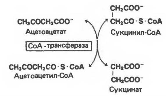
In extrahepatic tissues, two reactions lead to the activation of acetoacetate into acetoacetyl-CoA. One of these reactions utilizes succinyl-CoA and is catalyzed by succinyl-CoA:acetoacetate-CoA transferase. Acetoacetate reacts with succinyl-CoA, transferring the CoA moiety to acetoacetate to yield acetoacetyl-CoA and succinate.
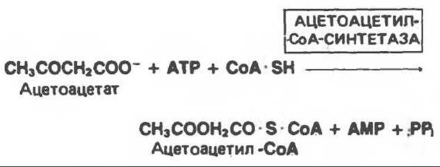
The other reaction is carried out by activating acetoacetate with ATP in the presence of CoA, a process catalyzed by acetoacetyl-CoA synthetase.
D(—)-3-Hydroxybutyrate can be directly activated by synthetase in extrahepatic tissues; however, the predominant pathway involves its conversion to acetoacetate, catalyzed by D(—)-3-hydroxybutyrate dehydrogenase in the presence of NAD+, followed by subsequent activation to form acetoacetyl-CoA. The acetoacetyl-CoA generated through these reactions is cleaved by thiolase to yield acetyl-CoA; the latter is then oxidized in The Citric Acid Cycle (Fig. 28.4).
Ketone bodies are oxidized in extrahepatic tissues in direct proportion to their concentration in the blood; they are oxidized preferentially over glucose and free fatty acids (FFAs). As blood ketone body levels rise, their oxidation increases until they saturate the oxidative machinery at a concentration of approximately 70 mg/100 mL. Under this condition, it appears that the majority of the oxygen consumed by the animal is dedicated to the oxidation of ketone bodies.

Fig. 28.4. The pathway of hepatic ketogenesis. FFA — free fatty acids; HMG — 3-hydroxy-3-methylglutaryl.

Fig. 28.5. Formation of acetoacetate from acetoacetyl-CoA (with HMG-CoA formed as an intermediate stage).
Most evidence indicates that ketonemia is caused by an increased production of ketone bodies in the liver rather than their insufficient utilization in extrahepatic tissues. At the same time, experiments on pancreatectomized rats show that in severe diabetes, ketosis can be exacerbated by a reduced capacity of the Organism for ketone body Catabolism. In moderate ketonemia, only a few percent of the total amount of produced ketone bodies are excreted in the urine.
Since threshold-like effects (which are, however, not true threshold effects) are observed during the renal excretion of ketone bodies and vary among species and individual organisms, the severity of ketogenesis should be judged by the blood ketone level rather than urinary excretion.
Acetoacetate and D(—)-3-hydroxybutyrate are readily oxidized in extrahepatic tissues, whereas the in vivo oxidation of acetone is difficult.
Regulation of Ketogenesis
Three stages are distinguished at which relevant factors can regulate ketogenesis. (1) Ketosis does not occur in vivo until there is an increase in the blood level of free fatty acids resulting from triacylglycerol lipolysis in adipose tissue. Fatty acids serve as precursors for ketone bodies in the liver. In both fed and starved animals, the liver has the capacity to take up 30% or more of the free fatty acids passing through it; therefore, at high concentrations of these acids, their uptake is quite substantial. Consequently, factors controlling the mobilization stage of free fatty acids from adipose tissue are crucial for The regulation of ketogenesis (Fig. 28.6). (2) Two pathways are possible for the utilization of free fatty acids after they enter the liver and are converted into active acyl-CoA derivatives, namely Esterification—yielding predominantly triacylglycerols and Phospholipids—and ß-oxidation to acetyl-CoA. (3) In turn, acetyl-CoA can either be oxidized in The Citric Acid cycle or enter the ketogenesis pathway to form ketone bodies.

Fig. 28.6. Regulation of ketogenesis. 1–3 — three KEY STAGES IN the free fatty acid (FFA) metabolic pathway that determine The amount of ketone bodies produced.
One of the possible antiketogenic regulatory factors is the esterification of free fatty acids, which depends on the availability of precursors in the liver ensuring the formation of sufficient glycerol 3-phosphate. However, in experiments with the perfused liver taken from a starved animal, the availability of glycerol 3-phosphate did not limit esterification. It remains unclear whether hepatic glycerol 3-phosphate availability always limits The rate of esterification; nor is there convincing evidence that in vivo the rate of esterification is limited by The activity of the corresponding enzymes. This is unlikely, as neither free fatty acids nor their esterification intermediates along the triacylglycerol synthesis pathway accumulate in the liver (Fig. 25.1). The activity of phosphatidate phosphohydrolase in the liver increases under conditions of accelerated triacylglycerol synthesis.
Experiments with the perfused liver demonstrated that the liver of fed rats esterifies significantly more 14C-free fatty acids than does the liver of starved animals; in the latter, the corresponding fraction of non-esterified fatty acids is oxidized to 14CO2 or 14C-ketone bodies. These results can be explained by the fact that The transport of long-chain acyl groups into the mitochondria, where ß-oxidation takes place, is regulated by carnitine palmitoyltransferase I located in the mitochondrial membrane (Fig. 23.1); this enzyme has low activity in fed animals, in whom Fatty acid oxidation is suppressed, and high activity during starvation, which is accompanied by enhanced fatty acid oxidation. McGarry and Foster (1980) showed that malonyl-CoA—an initial intermediate in fatty acid synthesis (see Fig. 23.5) whose concentration increases in the fed state—inhibits carnitine palmitoyltransferase and halts ß-oxidation. Thus, in the fed state, active Lipogenesis occurs, and a high concentration of malonyl-CoA is achieved, which inhibits carnitine palmitoyltransferase I (Fig. 28.7). If the concentration of free fatty acids in liver Cells is low, they are almost entirely converted via esterification into acylglycerols and exported from the liver as VLDL. However, at the onset of starvation, when the concentration of free fatty acids rises, acetyl-CoA carboxylase is inhibited and the malonyl-CoA concentration decreases; the inhibition of carnitine palmitoyltransferase is thus lifted, creating conditions for enhanced acyl-CoA oxidation. These processes are further promoted during starvation by a decreased [Insulin]/[Glucagon] ratio, which accelerates lipolysis and the release of free fatty acids in adipose tissue while inhibiting acetyl-CoA carboxylase in the liver.
When serum free fatty acid levels rise, a proportionally larger amount of free fatty acids is converted into ketone bodies and, correspondingly, less is oxidized to CO2 in the citric acid cycle. Through this regulation, acetyl-CoA is partitioned between the ketogenesis pathway and oxidation to CO2 in such a way that the Free energy stored as ATP during free fatty acid oxidation remains constant. Complete oxidation of 1 mole of palmitate via ß-oxidation and subsequent formation of CO2 in the citric acid cycle generates 129 moles of ATP (see Chapter 23); however, if the end product is acetoacetate, only 33 moles of ATP are produced, and if it is 3-hydroxybutyrate, only 21 moles. Consequently, ketogenesis can be viewed as a mechanism enabling the liver to oxidize large amounts of fatty acids using Reactions of the Oxidative Phosphorylation system while generating relatively few high-energy bonds.
Several other hypotheses have been proposed regarding the metabolic shift of fatty acid oxidation toward ketogenesis. Theoretically, a decrease in mitochondrial oxaloacetate concentration should reduce the capacity for acetyl-CoA METABOLISM in the citric acid cycle. The reduction in oxaloacetate concentration may be caused by an increased [NADH]/[NAD+] ratio during enhanced ß-oxidation. Krebs suggested that since oxaloacetate is also on the main gluconeogenic pathway, an enhancement of this process leading to lowered oxaloacetate levels could account for severe forms of ketosis, particularly in diabetes and bovine ketosis. Utter and Keech showed that pyruvate carboxylase, which catalyzes The conversion of pyruvate to oxaloacetate, is activated by acetyl-CoA. Consequently, when acetyl-CoA is abundant, a sufficient amount of oxaloacetate is required to trigger the condensation reaction in the citric acid cycle.
In Conclusion, ketosis results from a deficiency of available CARBOHYDRATES, a circumstance that promotes ketogenesis in the following ways (Figs. 28.6 and 28.7). (1) It leads to an imbalance between esterification and lipolysis in adipose tissue, causing free fatty acids to enter the bloodstream. These acids are the primary substrate for hepatic ketone body production; therefore, all metabolic and endocrine Factors influencing the release of free fatty acids from adipose tissue also affect ketogenesis. (2) Once free fatty acids enter the liver, their partitioning between esterification and oxidation pathways is regulated by carnitine palmitoyltransferase I, whose activity depends (indirectly) on free fatty acid concentration and the hormonal status of the liver. (3) As the amount of oxidized fatty acids increases, the proportion of produced ketone bodies rises while the proportion of substrate catabolized to CO2 decreases, with regulatory mechanisms maintaining a constant overall ATP yield.
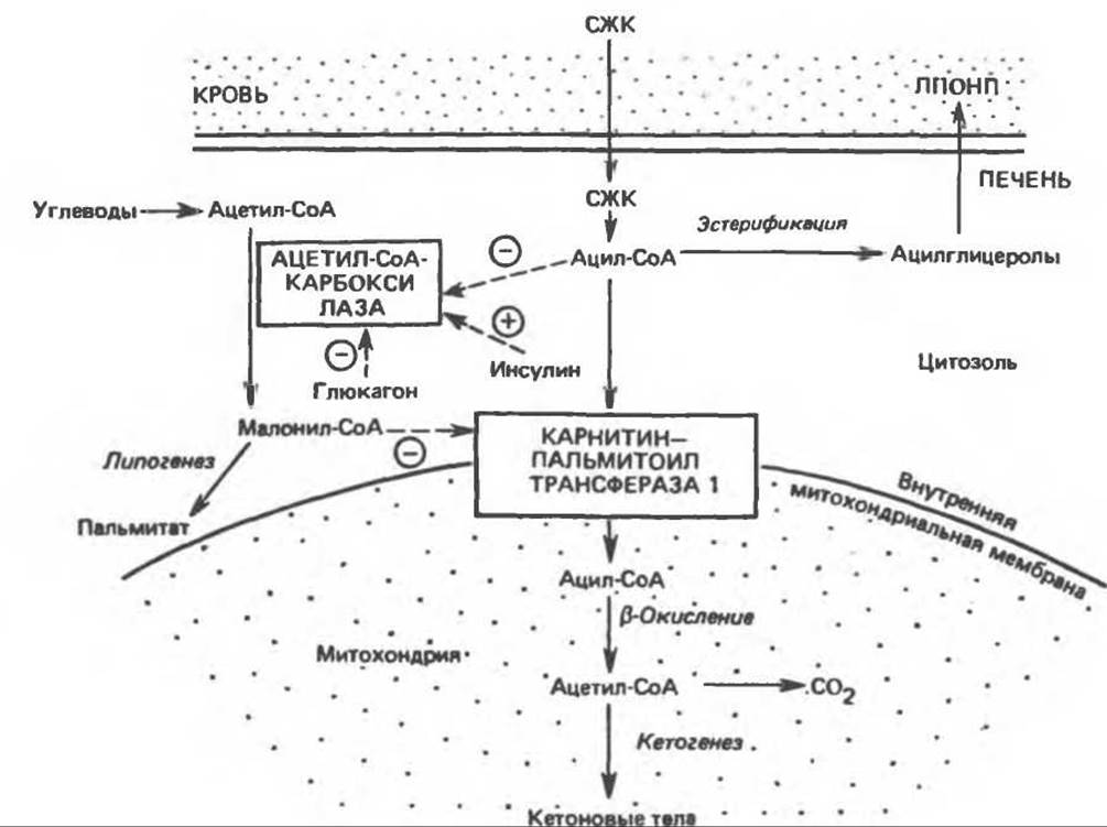
Fig. 28.7. Regulation of long-chain fatty acid oxidation in the liver. FFA — free fatty acids, VLDL — very low-density Lipoproteins. Dashed lines indicate positive (⊕) and negative (⊝) regulatory effects, and solid lines represent substrate flux.
Ketosis In Vivo
Ketosis observed during starvation and high-fat diets is relatively mild compared to the ketosis that develops in uncontrolled diabetes, pregnancy toxemia in sheep, or Lactation ketosis in cattle. The main reason for this appears to be that in these pathological conditions, the amount of carbohydrates available to tissues is significantly lower than during starvation or high-fat feeding. Thus, in moderate diabetes, high-fat diets, and chronic starvation, hepatic Glycogen is preserved (in varying amounts), and the decrease in free fatty acid levels is less pronounced. This probably accounts for the less severe ketosis observed in these cases. In ruminants, ketosis occurs against the Background of a marked drop in blood glucose, associated with the metabolic demands of the fetus or Mammary Glands during intense lactation (Fig. 28.8). As a result, ruminants may develop acute hypoglycemia, in which virtually no glycogen remains in the liver. Under these conditions, ketosis takes a severe form. As hypoglycemia progresses, insulin secretion decreases, which not only diminishes glucose utilization but also enhances lipolysis in adipose tissue.
In diabetes mellitus, insulin deficiency or absence likely affects primarily the metabolism of adipose tissue, which is extremely sensitive to this hormone. As a result of the release of large amounts of free fatty acids, their plasma concentration can be more than twice as high as that of a healthy starved individual. Changes in the activity of several hepatic enzymes are also observed, resulting in elevated rates of Gluconeogenesis and glucose output into the bloodstream (despite high blood glucose concentrations).
Last update: 06/08/2026
Editorial and Educational Adaptation: This material has been compiled based on the primary/original source text. The project team performed an editorial review, corrected technical inaccuracies, structured sections, and adapted the content for an educational format.
What was processed:
- elimination of formatting defects (OCR errors, structural breaks, corrupted characters);
- editorial organization of content;
- standardization of terminology in accordance with academic sources;
- verification of factual statements against the original source text.
All mentions of the author, publication year, and origin of the primary text have been preserved in accordance with the source.