Principles of Biochemistry Volume 1 - A. Lehninger 1985
Biomolecules
Globular Proteins: Structure and Function of Hemoglobin
X-ray crystallography of myoglobin - a landmark achievement in protein research
The answer to the questions posed above was provided by one of the most powerful Methods—X-Ray Diffraction Analysis, which, as we have already seen, has successfully resolved The Structure of several Fibrillar Proteins. However, the X-ray analysis of Globular proteins is a much more challenging task than that of fibrillar proteins, which are stretched along a single axis and typically exhibit a periodic structure. Determining the three-dimensional Introduction/12.html">Structure of Globular proteins from their X-ray diffraction patterns requires extensive calculations using powerful computing resources.
The first major breakthrough in resolving the three-dimensional structure of globular proteins came from X-ray diffraction studies of Myoglobin, carried out in England in the 1950s by John Kendrew and his colleagues. Myoglobin is a relatively small oxygen-binding protein (molecular weight 16,700) found in Muscle Cells. Its function is to store bound oxygen and facilitate its transport to Cell/35.html">Mitochondria, which consume oxygen during The oxidation of nutrients entering The Cell. The myoglobin molecule consists of a single polypeptide chain of 153 amino acid residues with a known sequence, and a single heme group—a complex of protoporphyrin and iron (Fig. 8-2), which is also found in Hemoglobin, the oxygen-binding protein of red Blood Cells. The presence of the heme group accounts for the deep red-brown color characteristic of both Myoglobin and hemoglobin. Myoglobin is particularly abundant in the Muscles of marine mammals, such as whales, seals, and dolphins, where its high concentration gives their muscles a dark brown color. Myoglobin allows these animals to store sufficient oxygen in their muscles during prolonged dives.
Class="center">
Fig. 8-2. The heme group present in myoglobin, hemoglobin, and many other Hemoproteins. It is a complex polycyclic structure called protoporphyrin, bound to an iron atom in its [Fe (II)] state (ferrous form). The iron atom has six coordination bonds, four of which lie in the plane of, and are bound to, the flat porphyrin ring system, while the other two are perpendicular to it. In myoglobin and hemoglobin, one of these two perpendicular bonds is occupied by a nitrogen atom from a Histidine residue. The other bond is vacant and serves to bind an oxygen molecule, as shown in the side view at the bottom right. In myoglobin and hemoglobin, carbon monoxide (CO) can compete with O2 for this vacant site, binding to the iron atom 200 times more tightly than O2. In Carbon monoxide poisoning, a significant portion of hemoglobin is converted to carboxyhemoglobin, preventing The transport of O2 from the Lungs to the Tissues.
The X-ray diffraction patterns used by Kendrew to study the structure of sperm whale myoglobin (Fig. 8-3) were highly complex, containing nearly 25,000 reflections. The calculations required to analyze the intensities of all these diffracted X-rays were carried out in successive stages. In The First stage, completed in 1957, the three-dimensional structure of myoglobin was resolved at 0.6 nm resolution. Although this level of resolution was insufficient to determine the exact positions of individual atoms, it revealed how the polypeptide chain is folded. It turned out to be folded in a rather complex and irregular manner, so that the outline of myoglobin's tertiary structure resembled a folded sausage (Fig. 8-4). Because the R-groups are omitted from the diagram, the molecular structure appears much more open than it actually is. The figure also shows the flat heme group, which is tightly nestled against the polypeptide chain, though not covalently bound to it. In the second stage, the X-ray analysis of myoglobin was performed at 0.2 nm resolution, which was high enough to identify most of the R-groups. In the Third Stage, all amino acid residues were identified at a resolution of 0.14 nm. The resulting Amino Acid Sequence agreed remarkably well with chemical analysis data.
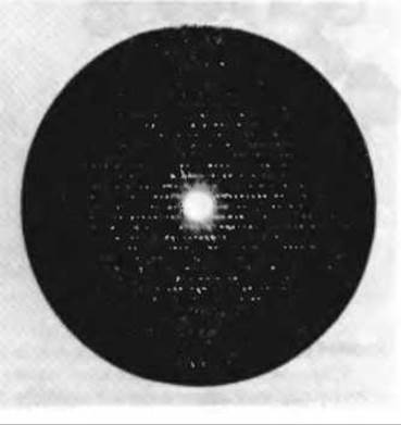
Fig. 8-3. An X-ray diffraction photograph of crystalline sperm whale myoglobin. The precise three-dimensional structure of myoglobin was calculated from the positions and intensities of the diffraction spots resulting from the interaction of the X-ray beam with the myoglobin atoms in the crystal.
Figure 8-4 shows the detailed, residue-by-residue Secondary structure of the myoglobin polypeptide backbone, depicted within the general sausage-like outline of the molecule, as well as its tertiary structure—the three-dimensional folding of the entire chain. The backbone of the myoglobin molecule consists of eight relatively straight segments interrupted by bends in the polypeptide chain. Each straight segment is an α-Helix; the longest contains 23 amino acid residues, and the shortest has only 7. All of these helical segments proved to be right-handed α-helices. Approximately 80% of The amino acid residues in the myoglobin molecule reside in these α-helical regions. X-ray diffraction analysis also revealed the precise Location of each of the R-groups, which project outward from the contours shown in the figure and fill virtually all the space between the loops of the molecule.
Several other important Conclusions were drawn from the precise models of the myoglobin molecule constructed from the X-ray data.
1. The myoglobin molecule is so compact that only four Water molecules can fit inside it.
2. All polar R-groups, with only two exceptions, are located on the outer surface of the molecule, and all of them are hydrated.

Fig. 8-4. The Tertiary Structure of sperm whale myoglobin as resolved by X-ray diffraction. The backbone structure shown was obtained at 0.2 nm resolution. The polypeptide chain, showing only the α-carbon backbone, has a sausage-like contour. The space between the loops of the chain is not empty but is filled with R-groups (not shown). The myoglobin molecule contains eight α-helical segments. The porphyrin ring of the heme group is highlighted in red.
3. Most of the hydrophobic R-groups are located in the interior of the myoglobin molecule and are thus shielded from contact with water (see Table 8-1, which lists the Amino Acids with the most hydrophobic and most hydrophilic R-groups, as well as those with R-groups of intermediate character).
4. Each of the four Proline residues in the myoglobin molecule is located at a bend in the polypeptide chain (recall that the rigid R-groups of proline cannot be accommodated in an α-helix; Section 7.7). Other bends or turns in the chain contain residues of Serine, Threonine, and asparagine—amino acid residues that do not favor α-helix formation when clustered together (Section 7.7).
Table 8-1. Classification of amino Acids According to Their Polarity and Location in Globular Protein Molecules
|
Highly hydrophilic amino acids, almost always located on the outer surface of globular protein molecules |
|
|
Aspartic acid |
|
|
Glutamic acid |
|
|
Asparagine |
Histidine |
|
Glutamine |
|
|
Highly hydrophobic amino acids, located mainly in the interior of globular protein molecules |
|
|
Phenylalanine |
|
|
Leucine |
Valine |
|
Isoleucine |
|
|
Amino acids of intermediate polarity; they can be found both in the interior and On the surface of globular proteins |
|
|
Proline |
|
|
Threonine |
|
|
Serine |
|
5. All peptide groups have a planar trans configuration (Section 7.5).
6. The flat heme group lies in a pocket near The surface of the molecule. The iron atom at the center of the heme group has two coordination bonds perpendicular to the plane of the heme. One of these is bound to the R-group of histidine residue 93, and the other serves to bind the O2 molecule.
Last update: 06/08/2026
Editorial and Educational Adaptation: This material has been compiled based on the primary/original source text. The project team performed an editorial review, corrected technical inaccuracies, structured sections, and adapted the content for an educational format.
What was processed:
- elimination of formatting defects (OCR errors, structural breaks, corrupted characters);
- editorial organization of content;
- standardization of terminology in accordance with academic sources;
- verification of factual statements against the original source text.
All mentions of the author, publication year, and origin of the primary text have been preserved in accordance with the source.