Fundamentals of Biochemistry - A. A. Anisimov 1986
Carbohydrates
Carbohydrate interconversions, enzymatic synthesis, and degradation
6.6.1. Enzymatic Interconversions of Monosaccharides
The primary simple CARBOHYDRATES formed during Photosynthesis are phosphotrioses—glyceraldehyde-3-phosphate and dihydroxyacetone phosphate—which give rise to all monosaccharides and other carbohydrates. Upon entering Condensation Reactions Catalyzed by aldolase, they form fructose-1,6-bisphosphate. The latter, by partially or fully losing its phosphate groups, can be converted into other hexoses (glucose, mannose, and their sugar phosphates) or participate in the synthesis of sucrose and starch.
Monosaccharide interconversions occur through the action of specific Enzymes at the level of sugar phosphates and nucleoside diphosphate sugars. Under the action of hexokinase, glucose is converted into glucose-6-phosphate:
Class="center">
Similar reactions are known for other classes of carbohydrates. The interconversion of monosaccharides itself is carried out by isomerases. For example, the enzyme glucose-phosphate isomerase catalyzes the reversible conversion of glucose-6-phosphate into fructose-6-phosphate, while mannose-phosphate isomerase catalyzes The conversion of mannose-6-phosphate into fructose-6-phosphate. Both enzymatic actions proceed via an enediol intermediate, and both isomerases are stereospecific.
The formation of free monosaccharides from their sugar phosphates is mediated by Phosphatases, which are extremely widespread in plants, microorganisms, and animals.
Plants contain isomerases that catalyze the interconversion of uronic acids (e.g., UDP-glucuronic acid ⇄ UDP-galacturonic acid) and certain pentoses (e.g., UDP-xylose ⇄ UDP-arabinose).
Galactose is converted into glucose-1-phosphate According to the following pathway:
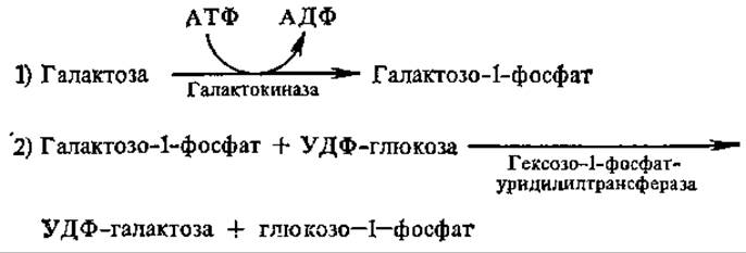
The reactions involved in the conversion of galactose to glucose have drawn significant attention due to a hereditary disorder known as galactosemia, which is caused by a genetic deficiency in hexose-1-phosphate uridylyltransferase activity. As a result, an infant's Organism1 loses The ability to metabolize galactose derived from milk lactose. The activity of another enzyme found in the Liver of adults—galactose-1-phosphate uridylyltransferase, which directly synthesizes UDP-galactose from UTP and galactose-1-phosphate—is detected only in trace amounts in infants, which is insufficient for Galactose METABOLISM.
The reverse process, the conversion of glucose to galactose, is of great interest with regard to lactose synthesis in the mammary gland, since the gland receives solely glucose via the Blood supply. It proceeds via the following pathway:
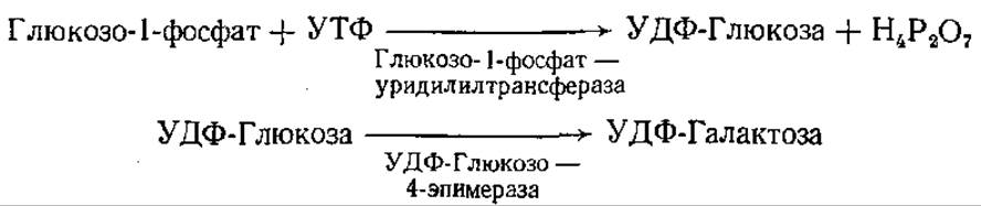
The formation of pentoses within The Cell can occur through various pathways. In many cases, the primary mechanism of pentose Biosynthesis is the Pentose Phosphate Pathway of Carbohydrate Oxidation (see Section 6.9.3), while in plants, the photosynthetic Calvin cycle serves as an additional mechanism.
Furthermore, pentoses can be formed from UDP-uronic acids via decarboxylation. For instance, xylose is derived from glucuronic acid, and arabinose from galacturonic acid. Plants are particularly characterized by transformations following this scheme:

It is known that plant aldolase exhibits broad substrate Specificity and can catalyze The biosynthesis of both hexoses (fructose) and pentoses. Because of its activity, dihydroxyacetone phosphate can condense not only with glyceraldehyde but also with a series of other aldehydes (such as acetaldehyde and glycolaldehyde), thereby producing pentoses.
6.6.2. Biosynthesis of Oligo- and Polysaccharides. The Role of Nucleoside Diphosphate Sugars (NDPS) in Polysaccharide Biosynthesis. Since the Hydrolysis of a single glycosidic bond in an oligo- or polysaccharide molecule is accompanied by the release of ~16.8 kJ of energy, and even 29.3 kJ for sucrose, the formation of a glycosidic bond is only possible with an input of the required energy. Therefore, the synthesis of oligo- and polysaccharides in living Cells cannot be explained simply by the Reversal of the action of corresponding Hydrolases.
Experimental confirmation has been established for those pathways of Polysaccharide synthesis in which the reacting species are not free monosaccharides, but rather their derivatives with an elevated level of Free energy, where reactions follow a substitution (transfer) mechanism rather than simple molecular addition. During group transfer and substitution reactions, no significant changes in free energy typically occur, allowing these reactions to proceed readily in both directions. This explains the reversibility in certain cases of oligo- and polysaccharide phosphorolysis: the products of the Cleavage action of Glycosyltransferases—sugar phosphates—possess a sufficiently high ester bond Free energy of ~15–20 kJ/mol.
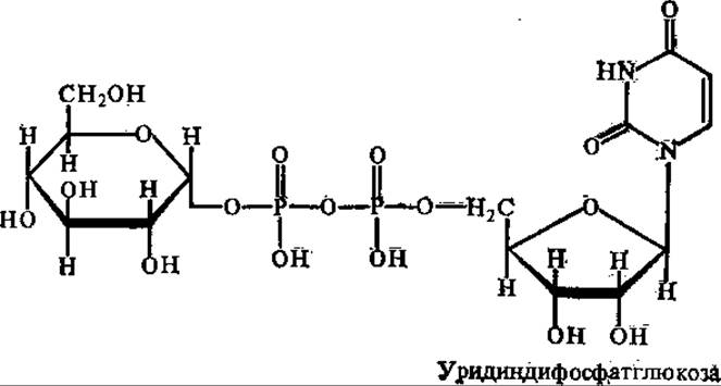
Through the work of Argentine biochemist L. Leloir and his coworkers in the 1960s, it was established that NDPS serve as characteristic Donors of glycosyl residues for polysaccharide synthesis: UDP-glucose, ADP-glucose, UDP-glucosamine, UDP-xylose, etc.
The free energy of the bond between glycosyl residues and nucleoside diphosphates (NDP) is relatively high; in UDP-glucose, for example, it exceeds 30 kJ/mol, which is entirely sufficient for the synthesis of glycosidic bonds in polysaccharides.
NDPS are formed from nucleoside 5'-triphosphates and sugar 1-phosphates through the action of enzymes collectively known as nucleotidyltransferases.
Nucleoside triphosphate (NTP) + Sugar-1-phosphate NDPS + H4P2O7
No significant changes in free energy occur during this reaction; however, due to the subsequent hydrolysis of H4P2O7 by inorganic pyrophosphatases (which are virtually always highly active in cells), the process becomes irreversible.
Further polysaccharide synthesis proceeds according to the scheme: NDPS + acceptor → NDP + sugar-acceptor. Reactions of this type are catalyzed by enzymes belonging to the glycosyltransferase subclass, whose trivial names are derived from the carbohydrate being synthesized: sucrose synthase, Glycogen synthase, etc., with NDPS acting as their Coenzymes. If a monosaccharide Functions as the acceptor, a disaccharide is synthesized; however, the acceptor can be any other carbohydrate, and its interaction with NDPS increases the molecular size by 1 glycosyl residue. With repeated iterations of the reaction, the acceptor molecule—or "primer"—is elongated by 1 glycosyl unit each time.
The recurrence of the reaction is sustained by the regeneration of NDP into NDPS:
NDP + ATP → NTP + ADP
NTP + Sugar-1-phosphate → NDPS + H4P2O7
As a result of this entire reaction chain, NDPS is regenerated; as a component of the catalyst (enzyme), it is not consumed and does not become part of the end products—these properties of all catalysts remain uncompromised.
It is currently believed that the examined reaction system involving NDPS serves as the primary pathway for the biosynthesis of oligo- and polysaccharides.
Sucrose biosynthesis. The biosynthesis of sucrose proceeds via two main pathways.
1. UDP-glucose + Fructose-6-phosphate → UDP + Sucrose phosphate. This reaction is catalyzed by sucrose phosphate synthase. Subsequently, the resulting sucrose phosphate yields sucrose and H3PO4 under the action of phosphatase.
2. UDP-glucose + Fructose ⇄ UDP + Sucrose. Another enzyme, sucrose synthase, takes part in this reaction.
It has been established that deoxyuridine diphosphate glucose is utilized just as effectively as UDP-glucose, whereas ADP-glucose is utilized significantly less effectively.
The Physiological Role of the two aforementioned enzymes differs: sucrose phosphate synthase is characteristic of chlorophyll-containing Tissues, whereas sucrose synthase is found in non-chlorophyllous tissues, where its activity is directed more toward the formation of UDP-glucose than of sucrose. The synthesis of sucrose involving sucrose phosphate synthase requires a substantial energy input, rendering this reaction irreversible. Energy is required to activate the reacting glucose and fructose according to the following series of reactions:
ATP + Glucose → Glucose-6-phosphate + ADP
Glucose-6-phosphate ⇄ Glucose-1-phosphate UTP + Glucose-1-phosphate → UDP-Glucose + PPi
ATP + Fructose Fructose-6-phosphate + ADP
Thus, the formation of a single glycosidic bond in sucrose requires three high-energy bonds of NTP. On the other hand, owing to the strongly exergonic nature of this reaction, the accumulation of high concentrations of sucrose is possible in the sap of certain plants (sugarcane, sugar beet) despite a very low content of its precursors. Establishing the fact that UDP-glucose participates in sucrose synthesis is of great importance, as it plays a paramount role in the synthesis of plant Glycosides in general as well.
Certain Bacteria contain the enzyme sucrose glucosyltransferase (sucrose phosphorylase), which catalyzes the reversible reaction:
Sucrose + Pi ⇄ Glucose-1-phosphate + Fructose
If inorganic phosphate is rapidly removed from the reaction medium, this reaction can lead to sucrose synthesis; however, under intracellular conditions, sucrose degradation typically takes place.
Lactose biosynthesis. The disaccharide lactose is formed in the Mammary Glands of mammals through the following reaction: UDP-galactose + D-Glucose → UDP + Lactose. The enzyme lactose synthase, which catalyzes this reaction, is complex and consists of two subunits: protein A, found not only in the mammary gland but also in The Liver and Small Intestine, and protein B, which lacks catalytic activity but alters the Specificity of protein A such that protein A can use exclusively D-glucose as a galactose acceptor rather than N-acetyl-D-glucosamine, as occurs in the absence of protein B.
Biosynthesis of glycogen and starch. Excess sugars synthesized within the organism or ingested with food form reserve deposits, primarily in the form of starch in plants and glycogen in animals.
Starch synthesis in plants is catalyzed by several enzymes. Starch synthase (UDPG- or ADPG-α-glucan glucosyltransferase), utilizing ADP-glucose (less frequently UDP-glucose) and an acceptor primer consisting of four or more glucose residues (i.e., a dextrin), synthesizes linear amylose chains and the unbranched regions of amylopectin.

L. Leloir (1964) established that the most active donor of glucosyl residues for starch synthesis in plant storage Organs is ADP-glucose. It is known that starch accumulating in leaves during photosynthesis can easily be converted into sucrose—the primary transport form of carbohydrates in plants. In this form, the synthesized carbohydrates flow into seeds, tubers, and bulbs, where they are stored again as starch or Fructans (inulin, levulosans). Amylases do not take part in this process; the primary role belongs to transglycosylation reactions involving NDPS. Sucrose can be converted into starch via two pathways:
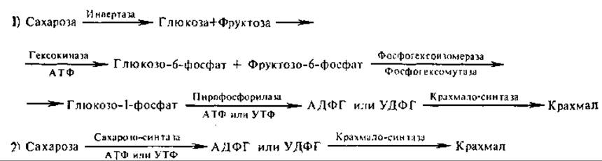
The synthesis of starch via the first pathway consumes 4 moles of ATP, whereas the second pathway consumes only 2, making it a more economical and probable route for starch synthesis from sucrose. The conversion of sucrose into levulosans proceeds just as readily. In bacteria, various dextrans (glucans) and levans (fructans) are synthesized from sucrose in a similar manner.
In vitro, the formation of linear amylose chains with α(1→4)-linkages in the presence of a primer dextrin can also occur via the reversal of phosphorolysis according to the reaction: Glucose-1-phosphate + Acceptor ⇄ α(1→4)-Glucosyl-acceptor + Orthophosphate. However, the feasibility of such synthesis in vivo involving the enzyme phosphorylase has not been proven. In plants, starch phosphorylase can catalyze only the synthesis of short primer dextrin chains, which are subsequently elongated into amylose molecules by the action of ADPG-α-glucan glucosyltransferase (starch synthase).
Plant phosphorylases differ significantly from animal ones. For instance, unlike the Muscle enzyme, potato phosphorylase lacks Serine phosphate and does not require AMP as a cofactor. Neither starch synthase nor phosphorylase is capable of catalyzing the formation of a(1→6) bonds in amylopectin, which are characteristic of branching points. This reaction is carried out by enzyme Q (branching factor), a transglycosylase that transfers the terminal oligosaccharide of an amylose chain to the sixth hydroxyl group of a glucose residue in the same or another amylose molecule, thereby creating a branch (Fig. 6.9).
In the biosynthesis of another reserve polymer, glycogen, the primary role also belongs to NDPS. In animals, uridine diphosphate typically serves as the glycosyl group carrier, whereas in microorganism cells this function is performed by ADP, and occasionally by CDP and GDP. The reaction proceeds in several stages. In The First stage, catalyzed by glucose-1-phosphate uridylyltransferase, UDP-Glucose is formed via the reaction: a-Glucose-1-phosphate + UTP ⇄ UDP-Glucose + Pyrophosphate.
In the second stage, the glucosyl group of UDP-glucose is transferred to the terminal residue of the amylose chain at the non-reducing end, forming a(1→4) bonds. This reaction is catalyzed by glycogen synthase (UDPG-a-glucan glucosyltransferase): UDP-Glucose + (Glucose)n → UDP + (Glucose)n+1. The reaction equilibrium is shifted toward the preferential synthesis of glycogen.
For glycogen synthase to act, an acceptor chain consisting of at least four glucose residues is required. As the chain lengthens, the enzyme's activity increases. The branching of glycogen chains is carried out by transglycosylase, exactly as in the case of amylopectin.
Cellulose biosynthesis. It has been established that in the cotton plant—the primary producer of cellulose—the biosynthesis of this polysaccharide occurs through The transfer of glucose residues from GDPG to a primer acceptor. An enzyme catalyzing this reaction has been isolated. Similar data exist for cellulose synthesis in a species of bean. At the same time, there is evidence that in oat coleoptiles and certain bacteria, cellulose biosynthesis proceeds with the participation of UDPG. The possibility of UDPG functioning as a glucose residue donor during cellulose formation in cotton plants is also not ruled out.
Biosynthesis of carbohydrate components of Glycoproteins. The carbohydrate components of glycoproteins are characterized by a high content of aminosugars, acetylglucosamine, and sialic acids. In humans and most animals, aminosugars are formed by transferring the amide group of glutamine to fructose-6-phosphate with the subsequent cleavage of H3PO4 by phosphatase:
Fructose-6-phosphate + Glutamine → Glucosamine-6-phosphate +
+ Glutamic acid
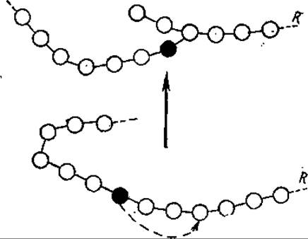
Fig. 6.9. Action of amylo(1,4→1,6)-transglycosylase (branching factor, Q-enzyme).
The dark circle indicates the glucose molecule whose aldehyde carbon has been transferred with the formation of an a(1—6) bond at the branch point; R represents a fragment of the glycogen molecule
In bacteria and insects that form chitinous integuments from glucosamine, NH3 is used instead of glutamine for the synthesis of glucosamine-6-phosphate. Acetylglucosamine is formed as a result of the transfer of an acetyl group from acetyl-CoA to glucosamine-6-phosphate. The interaction of acetylglucosamine phosphate with UTP yields UDP-acetyl-glucosamine (with the release of pyrophosphate), and the subsequent isomerization of the latter yields UDP-acetylmannosamine. As a result of The addition of phosphoenolpyruvate to acetylmannosamine via aldol condensation, phospho-N-acetylneuraminic acid is formed, and from it, N-acetylneuraminic acid.
The formation of polymeric carbohydrate chains in glycoproteins is carried out, as with all polysaccharides, with the participation of NDPS. For example, during hyaluronic acid biosynthesis, the donors of monosaccharide residues are UDP-glucuronic acid and UDPG. UDP-xylose plays an important role in the formation of heparin and chondroitin sulfate, since xylose serves precisely as the linking unit in the complex of these carbohydrates with Proteins. The synthesis of these carbohydrates begins with the transfer of xylose from UDP-xylose to the serine hydroxyl in the protein molecule. The addition of sulfate residues to the components of heparin and chondroitin sulfate is catalyzed by a specific enzyme, mucopolysaccharide sulfotransferase, with 3-phosphoadenosine-5-phosphosulfate acting as the sulfo group donor.
During the biosynthesis of bacterial Teichoic Acids, the main chain is constructed by transferring ribitol phosphate residues from CDP-ribitol and acetylglucosamine residues from UDP-acetylglucosamine. The elongation of the carbohydrate component occurs through the sequential action of various glycosyltransferases with their NDPS coenzymes: galactosyltransferase, fucosyltransferase, sialyltransferase, etc. The question of the mechanism determining the order of succession of monosaccharide residues during synthesis remains insufficiently clear. The widespread participation of NDPS in the synthesis of all oligo- and polysaccharides is combined with the selective "specialization" of donor NUCLEOTIDES. Thus, the donors of galactose, glucosamine, and muramic acid residues in animals are UDP-nucleotides, the donors of L-fucose and D-manose are GDP, those of glycerol and tyvelose are CDP, and that of N-acetylneuraminic acid is CMP.
Involvement of polyprenol phosphates in polysaccharide biosynthesis. In the 1960s and 1970s, a new type of carbohydrate residue carrier in polysaccharide biosynthesis—polyprenol phosphates—was discovered and investigated. They are lipid in nature, play a paramount role in glycoprotein biosynthesis, and are found in All living organisms. Their discovery was a major milestone in recent Carbohydrate Biochemistry.
Polyprenoid carriers accept a carbohydrate residue (mono- or oligosaccharide) from NDPS and transfer it to an acceptor (e.g., a bacterial antigen). In some cases, the Synthesis of the oligosaccharide occurs directly on polyprenol phosphate (PPP).
In the scheme, the process proceeds as follows:
NDPS + PPP → PPP-sugar + NDP
PPP-sugar + Acceptor → PPP + sugar-acceptor
Based on their Structure, polyprenoid carriers are divided into two groups: polyprenol phosphates and polyprenol pyrophosphates. The polyprenol pyrophosphate group is more numerous; unlike the former, they transfer not only monosaccharide residues but also di-, tri-, and tetrasaccharide residues. Their usual function is Participation in the biosynthesis of oligosaccharide components of bacterial cell walls.
Polyprenol phosphates have the following schematic structure:
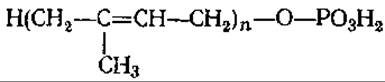
The isoprene unit bearing the hydroxyl group through which phosphate attachment occurs is typically hydrogenated and saturated. In bacteria, the most frequently encountered is undecaprenol (C55), which possesses 11 isoprene residues. Higher plant polyprenols contain from 5 isoprene residues (birch wood) to 16 (conifer needles). Animal PPPs are of even higher molecular weight (n = 17–21). The most widespread among them are dolichols, which comprise those polyprenols in which the a-isoprene residue bearing the hydroxyl group is saturated:
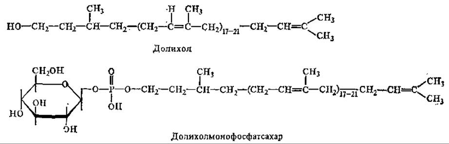
With the participation of dolichol phosphates, the oligosaccharide "core" is formed and incorporated into glycoproteins via N-glycosidic bonds. The cores are characterized by a high content of mannose and acetylglucosamine. The synthesis of the oligosaccharide is completed by glycosyltransferases without the further participation of dolichol phosphate. Protein glycosylation takes place in the Golgi apparatus. If, however, a protein is synthesized on free rather than membrane-bound Polysomes, it typically does not undergo glycosylation.
There is evidence that Vitamins A and K can function as carriers of carbohydrate residues, similarly to polyprenols.
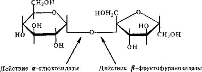
6.6.3. Breakdown of oligo- and polysaccharides. The breakdown of oligo- and polysaccharides proceeds via hydrolysis and phosphorolysis. Carbohydrate-hydrolyzing enzymes are characterized by stereospecificity. For instance, the hydrolysis of sucrose is catalyzed by ß-fructofuranosidase, also known as sucrase (invertase), which cleaves the bond located at the ß-glucosidic C-atom of the fructose residue. Invertase is found in higher plants, microorganisms, and animal digestive juices, and is particularly active in Yeast. The cleavage of sucrose can also be mediated by a-glucosidase, but in this case, the bond at the a-glucosidic C-atom of the glucose residue is broken:
Sucrose degradation can also occur through the action of sucrose synthase as a result of reaction reversal (see p. 347). It is precisely in this manner that sucrose is incorporated into the metabolism of sugar beet roots. Reaction reversibility ensures the economical utilization of sucrose: if the products of its breakdown are not consumed rapidly enough, the same enzyme converts their excess back into sucrose, thereby implementing a self-regulation mechanism.
Finally, in microorganisms, sucrose breakdown can proceed via phosphorolysis (reaction 3).
The hydrolysis of lactose into glucose and galactose is mediated by lactase (ß-galactosidase). It is present in the mammary glands of animals, in lactose-fermenting Yeasts responsible for the Fermentation of various dairy products, as well as in bacteria and Molds.
The disaccharide maltose undergoes hydrolysis through the action of a-glucosidase, or maltase, which is found in plant tissues, molds, yeast, bacteria, and the digestive juices of humans and animals. Maltase is particularly active in malt. In bacteria, maltose can also be cleaved by maltose phosphorylase.
The hydrolysis of cellobiose is catalyzed by ß-glucosidase.
The breakdown of starch and glycogen can likewise be both hydrolytic and phosphorolytic. Hydrolysis is carried out by amylases.
Currently, Three types of amylases have been identified: a-amylase, ß-amylase, and glucoamylase, which differ in their properties, natural occurrence, and mode of action on starch (glycogen) (Fig. 6.10).
a-Amylase is an endoamylase. It randomly cleaves a(1→4)-bonds within the chains of amylose, amylopectin, and glycogen. The reaction yields A large number of dextrins of varying molecular weights. Small amounts of maltose, maltotriose, and glucose are also formed among the final products. a-Amylase is found in saliva, pancreatic juice, germinated cereal seeds, molds, and bacteria. It is extremely rare in ungerminated seeds. It is sensitive to acidification but thermostable.

Fig. 6.10. Action of various amylases on linear regions of starch
ß-Amylase hydrolyzes amylose, amylopectin, and glycogen from the non-reducing end, releasing maltose residues. Under the action of ß-amylase, amylose is 100% converted into maltose. In amylopectin and glycogen, it attacks only the outer chains, resulting in the formation of maltose and a ß-limit dextrin. ß-Amylase is present in ungerminated seeds of wheat, rye, and barley.
The amylase-catalyzed Hydrolysis of Starch is of great importance in the technology of bread-making, brewing, alcohol production, and the textile industry.
Glucoamylase hydrolyzes starch to yield glucose and small amounts of dextrins. Glucoamylase preparations are isolated from molds and used to produce glucose syrup and crystalline glucose from starch.
None of the aforementioned amylases are capable of cleaving a(1→6)-bonds. The cleavage of these bonds is carried out by isoamylase (R-enzyme).
The second pathway for the breakdown of starch and glycogen is phosphorolysis.

In this reaction, a single glucose residue is cleaved from the non-reducing end and combines with phosphoric acid to form glucose-1-phosphate. The process is repeated multiple times until the entire starch or glycogen molecule is degraded down to the branch points. This pathway of dissimilation is energetically more advantageous than hydrolysis, as glucose is already in an activated form (glucose-1-phosphate) and readily enters into various reactions. a-Glucan phosphorylase acts exclusively on a(1→4)-bonds, and the reaction halts as soon as the enzyme reaches a branch point, yielding a limit dextrin. For degradation to resume, another enzyme is required—isoamylase—which cleaves the 1,6-bonds and thereby exposes a new segment of the polysaccharide chain to the action of starch (glycogen) phosphorylase.
The hydrolytic breakdown of cellulose (plant fiber) yielding cellobiose is catalyzed by the enzyme cellulase. The latter is a complex of two enzymes: endoglucanase and exoglucanase. They are found in germinating grain, as well as in certain bacteria and wood-decaying molds.
Hemicellulases—enzymes that hydrolyze various hemicelluloses into individual monosaccharides—have been found in germinating seeds and molds. For example, the mold Aspergillus niger contains xylanase, which breaks down xylans into xylose.
In addition to those listed, A number of other glycosidases are known that catalyze the cleavage of O-, N-, or S-glycosidic bonds in various glycosides and carbohydrate-containing compounds (glycoproteins, Glycolipids, Proteoglycans).
In Human and Animal tissues, glycosidases are localized primarily in Lysosomes. The absence or partial deficiency of any lysosomal glycosidase or one of its molecular isoforms leads to glycosidoses (lysosomal storage diseases), a group of hereditary disorders associated with impaired degradation of carbohydrate-containing compounds.
Last update: 06/08/2026
Editorial and Educational Adaptation: This material has been compiled based on the primary/original source text. The project team performed an editorial review, corrected technical inaccuracies, structured sections, and adapted the content for an educational format.
What was processed:
- elimination of formatting defects (OCR errors, structural breaks, corrupted characters);
- editorial organization of content;
- standardization of terminology in accordance with academic sources;
- verification of factual statements against the original source text.
All mentions of the author, publication year, and origin of the primary text have been preserved in accordance with the source.