Protein Chemistry - Part 1 - General Protein Chemistry - Ashmarin I. P. 1968
Protein Electrochemistry. Protein-Water Interaction
Protein Ionization. Binding of Hydrogen Ions
The molecular Structure of all Proteins is based on polypeptide chains, in which α-amino and α-carboxyl groups of various Amino Acids are linked by peptide bonds. Consequently, except for terminal groups, all α-carboxyl and α-Amino groups are non-ionizable under normal conditions and cannot be regarded as acidic or basic groups. The ionizable groups of proteins are predominantly located in the side chains of trifunctional amino acid residues. These include the free β- and γ-carboxyl groups of glutamic and aspartic acids, the imidazole group of Histidine, the ε-amino group of Lysine, the guanidino group of Arginine, the phenolic group of Tyrosine, and the sulfhydryl group of Cysteine. These side-chain ionic groups are the primary source of the electric charge on the protein molecule's surface. Depending on the ambient pH, protons associate with or dissociate from these side groups, thereby altering their ionization equilibrium state and the net charge of the protein molecule (the pK values for specific protein side groups are quite close to the pK3 values listed in Table 6). Since the proportions of these groups vary among different proteins, their isoelectric points correspond to different pH values. It should be emphasized that the isoelectric point of a protein molecule is generally defined as the pH at which its average net (effective) charge is zero.
A protein's isoelectric point should not be confused with its isionic point, as these two values do not always coincide. The isionic point of a protein is defined as the pH at which the number of protons bound by basic groups equals the number of protons released by dissociated acidic groups within the protein molecule. The isoelectric and isionic points coincide only when the protein solution contains no ions other than hydrogen and hydroxyl ions. In the presence of other anions and cations, these points diverge. To illustrate this, let us consider a protein solution whose pH equals the protein's isoelectric point and which contains no ions other than H+ and OH-.
In this case, the number of protons liberated during the dissociation of COOH and NH3 acidic groups will equal the number of protons bound by COO- and NH2 basic groups; that is, the isoelectric and isionic points of the protein coincide. When sodium chloride is added to such solution, a small amount of Cl- anions will be bound by acidic groups  and Na+ cations by COO- basic groups, which will induce additional dissociation of the COOH group and a decrease in the dissociation of the NH3 group.
Class="center">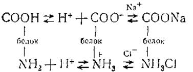
If the binding of anions by acidic groups occurred as intensively as the binding of cations by basic groups, the reduction in the number of protons split off by the group  would be compensated by enhanced dissociation of the carboxyl group, thereby preserving the isionic state. However, the binding of Cl- anions proceeds more vigorously than that of Na+ cations, disrupting isionia and reducing the total number of protons.
To restore the isionic state, it is necessary to further decrease the proton concentration—in other words, to alkalize the solution to enhance carboxyl group dissociation. Admittedly, some of the dissociated protons will be bound by NH2 basic groups, leading to an increase in the number of  groups.
Nevertheless, the ionization reaction of the carboxyl group proceeds faster than the proton-binding reaction, thereby restoring the isionic state. Thus, the relatively faster binding of anions by the protein, accompanied by a corresponding deceleration of proton binding, causes the protein's isionic point to rise in the presence of salts. It has been demonstrated, for instance, that the isionic point of serum albumin in the presence of 0.15 M sodium chloride increases from pH 4.8–4.9 to 4.27, and this shift becomes more pronounced as protein concentration increases. The Effect of sodium salts on the isionic point of serum albumin is illustrated in Fig. 43.
The Definition of the isionic point given above does not specify how to measure the corresponding pH experimentally. Protein isionic points can be determined using various Methods. The most common Procedure is as follows: A protein solution is subjected to exhaustive electrodialysis to remove all electrolyte ions except hydrogen and hydroxyl ions. The protein recovered from such a solution can be regarded as an isionic substance. When dissolved in Water, it imparts a definite pH to the solution, which can be taken as its isionic point. Thus, one of the experimentally grounded Definitions of the isionic point is: the isionic point is the pH of an aqueous solution of an isionic substance.
As for the isoelectric point, based on several experimental approaches used for its estimation, it can be defined as the pH at which the electrophoretic mobility of the protein is zero. Solubility, Membrane Potential, and osmotic pressure reach their minimum values at this point, whereas the viscosity and Dielectric Constant of the solution reach their maxima.
Thus, the electrical charge On the surface of a protein molecule and its isoelectric point are determined by the ionizable groups in The amino acid side chains, since these groups can either take up or release protons depending on the ambient pH. The quantity and type of these groups can be determined by electrometric titration, the curves of which show the dependence of the number of protein-bound protons on the ambient pH. However, it should be noted immediately that a protein molecule may contain a very large number of titratable groups, resulting in substantial overlap between the ionization ranges of different groups. This naturally complicates the calculation of the exact number of a specific group type from The titration curve, as well as differentiating the number of α-carboxyl groups from the β- and γ-carboxyl groups of dicarboxylic acids, or terminal α-amino groups from the guanidino groups of histidine.
Electrometric titration is performed by adding strong acids (usually Hydrochloric acid) or strong bases (such as sodium hydroxide) to an isionic protein solution. The pH of the titrated solution is measured directly, while the number of bound protons and hydroxyl ions must be calculated using special formulas that take into account The amount of isionic protein (g in grams) and the concentration of protons (hydroxyl ions) in the initial acid (alkali) solution and after the solution pH shifts from pH1 to pH2 (C1 and C2 in mol/L, respectively):

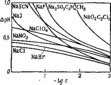
Fig. 43. Effect of sodium salts on the isionic point of human serum albumin (from Neurath and Bailey, 1956).
The branch of the titration curve in the acidic region is considered as a function of hydrogen ion binding, whereas the branch in the alkaline region is viewed as a function of hydroxyl ion binding. In reality, hydroxyl ions interact not with the protein itself, but with protons dissociated from it, forming water molecules. Thus, the ascending branch of the curve allows the calculation of the total number of side-chain and α-carboxyl groups, whereas the descending branch yields the total number of basic groups. Figure 44 shows the potentiometric titration curve of Ovalbumin.
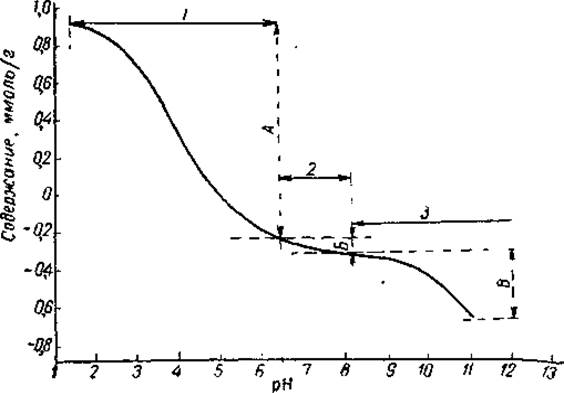
Fig. 44. Potentiometric titration of ovalbumin in water (after Kenchinton, 1963).
Titration ranges: 1 — carboxyl groups, 2 — imidazole and α-amino groups, 3 — ε-amino groups, tyrosine OH groups, and cysteine SH groups; contents: A — carboxyl groups, B — imidazole and α-amino groups, B — ε-amino groups, tyrosine OH groups, and cysteine SH groups.
Examination of the ovalbumin titration curve reveals well-defined inflection points at pH 3–4 and 9–10, which arise from the buffering action of carboxyl and amino groups, dividing the curve into a series of segments or titration intervals. Each interval corresponds to the ionization range of specific protein side groups, making it possible to calculate the number of these groups from the amount of protons bound or released within that particular titration segment. For instance, titration from pH ~6.5 to the point of maximum proton binding (around pH 1.5) is attributed to the free carboxyl groups of aspartic and glutamic acids and the α-carboxyl groups. Therefore, the total number of carboxyl groups equals the number of acid equivalents required for titration from pH ~6.5 to pH 1.5. In solutions of high Ionic strength, maximum proton binding occurs as early as pH 2–2.5. Furthermore, the potentiometric titration method does not permit the determination of CONH2 groups present in side chains.
Titration from pH 8.5 to pH 6.5 is driven by the interaction of protons with histidine imidazole groups and terminal α-amino groups, which are present in the protein in small quantities. The total number of these groups equals the number of acid equivalents required for titration in this pH range. Finally, the total number of lysine ε-amino groups, tyrosine hydroxyl groups, and cysteine sulfhydryl groups equals the number of base equivalents required for titration from pH 8.5 to pH 11–12. Arginine cannot be determined directly by titration because the dissociation constant of the guanidino group is so high (pK3 is somewhat above 13) that this group cannot convert into an ionized form in noticeable quantities at any pH accessible to accurate measurement. Consequently, the maximum base binding by the protein cannot be determined with sufficient precision.
From titration data obtained between the isionic point and the point of maximum proton binding near pH 1.5, one can determine the content of basic groups (imidazole, amine, and guanidino) regardless of which specific groups actually participate in the ionic equilibrium processes. At first glance, it may seem counterintuitive that the total number of basic groups is determined by titration in the acidic pH range. To clarify this, we must return to the definition of the protein's isionic point. As mentioned above, the isionic point corresponds to the pH at which the number of protons liberated by the acidic groups of the protein molecule equals the number of protons bound by its basic groups. This means that at the isionic point, the number of protons released by the protein molecule equals the maximum charge that the protein can acquire through maximal proton binding. Since all nitrogen-containing groups carry a positive charge at the pH of maximum acid uptake (the pH of maximum proton binding), while all other groups remain uncharged, the charge of the protein molecule reaches its maximum, and the number of nitrogen-containing groups can be determined from the amount of bound acid. In other words, the total number of basic groups equals the number of acid equivalents required for titration from the isionic point to pH 1.5.
For many proteins, electrometric titration results agree quite well with data obtained from chemical Amino acid analysis. For example, the total number of anionic and cationic groups determined electrometrically in ovalbumin, serum albumin, and lactoalbumin closely matched the number of acidic and basic amino acids found through corresponding chemical analyses. In other proteins, however, such as Insulin, a large excess of groups titratable in the alkaline region was detected. This latter finding can only be explained by the presence of A large number of terminal α-amino groups in insulin.
In Conclusion, it should be noted that interpreting protein titration curves involves several difficulties arising from a variety of circumstances. Proteins contain a very large number of ionizable groups that bind and release protons. Titration curves indicate that approximately 1 mmol of acid and 1 mmol of base are required per 1 g of protein. Given a Molecular Weight of around 100,000 for a protein, each protein molecule therefore contains roughly 100 acidic and 100 basic groups. However, precisely establishing the number of specific basic groups is difficult because the titration curve exhibits some overlap in the pH region between 8 and 12. Consequently, pH 8.5 is chosen somewhat arbitrarily as the endpoint for neutralizing α-amino and imidazole groups. The titration curve is also affected by interactions between proteins and ions other than hydrogen. Specifically, proteins form stable bonds with divalent ions such as calcium, magnesium, phosphate, and carbonate, as well as monovalent chloride ions. As already discussed, such interactions lead to a shift in the isionic point and alter the electrochemical Properties of the protein due to the neutralization of ionizable groups, thereby distorting the titration curve. The shift in the isionic point is particularly pronounced in the presence of phosphate ions, which bind most strongly to basic groups.
When discussing the neutralization of side groups, it must be kept in mind that this process can be driven not only by interactions with inorganic ions, but also by interactions between protein molecules themselves. Electrostatic (salt-like) bonds can form between the positive and negative groups of adjacent molecules, causing protein molecules to aggregate. This interaction not only complicates the interpretation of titration curves, but often makes it impossible to prepare an isionic protein solution via electrodialysis, because this process removes the neutral salts necessary to prevent protein aggregation and precipitation.
Finally, a major challenge in obtaining titration curves for certain proteins is their lability. Some proteins denature at pH values above 2 or below 11, making accurate determination of native protein ionization in these ranges impossible. To overcome this hurdle, titration is performed at low temperatures, or a series of samples of the isionic protein solution is prepared, to each of which a progressively increasing amount of acid or alkali is added. Following The addition of acid (or alkali), the pH is measured immediately. Because the neutralization of ionized groups occurs instantaneously, whereas Denaturation takes slightly longer, this technique successfully separates the two reactions. Based on the pH values obtained for each point, one can construct the titration curve for a given protein and calculate the number of ionized groups within the denaturation pH regions.
Last update: 06/08/2026
Editorial and Educational Adaptation: This material has been compiled based on the primary/original source text. The project team performed an editorial review, corrected technical inaccuracies, structured sections, and adapted the content for an educational format.
What was processed:
- elimination of formatting defects (OCR errors, structural breaks, corrupted characters);
- editorial organization of content;
- standardization of terminology in accordance with academic sources;
- verification of factual statements against the original source text.
All mentions of the author, publication year, and origin of the primary text have been preserved in accordance with the source.