ZOOLOGY OF INVERTEBRATES IN THREE BOOKS - BOOK 2 - G.I. Shcherbak - 1996
PHYLUM ARTHROPODA
SUBPHYLUM CHELICERATA
CLASS ARACHNIDA
Arachnids are distributed globally. The vast majority of species are free-living terrestrial animals, while only some mites and ticks are plant or animal parasites, or inhabitants of marine and fresh waters. Approximately 60,000 species have been described.
Arachnids share all the Characteristic Features of chelicerates, specifically: the division of the body into a prosoma (cephalothorax), bearing chelicerae, pedipalps, and four pairs of walking legs, and an opisthosoma (abdomen), which only rarely bears modified appendages (Fig. 189).
Class="center">
Fig. 189. Arachnids:
a — scorpion Euscorpius carpaticus; b — pseudoscorpion Chelifer cancroides; c — solifuge Galeodes araneoides; d — spider Latrodectus tredecimguttatus; e — ricinuleid Kenenia mirabilis; f — tick Ixodes ricinus;
The prosomal segments in most arachnids are fused into a solid mass covered by a single dorsal carapace. Only in solifuges, as well as schizomids and ricinuleids (small groups of arachnids, with the first distributed in the equatorial belt and the second in southern regions with high humidity), are only the four anterior prosomal segments fused into a single tagma, while the two posterior segments remain free, each covered by its own tergite. Abdominal segmentation varies significantly among different arachnid groups, and its Structure will be examined in detail for each specific group.
The first pair of appendages—the chelicerae—most commonly terminate in a chela (pincer), and much less frequently in a hook-like structure (e.g., in spiders). Typically, chelicerae are two- or three-jointed; the terminal segment Functions as the movable finger of the chela or as a hook. In scorpions and pseudoscorpions, the pedipalps also terminate in powerful chelae, whereas in solifuges they become entirely similar to walking legs. The subsequent four pairs of prosomal appendages consist of six to seven segments and function as walking legs.
Externally, the body of arachnids is covered by a multilayered cuticle, beneath which lies a layer of flattened hypodermal Cells. The Fine Structure of the cuticle varies among different arachnid groups. The degree of cuticular sclerotization differs across groups and species; however, as a rule, specific areas of the body—such as plates and leg segments—are particularly hard, while the areas between them are thinner and more elastic.
Derivatives of the hypodermal epithelium include numerous glands: scent glands (in harvestmen), silk glands (in spiders), venom glands (in scorpions and spiders), and others.
Arachnids, especially scorpions, possess a well-developed internal Skeleton similar to that of horseshoe crabs.
The Muscular System is highly complex. For instance, the body of a scorpion contains no fewer than 150 (partially paired) Muscles, excluding the limb muscles.
The Digestive System in arachnids, as in other Arthropods, consists of three sections, but their posterior (ectodermal) gut section is much shorter.
The structure of the digestive system varies somewhat among representatives of different groups (Fig. 190), particularly the posterior part of the endodermal gut. All arachnids feature a muscular Pharynx that functions mostly on THE PRINCIPLE OF a pump, through which semi-liquid food is sucked in. The pharynx leads into a slender Esophagus, which in some spiders features an additional expansion—the sucking Stomach. This is followed by the midgut; its anterior part often expands (sometimes becoming sac-like) to form a stomach from which lateral outpouchings (diverticula) of varying length and thickness branch off. This significantly increases the volume and surface area of the walls where Digestion and nutrient absorption take place. The ducts of a massive paired gland (the Liver, or midgut gland), whose functions correspond to the combined Functions of the vertebrate liver and Pancreas, open into the midgut of most arachnids. Food is partially or primarily digested within the liver (cavity and intracellular digestion), and its cells store nutrients. In scorpions, besides the liver, there is also a gastric gland located in the prosoma, consisting of numerous glandular lobules and opening into The Stomach via two ducts. Histologically and functionally, it does not differ from the liver. Intracellular digestion is very widespread in arachnids, carried out by cells of both the midgut and the liver. The posterior ectodermal gut resembles a short, straight tube.

Fig. 190. Diagram of the digestive system structure in arachnids: a — scorpion; b — mite Varroa jacobsoni
Extraintestinal digestion is also characteristic of arachnids. By piercing the prey's integument, they inject digestive juices containing potent digestive Enzymes produced in the midgut and partially in the Salivary Glands (if present). By breaking down Proteins, these enzymes transform the victim's body into a semi-liquid mass suitable for ingestion. As already mentioned, most arachnid groups are predators, and only less commonly do they feed on various plant debris (harvestmen, some mites), suck plant juices, or feed on the Blood of vertebrates, including humans (ticks).
The primary excretory Organs of arachnids are Malpighian tubules—blindly ending, sometimes branched tubes that open into the posterior part of the midgut (Fig. 191). They function in conjunction with the posterior region of the midgut, which often forms specialized expanded areas (the cloacal pouch in spiders, the rectal bladder in some mites). The histological structure of these areas differs little from that of the Malpighian tubules opening into them. The main excretory product in arachnids is guanine. This Water-insoluble substance, much like the uric acid of insects, enables arachnids to conserve water in their bodies. Within the Malpighian tubules, soluble nitrogenous waste products are converted into insoluble, spherical crystals of guanine. These crystals move along the tubules to the posterior section of the midgut, where reabsorption—the uptake of water and ions back into the hemolymph—takes place. As proven by studies on mites, this process is facilitated by the specialized STRUCTURE OF THE posterior gut epithelium.

Fig. 191. Excretory organs of arachnids:
a — Malpighian tubules of Araneus diadematus; b — coxal gland of a scorpion
Unlike analogous structures in insects, the Malpighian tubules of arachnids are of endodermal origin and are functionally connected not to the hindgut, as in insects, but to the midgut. This indicates that the Malpighian tubules of insects and arachnids are not homologous structures, but arose independently as an adaptation to life in conditions of moisture deficit.
Besides Malpighian tubules, the excretory function is performed by paired coxal glands, which vary in structural details, but in most cases consist of an end sac, a convoluted tubule (labyrinth), and a more or less straight efferent duct with a Urinary Bladder. The duct opens near the coxae of the third to fifth pairs of prosomal appendages. Coxal glands are well developed in embryos and juveniles, but they usually atrophy to a greater or lesser extent, persisting throughout life only in harvestmen. In palpigrades, coxal glands are believed to be the sole excretory organs.
Various Regions of the intestine also participate in excretion, even in forms with well-developed specialized excretory organs. Metabolic waste products accumulate particularly intensively in the epithelium of the diverticula (spiders, harvestmen, pseudoscorpions, etc.) and the posterior part of the endodermal midgut. In some trombidiform mites, this section performs the excretory function entirely and does not even connect with the anterior part of the midgut, which is blindly ending.
Guanine is concentrated in the intestinal cells and then released into the gut lumen, or the entire Cell may be sloughed off and eliminated to the exterior. Furthermore, in many arachnids, guanine is often deposited in Tissues and not excreted at all.
Specialized, relatively large cells known as nephrocytes, located in the cavities between organs, also take part in excretion by accumulating waste products.
The respiratory organs of arachnids include book Lungs (scorpions, spiders), tracheae (solifuges, harvestmen, pseudoscorpions, mites), or both combined (spiders). Each pulmonary sac opens to the exterior through a slit-like orifice, the stigma. Numerous parallel, leaf-like lamellae—thin, flattened folds of the lung wall arranged like the pages of a book—project into the cavity of the sac (Fig. 192, a). The wall of each pulmonary lamella is covered with a very thin cuticle. On its upper surface, each lamella bears small cuticular pillars that prevent adjacent lamellae from sticking together, thereby ensuring a continuous supply of air in the spaces between them. A hypodermal layer lies beneath the cuticle of the lamellae, while their interior contains narrow lacunar cavities filled with hemolymph. Gas exchange occurs through the thin walls of the lamellae. Scorpions possess four pairs of pulmonary sacs, whereas most spiders have one pair, and rarely two pairs.

Fig. 192. Respiratory organs of arachnids:
a — cross-section through a book lung of the cross spider; b — main tracheal trunks of a solifuge; c — diagram of the tracheal system structure in the mite Varroa jacobsoni;
Tracheae, as in other arthropods, constitute a system of variably branched tubes originating from specialized respiratory openings (stigmas). The shape, number, and Location OF THE stigmas, the thickness of the tracheal trunks, and the degree of their branching exhibit specific features in each group (Fig. 192, b, c). The tracheal system is best developed in solifuges. Their main tracheal trunks open via several pairs of spiracles on the prosoma and opisthosoma, and a single unpaired spiracle on the IV opisthosomal segment. The tracheae extending from the spiracles unite into powerful longitudinal trunks that are interconnected by transverse bridges and send numerous branches to all organs. The walls of solifuge tracheae feature cuticular spiral thickenings similar to insect taenidia.
Most spiders possess tracheae In addition to lungs. Typically, they have a single opisthosomal spiracle from which two pairs of blindly ending, unbranched tubes originate, lined with a delicate cuticle lacking spiral thickenings. The tracheae are bathed in hemolymph.
In small forms, particularly certain mites and palpigrades, Respiration occurs through the thin body integument, and specialized respiratory organs are absent.
The degree of Development of the Circulatory system correlates with the animal's size, body segmentation, and the structure of its respiratory organs. As the tracheal system develops, the circulatory system tends to become less developed.
The circulatory system is best developed in scorpions. Their Heart is a long tube extending along the dorsal side through almost the entire anterior part of the opisthosoma. It is enclosed in a thin-walled Pericardium and suspended within it by paired Connective Tissue strands (ligaments). On the DORSAL SIDE OF The Heart, there are seven segmentally arranged ostia. Through these ostia, equipped with specialized Valves, hemolymph flows in only one direction—from the pericardium into the heart. Anteriorly and posteriorly, the heart continues into the anterior and posterior aortae, from which numerous vessels branch out to supply blood to all organs and tissues. In addition to the aortae, nine pairs of lateral Arteries depart from the heart; eight of these plunge into the liver, forming numerous branches that permeate the entire organ (Fig. 193). The terminal branches of all vessels are open, and the hemolymph eventually enters a system of lacunae and sinuses, some of which possess their own connective tissue walls and may be referred to as Veins. In the prosoma and pre-abdomen, venous hemolymph collects into a pair of longitudinal ventral sinuses that expand near the lungs to form pulmonary sinuses, which bathe the pulmonary sacs. Upon entering the lacunae of the pulmonary lamellae, the hemolymph is oxygenated. From the pulmonary sinuses, hemolymph flows via seven pairs of Pulmonary veins into the pericardium, and from there through the ostia into the heart.

Fig. 193. Structure of the scorpion heart:
1 — anterior aorta; 2 — heart enclosed in the pericardium; 3 — ostium; 4 — lateral arteries; 5 — posterior aorta; 6 — ventral nerve cord; 7 — pulmonary veins; 8 — ventral sinus; 9 — lungs
In other arachnids, in accordance with their body structure, the heart becomes shortened, the number of ostia decreases (e.g., three to four pairs in spiders, one to two pairs in harvestmen), only the anterior aorta persists (spiders), the number of lateral vessels is reduced (three pairs in spiders), and in most mites, a distinct circulatory system is entirely absent, with hemolymph circulating freely within the body cavity. The hemolymph of arachnids contains the respiratory pigment hemocyanin.
Many arachnids are characterized by a high degree of concentration of The Nervous system, which is directly related to the reduction in their body length and the fusion of segments and tagmata (Fig. 194). The Brain consists of two sections: the anterior protocerebrum, which innervates the eyes, and the posterior tritocerebrum, which sends nerves to the first pair of appendages (chelicerae).

Fig. 194. Diagram of the nervous system structure in the scorpion Androctonus (a) and the spider Tegenaria (b):
1 — eyes; 2 — brain; 3 — subesophageal ganglion; 4 — ventral nerve cord; 5 — ganglia on the cord; 6 — esophagus; 7 — ventral nerve trunk; 8 — nerves innervating the spinnerets; 9 — lungs
The nervous system is least concentrated in scorpions (Fig. 194, a). It consists of supraesophageal and subesophageal ganglia connected by short, thick connectives, and a long ventral cord with seven ganglia. The subesophageal ganglion innervates 2–6 pairs of appendages. In spiders, solifuges, and certain other groups, the subesophageal ganglion and the ganglia of the ventral cord fuse into a single ganglionic mass (Fig. 194, b). In harvestmen, pseudoscorpions, and mites, all ganglia fuse into a unified ganglionic body (synganglion) surrounding the esophagus.
Visual organs are represented by simple eyes located on the dorsal side of the prosoma (Fig. 195). Their number varies among different arachnids: scorpions have one pair of median eyes and two to five pairs of lateral eyes; spiders most commonly possess eight eyes arranged in two rows, four of them positioned medially and the other two pairs laterally.
Lateral eyes have a simpler structure (Fig. 195, a). These are cup-shaped eyes comprising a convex lens, or crystalline lens, and a retina made up of elongated visual (retinal) cells. The latter are grouped together to form numerous retinulae with rhabdomes, which are not separated from one another by pigment. The basal ends of the visual cells converge into the Optic nerve. The hypodermis forms a continuous dark ring around the eye.
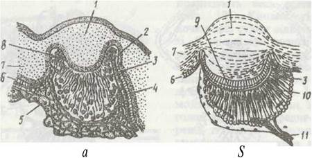
Fig. 195. Lateral (a) and median (b) eyes of a scorpion:
1 - lens; 2 - basal membrane; 3 - rhabdom; 4 - visual cells; 5 - connective tissue; 6 - hypodermis; 7 - cuticle; 8 - pigment cells; 9 - vitreous body; 10 - retina; 11 - optic nerve
The median eyes possess a lens, a vitreous body, and a retina composed of retinal and pigment cells (Fig. 195, 6). The retinal cells are grouped into retinulae, with five cells in each. The rhabdomeres of all five cells form the rhabdom. The processes of the visual cells form the optic nerve, which enters the brain.
Vision in most arachnids is limited: they perceive only changes in light intensity and movement, and only solifuges, wandering spiders, and scorpions are capable of analyzing the outlines of objects with their median eyes. Wandering spiders of the family Salticidae are distinguished by more advanced vision: specialized muscles move the eyes, enabling them to track prey while remaining motionless.
Numerous sensory hairs, varying in Structure and function, are distributed across the body and appendages. Alongside ordinary trichoid sensilla, trichobothria are very widespread in arachnids (Fig. 196, a). They are located on the pedipalps and legs or on the body (in some mites). A long Hair, sometimes thickened at the tip, is attached by a thin membrane at the bottom of a funnel-shaped depression in the cuticle; a group of sensory cells approaches its base. The slightest vibration of the air or substrate causes it to be displaced, which is perceived by the sensory cells.
Sensilla are often aggregated to form organs. Arachnids are characterized by so-called lyriform organs, located on the trunk and appendages. These are microscopic slits in the cuticle spanned by a thin membrane, which is contacted by The process of a sensory cell (Fig. 196, b, c). These organs are believed to be mechanoreceptors that perceive the degree of cuticular tension.

Fig. 196. Sense Organs of arachnids:
a - trichobothrium; b, c - lyriform organ from the surface and in cross-section, respectively; 1 - cuticle; 2 - hair; 3 - sensory nerve cell
In mites, a palpal organ has been described, located on the terminal segments of the pedipalps and consisting of several cone-shaped chemoreceptor sensilla. Functionally, this is primarily a gustatory organ, by means of which blood-sucking mites select a feeding site on the host's body. In addition, some of its sensilla act as olfactory receptors.
Ixodid ticks possess a specific Haller's organ, located on the dorsal surface of the tarsi of the first pair of legs; it plays a major role when the tick is searching for a host.
Arachnids are dioecious. The Gonads are located in the abdomen and can be paired or unpaired. The oviducts and sperm ducts are paired, but open externally through a single unpaired genital opening. Females typically have an expansion of the oviduct—the Uterus—and seminal receptacles that store sperm. The fine structure of the Reproductive System varies among representatives of different orders (Fig. 197). Fertilization is either spermatophoric or occurs via copulation. This issue will be discussed in greater detail below.

Fig. 197. Reproductive system of arachnids:
a - female of Araneus; b - mite Ixodes; c - male of Araneus; 1 - Ovary; 2 - seminal receptacle gland; 3 - seminal receptacle; 4, 5 - paired and unpaired PARTS OF THE oviduct; 6 - genital opening; 7 - seminal receptacle canal; 8 - external opening of the seminal receptacle; 9 - Vagina; 10 - accessory glands; 11 - uterus; 12 - Testis; 13 - sperm duct
Most arachnids are oviparous, although viviparous species also occur. Fecundity varies greatly—from a few hundred eggs to 30,000.
The eggs of most arachnids are large and rich in yolk; consequently, Cleavage is typically superficial and incomplete, as in other arthropods. The embryo develops primarily at the expense of the germ band. Segmentation is often more pronounced in the embryo, whose body possesses more segments than that of the adult animal. For example, in spider embryos, the abdomen consists of 12 segments, as in scorpions, with the 4–5 anterior segments bearing limb buds. In scorpion embryos, the rudiments of abdominal appendages are formed. In spiders and scorpions, one can trace how certain abdominal limb buds transform into lung books.
In almost all arachnids, development is direct, accompanied by growth and the further maturation of certain organs during molting.
The Classification of the class Arachnida is currently under revision. There is no consensus regarding either the number of taxa grouped within it or their rank. Furthermore, opinions have been expressed regarding the artificial Nature of the class as a whole. We will examine a series of arachnid groups that we recognize at the rank of subclass.
Last update: 13/08/2026
Editorial and Educational Adaptation: This material has been compiled based on the primary/original source text. The project team performed an editorial review, corrected technical inaccuracies, structured sections, and adapted the content for an educational format.
What was processed:
- elimination of formatting defects (OCR errors, structural breaks, corrupted characters);
- editorial organization of content;
- standardization of terminology in accordance with academic sources;
- verification of factual statements against the original source text.
All mentions of the author, publication year, and origin of the primary text have been preserved in accordance with the source.