Chordate Zoology - Textbook - I. V. Tsaryk - 2013
Chapter 2. SUBPHYLUM TUNICATA, OR LARVACEA, UNICATA, seu RQCHQRDATA
2.1. ORGANIZATIONAL FEATURES
2.1.1. Class Ascidiacea (Ascidians)
Ascidians are sessile benthic animals. They include both colonial and solitary forms. Their body size generally ranges from a few centimeters in diameter to a height of the same magnitude. However, some species can reach up to 40-50 cm, notably the cosmopolitan Ciona intestinalis or the deep-sea Ascopera gigantea (Fig. 2.1).
At the same time, there are very minute ascidians, measuring up to 1 mm.
Most solitary ascidians resemble irregular oblong sacs. They attach themselves by their lower part, known as the FOOT or base, to various solid objects.
There are also peculiar ascidians capable of swimming short distances above the sea floor. These are Representatives of the genus Octacnemus—semi-transparent animals up to 7 cm in diameter. Their delicate tunic forms eight broad, tentacle-like lobes around the oral siphons. Octacnemids are inhabitants of the deep ocean, found in tropical regions at depths of 2,000–4,000 m. Another interesting genus is Situla, individuals of which reach a height of up to 35 cm.
Class="center">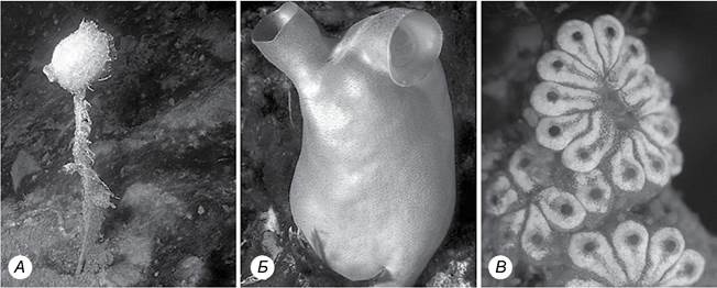
Fig. 2.1. Diversity of ascidians: A - Boltenia antarctica; B - Ascopera gigantea; C - Botryllus planus
The body of an ascidian is covered by a single-layered integument, the epidermis, which secretes a specialized thick outer layer known as the tunic. The tunic performs supportive and protective Functions. Ascidians are typically colored in shades of orange, reddish, brownish, or violet. The surface of the tunic frequently develops wrinkles and folds overgrown with Algae, hydroids, and other epiphytic organisms. Sometimes it is covered with sand grains or small pebbles, making the animal difficult to distinguish from its surroundings. Beneath the tunic lies the body wall proper, or mantle, which, In addition to the epidermis, contains Connective Tissue and Muscle fibers (Fig. 2.2).

Fig. 2.2. Anatomy of an ascidian
On the upper side of the body, two apertures are visible, situated on distinct tubercles or relatively long body outgrowths resembling the neck of a bottle. These are the siphons. One is the oral siphon, through which the ascidian draws in Water, and the other is the atrial (cloacal) siphon. The entrance to the siphons is guarded by folded or finger-like outgrowths—tentacles, which are usually more numerous in the oral siphon. The siphon lumens are also regulated by sphincter Muscles. Near the sphincters, the mantle fuses with the tunic; in other areas of the body, it lies loosely within the cavity formed by the tunic. The DORSAL SIDE OF the animal is located in the space between the siphons.
The digestive tract of an ascidian begins with the Mouth located inside the corresponding siphon. Extending from the mouth is a large Pharynx that occupies almost the entire space within the mantle. The pharynx forms a complex filtering and respiratory apparatus. It is perforated by a multitude of slits—stigmas (gill slits)—the lumens of which are lined with ciliated Cells. Externally and internally, the pharynx is covered by a dense network of Blood Vessels that facilitate gas exchange. Two thin-walled, sac-like outgrowths called epicards extend from the posterior wall of the pharynx. The epicards play an important role during asexual reproduction in ascidians. In some cases, they function as excretory Organs (Kidneys), accumulating Metabolic waste products.
The space between the pharynx and the mantle is called the atrial cavity (also known as the peribranchial cavity). Near the cloacal siphon, the atrial cavity expands into the cloaca, which communicates with the external environment via the cloacal siphon.
A dorsal lamina hangs into the pharyngeal cavity from the dorsal side of the pharynx. Running along the midline of the ventral side is a glandular-ciliated groove, the endostyle, The Structure of which is shown in Fig. 2.3.

Fig. 2.3. Cross-section of an ascidian endostyle
Water moves in an orderly fashion through the ascidian's body from the oral to the cloacal siphon via a system of cavities, driven by the cilia of the endostyle and pharyngeal walls. As in amphioxus, the glandular Cells of the endostyle produce mucus, which forms a sort of net that slowly flows along the pharyngeal walls, trapping fine food particles. Upon reaching the dorsal lamina, this mucus is twisted into a cord and passes into the short Esophagus. From there, food moves into The Stomach, and subsequently into the intestine, which terminates in the cloaca. Research has shown that the endostyle is functionally similar to the vertebrate Thyroid Gland, producing a hormone-like substance called iodotyrosine. The Digestive System of ascidians also includes the so-called Liver, which appears as lobes and outgrowths adjacent to the stomach and connected to it by a duct. However, a "liver" is absent in many species.
The Circulatory system OF ascidians is open. The tubular Heart lies on the ventral side and consists of the myocardium (inner muscular layer) and the Pericardium. Both ends of The Heart tube extend into large, branching vessels. The anterior (ventral) artery leads into the branchial artery and forms the so-called branchial basket in the pharyngeal walls from crisscrossing blood vessels. The posterior (dorsal) artery surrounds the Internal Organs, extends into the body walls and even the tunic, and forms lacunae around the organs. These vessels are conventionally called Arteries because the heart pumps blood alternately in one direction or the other until the cardiac pressure is balanced by the pressure in the distal segments (vessels or lacunae) of the system. Then, the blood begins to flow in the reverse direction through the same vessels. Such a tidal (pendulum) mode of Circulation, especially given the absence of respiratory pigments, is not highly efficient. However, for sessile or sluggish organisms such as ascidians, the minimal oxygen supply provided by simple diffusion into the blood within the branchial vessels is sufficient. Periodic reversal of blood flow, caused by Changes in the direction of heart contractions, is characteristic of all tunicates. Notable features of ascidian blood include a high concentration of sulfuric acid and an exceptionally high content of vanadium in the Blood Cells.
The Nervous system of ascidian larvae is more complexly organized than that of amphioxus: the neural tube is expanded at its anterior end and contains larval Sense Organs—pigment ocelli, statocysts, and baroreceptors. In adults, it is reduced to a suprapharyngeal ganglion located on the dorsal side (between the siphons) and several pairs of nerves that branch out into the pharynx and around the internal organs. Between the ganglion and the dorsal side of the pharynx lies a small gland, the duct of which opens into the pharynx at the bottom of a ciliated pit. This gland is sometimes considered homologous to the Pituitary Gland (hypophysis) of vertebrates. Sense organs are absent, but receptor arcs are retained, ensuring the Organism's response to changes in Temperature, chemical composition, and other environmental factors.
Ascidians lack specialized excretory organs. Most commonly, the walls of the digestive tract perform the excretory function. However, many ascidians possess accumulation kidneys composed of specialized cells called nephrocytes. Metabolic waste products accumulate in these nephrocytes and are released into the external environment after the animal's death. These cells are frequently clustered near the intestinal loop or Gonads. The coloration of many ascidians depends on the excretory products accumulated in these cells. Occasionally, concretions containing uric acid accumulate In the second loop of the intestinal bend.
In members of the family Molgulidae, accumulation kidneys are modified into an isolated sac, the cavity of which contains concretions. This sac also harbors symbiotic Fungi that feed on urates. How these symbiotic fungi are transmitted from one individual to another remains unknown. It is believed that juveniles become "infected" with the fungi from seawater.
Ascidians are hermaphrodites. The gonads are located within the intestinal loop, and their ducts open into the cloaca. Self-Fertilization is prevented by differences in the maturation timing of oocytes and spermatozoa within each individual. Fertilization and the Cytology/cytology/16.html">Early stages of Embryogenesis take place in the atrial cavity (rarely in the external environment).
In colonial forms, individual zooids are embedded in a common tunic (Fig. 2.4). Such colonies vary in shape and colonize the surface of rocks and underwater objects. Notable structural features include the shape and position of the heart, as well as the presence of a common cloaca shared by adjacent individuals.

Fig. 2.4. Colonial ascidians:
A - Clavelina lepadiformis; B - Apli- dium breviventer, C - Halocynthia roretzi; D - structure of colonial ascidians
The larva recapitulates (reproduces) the progressive traits of its free-living ancestors. Its Morphology undergoes rapid changes within a short period of time.
Provisional (larval) organs are most fully developed during the free-swimming larval stage (Fig. 2.5). This stage lasts only a few hours. The body is tadpole-shaped, with an adhesive disk at the anterior end. The mouth opening is located on the dorsal side and leads into the pharynx. Initially, the pharynx lacks stigmata (which develop During the first hours after settlement on the substrate), but a formed endostyle is present. The anterior part of the nerve tube contains a cerebral vesicle, which houses a pigment ocellus, a statocyst, and a baroreceptor. The notochord originates behind the stomach and is formed by a chain of vacuolated cells covered with muscle cells. After the larva settles, paired invaginations develop in the dorsal region of the body posterior to the cerebral vesicle; these expand, surround the pharynx (by which time 2-6 gill slits are already present), and begin to form the atrial cavity. Subsequent stages of metamorphosis involve the reduction of locomotory and nervous systems, enlargement of the pharynx, Differentiation of the gut, and Development of the circulatory and reproductive systems.

Fig. 2.5. STRUCTURE OF THE ascidian larva: A - free-swimming; B - settlement on the substrate; C - metamorphosis into the adult stage
Ascidians are also characterized by Vegetative Reproduction, during which one or more "budding stolons" develop in the "sole" region of the young ascidian —
these are outgrowths containing extensions of all internal organs. Buds form at the tip of the stolon, developing into daughter ascidians. They may either detach from the parent organism and settle on the bottom nearby, or remain closely connected and form a colony.
There are four types of budding: vascular, pyloric, stolonial, and palleal.
Vascular budding is the most common type. New zooids form within the tunic vessels. If the zooids are not connected by a common circulatory system after development is complete, true colonies are not formed (Clavelina lepadiformis), although the newly formed individuals still live in groups (Fig. 2.4, A). Otherwise, when the connection is maintained, true colonies are formed (Ecteinascidia tortugensis).
In pyloric budding, which occurs exclusively in species of the family Didemnidae, two unconnected buds develop in the abdominal region of the maternal ascidiozooid (Fig. 2.4, B). Both buds contain the integuments, epicardium, and gut of the parent organism. However, subsequent development of the buds leads to The formation of different organs characteristic of the parent: one bud develops the structures of the thoracic region (the pharynx and nervous system), while the other develops the abdominal structures (the gut and heart). When the parent organism divides in two, the "old" pharynx and ganglion combine with the newly budded heart and gut, and vice versa. This results in the formation of two "semi-new" individuals.
Buds can also develop on stolons formed from the outer epidermis of the parent organism, into which outgrowths of the posterior pharyngeal wall (the epicardium) and numerous mesenchymal cells extend. This type of budding is called stolonial budding and is characteristic of species in the family Polycitoridae.
Palleal, or mantle, budding is observed in highly evolved ascidians, including solitary forms. Adult individuals undergo budding. In this case, long cylindrical epidermal outgrowths form on the sides of the animal's body, containing tubular extensions of the outer wall of the peribranchial cavity, with individual cells accumulating between them (Fig. 2.4, C). Small ascidians develop on these walls and maintain a connection with the parent organism. The stolon stalks transform into blood vessels. Occasionally, a bud may detach from the parent, giving rise to a solitary ascidian organism.
Ascidian budding Methods are highly diverse. Even closely related species within the same genus may exhibit different budding strategies. Some ascidians can form "resting" buds that enable them to overwinter.
The order Pyrosomida comprises exclusively colonial forms.
Pyrosomes are pelagic tunicates. Based on how their colonies are formed, pyrosomes are divided into two groups: Pyrosomata fixata and Pyrosomata ambulata. The order is represented by only two genera and ten species. Of all tunicates, pyrosomes are structurally the closest to ascidians. With the exception of a single species, they are pelagic planktonic organisms. Each colony consists of hundreds of individual, geometrically arranged zooids known as ascidiozooids, which share a common dense tunic (Fig. 2.6). All zooids are equivalent and independent in feeding and reproduction. The colony has the shape of an elongated cylinder with a pointed end, containing an internal cavity and opening at the wide posterior end. Mechanical fibers run through the tunic from one individual to another, connecting their motor muscles and synchronizing their contractions.

Fig. 2.6. Pyrosome colony:
A - general view; B - cross-section; C - structure of ascidiozooids; D - pyrosome colony (East Timor)
Working together, the small zooids push water through the colony's cavity, generating jet propulsion. As a result, the entire rocket-shaped colony moves forward.
The tunic of pyrosomes is transparent and contains up to 99% water, making the colony nearly invisible in the water Column. Some colonies are pinkish in color. These pyrosomes can occasionally reach lengths of up to 4 m and a diameter of 20-30 cm. In 1969, a specimen of *Pyrostremma spinosum* over 20 m long and 1.2 m in diameter was photographed near New Zealand.
Last update: 13/08/2026
Editorial and Educational Adaptation: This material has been compiled based on the primary/original source text. The project team performed an editorial review, corrected technical inaccuracies, structured sections, and adapted the content for an educational format.
What was processed:
- elimination of formatting defects (OCR errors, structural breaks, corrupted characters);
- editorial organization of content;
- standardization of terminology in accordance with academic sources;
- verification of factual statements against the original source text.
All mentions of the author, publication year, and origin of the primary text have been preserved in accordance with the source.