MICROBIOLOGY - M.H. Serhiichuk - 2008
Chapter 5. MICROBIAL METABOLISM
Catabolism of Aerobic Chemolithotrophic Microorganisms
Lithotrophic microorganisms utilize Reduced Inorganic Compounds as an electron source (donor). Depending on the electron donor, aerobic chemolithotrophs are classified into:
- hydrogen Bacteria (oxidize molecular hydrogen);
- nitrifying bacteria (oxidize ammonium and nitrite);
- colorless sulfur bacteria (oxidize sulfide or sulfur);
- thiosulfate/thionate bacteria (oxidize sulfide);
- carboxydobacteria (oxidize carbon monoxide);
- iron bacteria (oxidize iron(II)).
The METABOLISM of aerobic chemolithotrophs is characterized by four key features:
1. ATP is generated via Oxidative Phosphorylation, i.e., within the Respiratory Chain.
2. Substrate-Level Phosphorylation Reactions are absent (with the exception of thionic bacteria).
3. Electrons enter the respiratory chain at the level of Cytochromes (with the exception of hydrogen bacteria).
4. Electron transport along the respiratory chain proceeds in both forward and reverse directions.
Since chemolithotrophs use inorganic compounds as their electron source, their oxidation occurs without employing the glycolytic, Entner-Doudoroff, or Pentose Phosphate Pathways, or the Krebs cycle. Consequently, substrate-level phosphorylation reactions characteristic of chemoorganotrophs are absent in these microorganisms. Electrons from oxidized inorganic compounds are fed directly into the respiratory chain. Their entry point is determined by the redox potential (ORP) of the donor oxidation reaction. Table 5.4 lists the ORP values for the Main reactions of chemolithotrophic metabolism.
Class="center">Table 5.4. Oxidation-reduction potentials (ORP) of the main reactions in chemolithotrophic metabolism
Reactions |
ORP, mV |
Н2→ 2Н+ + 2е- |
-410 |
НАДН + Н+ → НАД+ + 2е- + 2Н+ (oxidation reaction of НАДН2 in the respiratory chain during chemoorganotrophic metabolism) |
-320 |
H2S → S + 2Н++ 2е- |
-250 |
S + 3Н2О → SO32- + 6Н++ 4е- |
+5 |
SO32-+ Н2О → SO42- + 2Н++ 2е- |
-280 |
NH4++ 2Н2О → NO2- + 8Н++ 6е- |
+440 |
NO2- + H2O → NO3- + 2Н++ 2е- |
+350 |
O2 + 4Н++ 4е- → 2H2O (oxygen reduction reaction in the respiratory chain during aerobic Respiration) |
+810 |
Upon separating from the donor, the electron is transferred to a respiratory chain carrier whose ORP is higher than that of the donor oxidation reaction. Thus, in all chemolithotrophs except hydrogen bacteria, electrons enter the respiratory chain not at the beginning (flavin nucleotide level), but rather midway (at the cytochrome level) (Fig. 5.14).
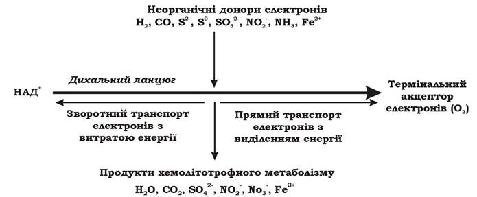
Fig. 5.14. General scheme of chemolithotrophic metabolism in microorganisms
In chemolithotrophic microorganisms, electrons can move along the respiratory chain in two directions: forward and reverse. During forward transport, electrons flow toward the terminal acceptor from carriers with lower ORP values to those with higher ones. This electron transfer is coupled with the generation of a proton gradient and, consequently, ATP synthesis. Because the forward electron pathway in aerobic chemolithotrophs is significantly shorter than in aerobic chemoorganotrophs, the number of phosphorylation sites is smaller (typically one), resulting in a much lower ATP yield. During reverse transport, electrons move from their entry point in the respiratory chain toward Flavoproteins (FMN), ultimately reducing НАД+. The purpose of reverse electron transfer is to generate reduced equivalents necessary for cellular Biosynthesis, which cannot be produced via other pathways in chemolithotrophic metabolism. Unlike chemoorganotrophs, where НАДН2 is formed without ATP expenditure, this process is energetically costly in chemolithotrophs. The energy required to drive electrons backward along the respiratory chain from carriers with higher ORP to those with lower ORP is provided by the Hydrolysis of ATP molecules previously synthesized during forward electron transport. For instance, Nitrobacter winogradskyi consumes 5 moles of ATP to synthesize 1 mole of НАДН2, whereas Thiobacillus consumes 2–3 moles of ATP.
Most chemolithotrophic bacteria are autotrophs. The assimilation of СО2 also requires substantial energy investments. For example, the synthesis of a single glucose molecule via The Calvin Cycle requires The Cell to expend 12 molecules of reduced equivalents and 18 ATP molecules.
Thus, unlike chemoorganoheterotrophs, chemolithotrophs generate ATP solely through oxidative phosphorylation via forward electron transfer along the short terminal segment of the respiratory chain, while additionally expending ATP on autotrophic biosynthesis and НАД+ reduction during reverse electron transfer.
Chemolithotrophs address their energetic challenges in the following manner:
1. They feature a highly branched terminal segment of the Respiratory Chain and a high concentration of cytochromes, which enables them to transfer A large number of electrons to the terminal acceptor. Consequently, most chemolithotrophs possess a well-developed system of internal Cytoplasmic membranes.
2. The oxidation of inorganic electron Donors by chemolithotrophs proceeds quite rapidly. They process large quantities of inorganic compounds and release substantial amounts of end metabolites into the environment, which plays a crucial role in global biogeochemical cycles.
Hydrogen (hydrogen-oxidizing) bacteria. These are aerobic chemolithoautotrophic prokaryotes capable of obtaining energy and electrons through the OXIDATION OF MOLECULAR hydrogen, while synthesizing all cellular components from CO2 carbon. Molecular hydrogen is also utilized in anabolism to generate reducing equivalents. The metabolic equations for hydrogen bacteria are as follows:

Hydrogen bacteria do not constitute a taxonomic group; rather, they are organisms united solely by a few shared physiological characteristics. Representatives of hydrogen bacteria span 20 genera, exhibiting diverse morphologies, Gram-staining properties, motility, and endospore formation. With the exception of thermophilic bacteria of the genus Hydrogenobacter, which are obligate chemolithoautotrophs, all other hydrogen bacteria are facultative forms. They can also utilize various Organic compounds—including single-carbon molecules such as carbon monoxide, methanol, and formate—as sources of carbon and energy.
The ability to oxidize molecular hydrogen is associated with the presence of hydrogenases that catalyze the reaction: Н2 → 2Н+ + 2е-. Hydrogen bacteria possess Two Types of this enzyme (Fig. 5.15):
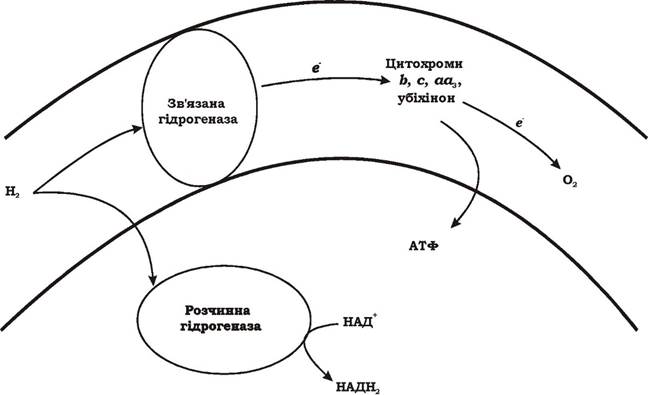
Fig. 5.15. Functions of the two hydrogenases in hydrogen bacteria
- soluble Hydrogenase, located in the Cytoplasm, which catalyzes the reduction of NAD+. The reaction product, NADH2, is subsequently used for biosynthesis;
- membrane-bound hydrogenase associated with subcellular particles. It is incapable of reducing NAD+ and directly transfers electrons to the respiratory chain at the level of flavoproteins, Quinones, or cytochrome b, thus affecting Energy Metabolism exclusively.
Most hydrogen bacteria contain only a single form of hydrogenase, which is membrane-bound. Certain species possess both forms, while others contain only the soluble form. When hydrogen bacteria harbor both hydrogenase variants, their functions are clearly demarcated. In the absence of soluble hydrogenase, The Challenge of obtaining reducing equivalents is met through Reverse Electron Transport to NAD+. When only the soluble hydrogenase is functional, it performs both roles: a portion of the reducing equivalents from NADH2 is fed into the respiratory chain, while the remainder is directed into constructive metabolism.
Thus, among all chemolithotrophic bacteria, only hydrogen bacteria are capable of directly reducing NAD+ via a specific hydrogenase form while oxidizing an inorganic substrate. This is directly reflected in the energetic yield of the process: the respiratory chain contains either three or two phosphorylation sites.
Nitrifying bacteria. Nitrification is the microbial oxidation of ammonium to nitrate. Certain chemoorganoheterotrophic microorganisms (Fusarium, Nocardia, Alcaligenes, Corynebacterium, Pseudomonas) can convert ammonium to nitrite as a byproduct of enzymatic activity, though such oxidation does not serve as a cellular energy source. In contrast, There are two distinct physiological groups of aerobic bacteria that carry out nitrification for energy generation—meaning they possess a chemolithotrophic metabolism. It is these organisms that are referred to as nitrifying bacteria.
The nitrification process consists of two phases executed by different physiological groups of bacteria via distinct mechanisms, designated as the First and Second phases of nitrification.
During the first phase of nitrification, ammonium (NH4+) is oxidized to nitrite (NO2-), effectively converting N3- to N3+. This process proceeds in two steps:
1. Ammonium → hydroxylamine (2NH4+ + O2 → 2NH4OH + 2H+)
This reaction is endergonic (ΔG0' = +26.8 kcal) and is catalyzed by the enzyme ammonia monooxygenase (AMO) in the presence of molecular oxygen. One atom of molecular oxygen is reduced to Water, while the other is incorporated into ammonium to yield hydroxylamine.
2. Hydroxylamine → nitrite (NH2OH + O2 → NO2 + H2O + H+)
Hydroxylamine is oxidized to nitrite by hydroxylamine oxidoreductase. This reaction is exergonic (ΔG0' = -59.3 kcal) and proceeds through a series of unstable hypothetical intermediates (hyponitrite, nitrohydroxylamine). The electrons released during hydroxylamine oxidation are channeled into the respiratory chain at the level of cytochrome c. Consequently, the true energetic substrate of the first nitrification phase is hydroxylamine.
The formation of hydroxylamine is necessitated by the high redox potential (+440 mV) of the reaction NH4+ + 2H2O → NO2- + 8H+ + 6e-. The respiratory chain of first-phase nitrifiers lacks a carrier capable of accepting an electron directly from ammonium. Therefore, the bacterium must expend energy to deliberately synthesize an intermediate product, hydroxylamine, which has a Redox Potential of -40 mV. The electrons generated during hydroxylamine oxidation are accepted by cytochrome c, which has a redox potential of +245 mV. Figure 5.16 illustrates the scheme of ammonium oxidation to nitrite during The first phase of nitrification.

Fig. 5.16. Scheme of ammonium oxidation to nitrite in the first phase of nitrification: 1, 2 - process stages; NH2OH - hydroxylamine; NOH - hypothetical intermediate products
The first phase of nitrification is carried out by Nitrosomonas europaea, Nitrosococcus oceanus, Nitrosococcus nitrosus, Nitrosolobus multiformis, Nitrosospira briensis, and Nitrosovibrio tenuis.
During the second phase of nitrification, nitrite (NO2-) is oxidized to nitrate (NO3-), converting N3+ into N5+.
This process proceeds According to the equation: 2NO2- + O2 → 2NO3- and is energetically favorable (ΔG0' = -36.4 kcal). The key enzyme of second-phase nitrifiers is nitrite oxidoreductase, which is membrane-bound. The electrons liberated during nitrite oxidation are transferred to the respiratory chain at the level of cytochrome a1. The second phase of nitrification is performed by Nitrobacter winogradskyi, Nitrospina gracilis, Nitrococcus mobilis, and Nitrospira marina.
The nitrifying bacteria of the first and second phases are characterized by a low ATP yield, the presence of reverse electron transport, autotrophic anabolism, oxygen requirements, and a narrow physiological pH range. Because nitrite is a chemically unstable compound that does not accumulate in nature, the nitrifiers of both phases inhabit the same niches.
Microorganisms oxidizing reduced sulfur compounds. Bacteria are capable of oxidizing such sulfur compounds as sulfides (S2-, HS-), sulfites (SO32-), thiosulfates (S2O32-), trithionates (S3O62-), tetrathionates (S4O62-), elemental sulfur (S0), rhodanides (CNS-), dimethyl sulfides (CH3SCH3), dimethyl disulfides (CH3SSCH3), and sulfide ores. They can be divided into three groups:
1. Bacteria with chemolithotrophic metabolism (thiobacilli).
2. Bacteria with chemoorganotrophic metabolism (colorless sulfur bacteria).
3. Bacteria with phototrophic metabolism (Purple and green bacteria).
The group of thiobacilli includes the genera Thiobacillus (T. acidophilus, T. albertis, T. aquaesulis, T. caldus, T. cuprinus, T. denitrificans, T. ferrooxidans, T. halophilus, T. hydrothermalis, T. neapolitanus, T. novellus, T. plumbophilus, T. prosperus, T. tepidarius, T. thiooxidans, T. thioparus, T. thyasiris, T. versutus), Thiomicrospira (T. denitrificans, T. thermophila, T. pelophila, T. chilensis, T. frisia, T. crunogena, T. kuenenii, T. psychrophila, T. thyasirae), Paracoccus (formerly Thiophaera) (P. pantotrophus), Thiodendron, and others. These are morphologically diverse, Gram-negative, non-spore-forming unicellular aerobes (Thiobacillus denitrificans is capable of nitrate respiration) possessing an obligate or facultative chemolithotrophic type of metabolism. Most thiobacilli are autotrophs (assimilating CO2 via the Calvin cycle), though heterotrophs also occur among them. Enzymes of Glycolysis, the Pentose Phosphate Pathway, and the Entner-Doudoroff Pathway have been identified in various representatives of this group capable of chemoorganotrophic growth. The operation of the Krebs cycle and the glyoxylate shunt has also been described. During chemolithotrophic metabolism, thiobacilli derive electrons from the oxidation of various sulfur compounds (Thiobacillus ferrooxidans also obtains energy from iron (II) oxidation). As a rule, they do not form molecular sulfur, but if it does appear as an intermediate product, it is never accumulated intracellularly.
Unlike other chemolithotrophs, thiobacilli obtain ATP during the oxidation of sulfur compounds As a result of both oxidative and substrate-level phosphorylation.
In oxidative phosphorylation, eight electrons, sequentially cleaved from sulfide, are fed into the respiratory chain at the level of cytochrome c. Molecular oxygen serves as the terminal electron acceptor in most cases.
Substrate-level phosphorylation occurs during The conversion of sulfide to sulfate. First, sulfide (S2-, HS-) is oxidized to sulfite (SO32-). This may proceed via an elemental sulfur-forming stage:

Subsequently, according to Peck's scheme (Table 5.5), sulfite is activated by AMP and oxidized to adenosine-5'-phosphosulfate (Fig. 5.17) by APS reductase. The electrons released in this process are channeled into the respiratory chain.
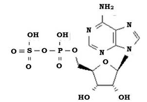
Fig. 5.17. Structural formula of adenosine-5'-phosphosulfate (APS)
Table 5.5. Scheme of substrate-level phosphorylation in thiobacilli according to Peck
No. |
Equation |
Enzymes |
1. |
2SO32- + 2 АМФ → 2 АФС + 4е- |
APS reductase |
2. |
2 АФС + 2 Фн → 2 АДФ + 2SO42- |
ADP sulfurylase |
3. |
2 АДФ → АМФ + АТФ |
Adenylate kinase |
Designations: APS – adenosine phosphosulfate, Pi – inorganic phosphate.
During the further phosphorylation of APS, sulfate (SO42-) is released into the medium, and the disproportionation of 2 ADP leads to the synthesis of ATP. Because this ATP molecule is formed outside the respiratory chain, it can be considered the result of substrate-level phosphorylation.
Thiobacilli obtain NADH2 via reverse electron transport along the respiratory chain.
Colorless sulfur bacteria closely resemble cyanobacteria but lack pigments. Based on their Morphology, they are divided into two groups: unicellular forms (genera Achromatium, Macromonas) and filamentous forms (genera Thiothrix, Thioploca, Thiospira, Beggiatoa). The only common feature of this group is that they deposit elemental sulfur in the periplasmic space of their Cells. It is currently uncertain whether these bacteria actually use reduced sulfur compounds as an electron source in Catabolism. They are known to possess low catalase activity, and 80–90% of the oxygen consumed during respiration is reduced only to H2O2. To inactivate this toxic hydrogen peroxide, these organisms require the presence of low concentrations of sulfides in their environment. The deposition of elemental sulfur in the periplasm is thus the result of the reaction between H2O2 and S2-. The capacity for autotrophic Nutrition in colorless sulfur bacteria has also not been definitively proven.
Purple and green bacteria will be discussed in detail below.
Carboxydobacteria are aerobes capable of growing by utilizing carbon monoxide (CO) as the sole source of electrons and carbon. Because they can also oxidize molecular hydrogen, some researchers classify carboxydobacteria as a group of hydrogen bacteria. At the same time, their ability to use a respiratory poison as a substrate points to a novel type of chemolithotrophic metabolism in carboxydobacteria. Unlike other bacteria, they are highly resistant to carbon monoxide, withstanding concentrations up to 20–95% by volume.
Carboxydobacteria oxidize CO according to the equation:

CO oxidation is carried out with the participation of at least one specific enzyme, CO oxidase, which is present in the cell in both soluble and membrane-bound forms. The bound CO oxidase is localized on the inner side of the cytoplasmic membrane. When carboxydobacteria grow on carbon monoxide, CO oxidase performs the following functions:
1. It oxidizes CO to CO2.
2. It transfers electrons to the respiratory chain.
3. It participates in NADH2 synthesis via reverse electron transport.
The respiratory chain of carboxydobacteria contains a single phosphorylation site.
Carbon dioxide, produced as a result of CO oxidation, serves as a carbon source and is assimilated via the Calvin cycle. Thus, the actual carbon source for carboxydobacteria is not CO, but CO2. The overall equation for CO utilization is as follows:

Fig. 5.18 shows the general scheme of CO assimilation by carboxydobacteria.

Fig. 5.18. Utilization of CO as a source of carbon and electrons by carboxydobacteria
One of the fascinating properties of carboxydobacteria is the very fact that they utilize CO, an inhibitor of terminal oxidases such as type a cytochromes. No unusual cytochromes have been found in the Electron Transport Chains of carboxydobacteria. The following mechanisms for their resistance to CO are considered possible:
- rapid detoxification of CO via CO oxidase;
- Induction of the synthesis of CO-insensitive terminal oxidases;
- enhanced synthesis of respiratory chain components;
- spatial Separation of the CO oxidation process from CO-sensitive cytochrome oxidases.
Carboxydobacteria are facultative chemolithoautotrophs. They are also capable of growing chemoorganoheterotrophically on organic substrates. Representative species of carboxydobacteria include Pseudomonas carboxydovorans, P. carboxydoflava, P. gazotropha, and Seliberia carboxydohydrogena.
Photosynthesis (Photophosphorylation) is the process by which organisms obtain energy from light, or the conversion of light energy into chemical bond energy.
Solar radiation reaching the Earth is divided into three groups (Fig. 5.19):
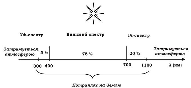
Fig. 5.19. Solar radiation reaching the Earth
1. Ultraviolet rays (λ = 300-400 nm). They account for 5% of light.
2. Visible spectrum (λ = 400-700 nm). Their share constitutes 75% of light.
3. Infrared rays (λ = 700-1100 nm). They account for 20% of light.
Last update: 13/08/2026
Editorial and Educational Adaptation: This material has been compiled based on the primary/original source text. The project team performed an editorial review, corrected technical inaccuracies, structured sections, and adapted the content for an educational format.
What was processed:
- elimination of formatting defects (OCR errors, structural breaks, corrupted characters);
- editorial organization of content;
- standardization of terminology in accordance with academic sources;
- verification of factual statements against the original source text.
All mentions of the author, publication year, and origin of the primary text have been preserved in accordance with the source.