Anatomy and Physiology of Children and Adolescents - M. R. Sapin 2007
Cardiovascular System
The Cardiovascular system comprises The Heart and Blood Vessels (Fig. 80, see color insert). This system Functions to transport blood, and along with it nutrients and energetic substrates, to the Organs and Tissues. From organs and tissues, Metabolic waste products are transported away via blood vessels by the bloodstream. Within the cardiovascular system, the heart acts as a muscular "pump," whose rhythmic contractions drive the movement of blood through the vascular network. Based on their Structure and function, blood vessels are divided into Arteries, microvessels (arterioles, venules), and Veins.
Arteries are the vessels that carry blood away from the heart to the organs and tissues. As they branch further away from the heart, the diameter of arteries progressively decreases down to the smallest arterioles, which transition into a capillary network deep within the organs. Arterioles, blood capillaries, and venules formed from capillaries constitute the microvasculature (microcirculatory bed), where exchange processes between the blood and tissues take place (Fig. 81).
Capillaries are the most numerous and finest vessels. Their diameter ranges from 3 to 11 µm. The Human Body contains approximately 40 billion capillaries, with a total length of about 100,000 km. Capillaries empty into venules, the fusion of which forms small veins. Veins are the vessels that carry blood back toward the heart. The total number of veins exceeds that of arteries, and the overall capacity of the Venous system surpasses that of the arterial bed.
Blood vessels are named after the organ they supply with blood (e.g., the renal artery) or the bone they lie adjacent to (e.g., the ulnar artery, etc.).
Class="center">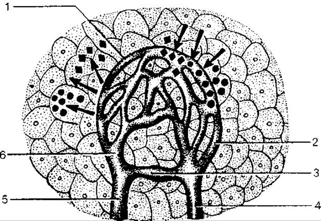
Fig. 81. Microcirculatory bed:
1 — capillary network (capillaries); 2 — postcapillary (postcapillary venule); 3 — arteriovenous anastomosis; 4 — venule; 5 — arteriole; 6 — precapillary (precapillary arteriole).
Arrows pointing away from capillaries indicate the delivery of nutrients to tissues; arrows pointing toward capillaries indicate the removal of metabolic waste from tissues.
Structure of Blood vessel walls. The walls of all arteries, much like those of veins, consist of three tunics (layers): internal, middle, and external (Fig. 82). Wall thickness and structural details vary across Different types of vessels. The internal tunic, or intima, is composed of flat endothelial Cells (endotheliocytes) resting on a basement membrane. The walls of most arteries contain a high concentration of elastic fibers that form the internal elastic membrane, imparting elasticity and resilience to the arteries. In small and medium-sized veins, the internal tunic forms crescent-shaped folds called Valves, which prevent the backflow of blood (Fig. 83). The middle tunic (media) consists of smooth Muscle cells. Compared to veins, the muscular tunic in arteries is better developed and thicker. The middle tunic also contains elastic fibers, which are particularly abundant in very large arteries such as the aorta and pulmonary trunk; these are known as elastic-type arteries. In the common carotid, subclavian, common iliac, and other medium-sized arteries, The ratio of smooth muscle cells to elastic fibers is roughly equal, classifying them as mixed muscle-elastic arteries. In strictly muscular arteries, the middle tunic is predominantly made up of smooth muscle cells. In small and medium-diameter arteries, elastic fibers form an external elastic membrane. The external tunic (adventitia) of blood vessels consists of loose Fibrous Connective Tissue, which houses the nerves and vasa vasorum that nourish the vessel walls.
Blood capillaries are present in all organs and tissues except for the Nails, Hair, the epithelial layer of the Skin, mucous membranes, and Cartilage. Having thin walls of varying structure (Fig. 84), blood capillaries mediate exchange processes between the blood and surrounding tissues. In the Lungs, capillaries facilitate gas exchange between blood and air. Typically, only 20–30% of capillaries are open in a resting human; the remaining capillaries are recruited into the Circulation during periods of heightened organ activity. The flow of blood into capillaries is regulated by smooth muscle sphincter cells located at the transition points from arterioles to capillaries.
Thus, the Circulatory system can be divided into three main components: the arterial segment, which transports blood from the heart and maintains constant blood pressure within the vessels; the microvasculature (arterioles, capillaries, venules), which ensures direct exchange between blood and tissues; and the venous bed, which provides the return of blood to the heart.

Fig. 82. STRUCTURE OF THE walls of a medium-sized artery (A) and a vein (B):
1 — endothelium; 2 — basement membrane; 3 — subendothelial layer; 4 — internal elastic membrane; 5 — myocytes; 6 — elastic fibers; 7 — Collagen fibers; 8 — external elastic membrane; 9 — loose fibrous connective tissue; 10 — blood vessels. Tunics: I — internal, II — middle, III — external.
On the external surface of the heart, the coronary (atrioventricular) and transverse sulci are visible, separating the atria from the ventricles, along with two longitudinal interventricular sulci (anterior and posterior) located at the boundary between the right and left ventricles. These sulci house the CORONARY ARTERIES AND cardiac veins. Above the coronary sulcus, flanking the aorta and pulmonary trunk, lie the pouch-like anterior extensions of the right and left atrial walls known as the right and left auricles.
The right and left atria are situated above the coronary sulcus and form the Base of the heart. Inferiorly, the atria communicate with the right and left ventricles, respectively, via the right and left atrioventricular orifices. During atrial contraction, blood is pumped through these openings into the ventricles (Fig. 86).
The SUPERIOR VENA CAVA empties into the superior part of the right atrium, returning deoxygenated blood from the HEAD, neck, upper limbs, and thoracic walls. Inferiorly, the INFERIOR VENA CAVA opens into this atrium, draining blood from the organs and walls of the thoracic and abdominal cavities, the pelvis, and the lower limbs. The coronary sinus of the heart also empties into the right atrium, returning venous blood from the heart muscle itself. The inferiorly located right atrioventricular orifice leads from the right atrium into the right ventricle.
Right ventricle. The internal surface of the right ventricle is uneven, marked by three cone-shaped papillary Muscles projecting into the lumen. Superiorly, the ventricle features two openings: the right atrioventricular orifice and the opening leading into the pulmonary trunk. The right atrioventricular orifice is guarded by the tricuspid valve. Thin chordae tendineae, originating from the papillary Muscles of the right ventricle, attach to the free margins of the three cusps (anterior, posterior, and septal) of this valve. The tricuspid valve permits blood flow from the right atrium into the right ventricle while, thanks to the tone of the papillary muscles, preventing the backflow of blood from the ventricles into the atria. At the Water/144.html">Origin of the pulmonary trunk lies a valve consisting of three semilunar cusps. This pulmonary valve allows blood to flow from the right ventricle toward the lungs and prevents backflow into the ventricle.
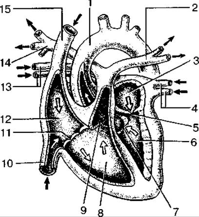
Fig. 86. Structure of the heart chambers and direction of blood flow (frontal section):
1 — aorta; 2 — left pulmonary artery; 3 — left atrium; 4 — left Pulmonary veins; 5 — left atrioventricular orifice; 6 — left ventricle; 7 — aortic valve; 8 — right ventricle; 9 — pulmonary valve; 10 — inferior vena cava; 11 — right atrioventricular orifice; 12 — right atrium; 13 — right pulmonary veins; 14 — right pulmonary artery; 15 — superior vena cava. Arrows indicate the direction of blood flow within the heart chambers.
Superiorly, the left atrium presents four openings through which the pulmonary veins empty into it (two from each lung). Inferiorly, the left atrium features the left atrioventricular orifice, which leads into the left ventricle.
Left ventricle. Two papillary muscles project from the inner surface of the ventricle and connect via thin chordae tendineae to the free margins of the two cusps (anterior and posterior) of the left atrioventricular (bicuspid or mitral) valve. Blood enters the left ventricle through the left atrioventricular orifice, which connects the left atrium to the left ventricle. Backflow of blood is prevented by the aforementioned bicuspid valve.
The aorta emerges from the left ventricle, with its opening located in the upper part of the left ventricle. The aortic orifice is equipped with a valve consisting of three semilunar cusps. This valve permits blood flow exclusively from the ventricle into the aorta and prevents any backward flow.
All heart valves open passively under METABOLISM/18.html">The Influence of blood flow. When the atrial musculature contracts, the cusps of the atrioventricular valves—which are folds of the inner lining of the heart wall, the endocardium—open, allowing blood to enter the ventricles. The chordae tendineae of the papillary muscles prevent the cusps from everting back toward the atria. Upon contraction of the ventricular musculature and their papillary muscles, the chordae tendineae tauten, keeping the valve cusps from prolapsing into the atria.
The cusps of the semilunar valves, which guard the orifices of the aorta and pulmonary trunk, freely allow blood to flow from the ventricles into the pulmonary trunk and aorta while preventing any backflow from these vessels into the ventricles.
Pericardium. In newborns, the pericardium is spherical in shape. The volume of the pericardial cavity is small, and it fits snugly around the heart. The dome of the pericardium is positioned high up, along the line connecting the sternoclavicular joints. The lower border of the pericardium runs at the level of the middle of the fifth intercostal spaces. A significant portion of the sternocostal surface of the pericardium is covered by the Thymus. The posterior region lies adjacent to the Esophagus, Trachea, Bronchi, aorta, and vagus nerves. The inferior wall of the pericardium is fused with the tendinous center and muscular part of the Diaphragm. By the age of 14, the BOUNDARIES OF THE pericardium and its anatomical relationships with the mediastinal organs correspond to those of an adult.
Last update: 10/08/2026
Editorial and Educational Adaptation: This material has been compiled based on the primary/original source text. The project team performed an editorial review, corrected technical inaccuracies, structured sections, and adapted the content for an educational format.
What was processed:
- elimination of formatting defects (OCR errors, structural breaks, corrupted characters);
- editorial organization of content;
- standardization of terminology in accordance with academic sources;
- verification of factual statements against the original source text.
All mentions of the author, publication year, and origin of the primary text have been preserved in accordance with the source.