Review of Medical Physiology - William F. Ganong 2002
Physiology of Nerve and Muscle Cells
Synaptic and Junctional Transmission
Synaptic Transmission - Electrical Events in Postsynaptic Neurons
A technique that allows researchers to penetrate a Spinal Cord Cell helps study electrical activity phenomena in a postsynaptic neuron. This is achieved by inserting a microelectrode into the ventral region of the spinal cord. Puncture of The Cell membrane is identified by the appearance of a stable potential difference (-70 mV) between the microelectrode and an extracellular electrode. Confirmation that the cell is a spinal motoneuron is obtained by stimulating the corresponding ventral ROOT and subsequently recording the cell's electrical activity. This stimulation generates an antidromic impulse (see Chapter 2) that propagates toward the cell body and stops at this point. Therefore, the presence of an Action Potential in the cell following antidromic stimulation proves that the impaled cell is indeed a spinal motoneuron rather than an interneuron. Activity in specific presynaptic terminals contacting the impaled cell can be initiated by stimulating the dorsal root (Fig. 4-7).
Class="center">
Fig. 4-5. The cycle of small synaptic vesicles in a presynaptic nerve terminal. Vesicles bud off from the early endosome and fill with neurotransmitter (NT; top left). They then move toward The Plasma Membrane, where docking and priming occur. Upon arrival of an action potential at the terminal, Ca2+ influx triggers fusion and exocytosis, releasing the vesicular contents into the synaptic cleft. Subsequently, the vesicle membrane is coated with clathrin and internalized via endocytosis. In the Cytoplasm, it fuses with the early endosome, and the cycle repeats (reproduced with permission from Sudhof TC: The synaptic vesicle cycle: a cascade of Protein-Structure/156.html">Protein Interactions. Nature 1995:375:645).
Excitatory Postsynaptic Potentials
Single stimuli applied to sensory nerves under the experimental conditions described above do not trigger a propagating action potential in the postsynaptic neuron. Instead, such stimulation causes either a brief partial depolarization or a brief hyperpolarization.
A depolarizing response evoked by a single stimulus of adequate strength begins 0.5 ms after the impulse reaches the spinal cord, peaks in 1.0-1.5 ms, and then decays exponentially. During this potential, the neuron's excitability to another stimulus increases, which is why it is termed an excitatory postsynaptic potential (EPSP). An EPSP is generated by the depolarization of the postsynaptic cell membrane adjacent to the presynaptic terminal. The excitatory transmitter opens Na+ or Ca2+ channels in the postsynaptic membrane, thereby producing an inward current. The region where this current is generated is so small that it does not propagate until positive charges depolarize the entire membrane. EPSPs resulting from activity in a single synaptic vesicle are small, but the depolarizations produced by each active synaptic vesicle summate.
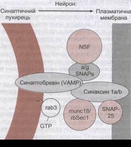
Fig. 4-6. Key interacting Proteins that mediate the tethering and fusion of synaptic vesicles in nerve terminals (reproduced with permission from Ferro-Novick S, John R: Vesicle fusion from Yeast to man. Nature 1994:370:191).
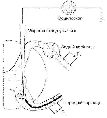
Fig. 4-7. Placement of recording and stimulating electrodes during The Study of synaptic activity in mammalian spinal motoneurons. Stimulation P2 is used to evoke antidromic impulses for cell identification, whereas stimulation P1 is used for orthodromic stimulation via the reflex arc.
Summation can be either spatial or temporal. If activity occurs simultaneously in more than one synaptic vesicle, spatial summation is observed, whereby The activity of one synaptic vesicle enhances that of another, bringing the membrane closer to the critical firing level of depolarization. Temporal summation occurs when repeated afferent stimuli elicit new EPSPs before the previous ones have decayed. Naturally, the longer an EPSP lasts, the greater the likelihood of summation. The phenomena of spatial and temporal facilitation are illustrated in Fig. 4-8. Thus, an EPSP does not obey the "all-or-none" law; rather, its amplitude is proportional to the strength of the afferent stimuli. If the EPSP is large enough upon reaching the critical level of depolarization in the cell, a full-fledged action potential is generated.
Synaptic Delay
A delay of 0.5 ms—known as the synaptic delay—occurs between the arrival of the impulse at the presynaptic terminal and the onset of the response in the postsynaptic neuron. Under maximal stimulation of the presynaptic neuron, this delay corresponds to the latency of the EPSP and is determined by the time required for the release of the synaptic transmitter and its action on the postsynaptic membrane. Consequently, the time required for impulse transmission through a neuronal pathway containing numerous synapses is longer than through a pathway with only a few synapses. Since the minimum time required for transmission across a single synapse is known to be 0.5 ms, measuring the transmission delay from the dorsal root through the spinal cord to the ventral root reveals whether a given reflex arc is monosynaptic or polysynaptic (i.e., contains more than one synapse).

Fig. 4-8. Spatial (A-C) and temporal summation (D-F) of EPSPs. Potential changes were recorded using a single microelectrode inserted into the postsynaptic cell. A-C: Afferent stimuli of increasing strength were applied. Here, an increasing number of synaptic vesicles are activated, ultimately reaching the critical level of depolarization (C) and generating an action potential. D-F: Two identical stimuli of equal strength were applied. In F, the time interval between them was decreased, which likewise achieves the critical level of depolarization and generates an action potential.
Inhibitory Postsynaptic Potentials
In some cases, stimulation that triggers EPSPs in certain Neurons causes hyperpolarization in others. Similar to EPSPs, these events peak 1.0-1.5 ms after stimulation and then decay exponentially with a time constant (the decay time corresponding to 1/e, or 1/2.718 of the maximum). The decay takes approximately 3 ms (Fig. 4-9). During this potential, the neuron's excitability in response to other stimuli is depressed, which is why it is called an inhibitory postsynaptic potential (IPSP). IPSPs exhibit both spatial summation—evidenced by an increase in response amplitude with stronger inhibitory afferent input—and temporal summation. This type of inhibition is referred to as postsynaptic, or direct, inhibition.
An IPSP can be caused by a local increase in membrane permeability to Cl-. When inhibitory synaptic vesicles are active, the release of the transmitter opens Cl- channels in the region of the postsynaptic membrane adjacent to the synaptic vesicle; Cl- moves down its concentration gradient, carrying a negative charge into the cell and increasing the Membrane Potential. The depression of nerve cell excitability during an IPSP results from shifting the membrane potential away from the critical level of depolarization. Consequently, a much greater excitatory (depolarizing) activity is required to reach the critical firing level. The fact that IPSPs are mediated by Cl- can be demonstrated in a repeated stimulation experiment where the resting membrane potential is altered and clamped using Voltage Clamp. When the resting membrane potential is held at the level of ECl, the inhibitory potential disappears (see Fig. 4-9), and at more negative membrane potentials, it reverses in sign (becomes positive).

Fig. 4-9. IPSP produced by increased Cl- influx resulting from stimulation at various membrane potential levels set by voltage clamping; RMP is the resting membrane potential of the neuron. Note that when the membrane potential reaches the level corresponding to ECl, the IPSP disappears, whereas at more hyperpolarized membrane potentials, it becomes positive.
An IPSP may also be generated by the opening of K+ channels, resulting in K+ efflux from the postsynaptic cell. Furthermore, IPSPs can also occur when Na+ and Ca2+ channels close.
Slow Postsynaptic Potentials
In addition to the fast EPSPs and IPSPs described above, slow EPSPs and IPSPs occur in autonomic ganglia, cardiac and smooth Muscle, as well as cortical neurons. These postsynaptic potentials have a latency of 100-500 ms and last for several seconds. Slow EPSPs are primarily caused by a decrease in K+ conductance, whereas slow IPSPs are caused by an increase in K+ conductance.
Late slow excitatory postsynaptic potentials with a latency of 1-5 s and a duration of 10-30 min have also been discovered in sympathetic ganglia. This potential is likewise mediated, at least in part, by a decrease in K+ conductance, and its transmitter is a peptide closely related to GnRH—a hormone produced by hypothalamic neurons that stimulates LH secretion (see Chapter 14).
Generation of the Action Potential in the Postsynaptic Neuron
The continuous reciprocal influence of excitatory and inhibitory activities on a postsynaptic neuron results in membrane potential fluctuations that correspond to the algebraic sum of hyperpolarizing and depolarizing effects. The neuronal soma thus acts as a specific type of integrator. If 10-15 mV are sufficient to reach the critical threshold of depolarization, an action potential spike is generated. However, ELECTRICAL PHENOMENA IN neurons are more complex. In motoneurons, the part of the cell with the lowest threshold for the initiation of a full-fledged action potential is the initial segment—the region of the axon located immediately distal to the axon hillock. This unmyelinated segment is depolarized or hyperpolarized by current sources arising in excitatory or inhibitory synaptic vesicles. This region of the neuron is activated first, and then the potential propagates in two directions: along the axon and in the reverse direction, toward the cell body. Retrograde impulses reaching the soma are likely important for "clearing" it of prior influences, which is necessary for the subsequent repetition of the reciprocal interaction of excitatory and inhibitory activities on the cell.
Function of Dendrites
For a long time, it was believed that dendrites are regions where impulses are generated that alter the membrane potential of the initial segment, meaning they are merely projections of the cell body that increase the integration area. If a neuron's dendritic tree is large and contains numerous presynaptic vesicular boutons, the potential for greater interaction between inhibitory and excitatory influences increases. Impulse streams arriving at and departing from dendrites wax and wane. The Role of dendrites in the generation of the Electroencephalogram (EEG) is discussed in Chapter 11.
Current data indicate that the function of dendrites in supporting neural activity is more complex. Action potentials can be recorded in dendrites. In many cases, they are initiated in the initial segment and propagate retrogradely; however, a propagatable action potential also arises within the dendrites themselves. Finally, There is a growing interest in microdomains present in the cytoplasm of many different cell types, particularly The formation of Ca2+ pools near individual dendritic spines, which enhances the potential for local Changes in the intensity of synaptic transmission and may thus be related to the processes of Learning and Memory.
Electrical Conduction
At synaptic contacts where electrical conduction occurs, an impulse arriving at the axon terminal generates an excitatory postsynaptic potential in the postsynaptic cell which, due to the presence of low-resistance bridges in the contact area, has a shorter latency period than the EPSP in synapses with chemical transmission. In mixed-type synapses, both this short-latency response and the long-latency, chemically mediated response occur.
It occurs As a result of spinal cord injury and in the case of multiple sclerosis, particularly when administered intrathecally via an implanted pump. Other Transmitters also mediate presynaptic inhibition through a G-protein-mediated action on Ca2+ and K+ channels.
Conversely, in the case of a prolonged action potential, presynaptic facilitation occurs (see Fig. 4-12), and Ca2+ channels remain open for a longer duration. The molecular mechanisms underlying presynaptic facilitation mediated by serotonin have been studied in detail in the marine mollusk Aplysia. The release of serotonin at axo-axonal terminals leads to an increase in the intracellular concentration of cyclic AMP, and the subsequent phosphorylation of a specific type of K+ channel causes these channels to close, slowing repolarization and prolonging the action potential.
Organization of Inhibitory Systems
Presynaptic and postsynaptic inhibition predominantly occur as a result of excitation in specific systems that converge on a particular postsynaptic neuron (afferent inhibition). Neurons can also inhibit via negative feedback. For example, each spinal motoneuron regularly gives off a recurrent collateral that forms a synaptic contact with an inhibitory interneuron terminating on the soma of the spinal neuron as well as on other spinal motoneurons (Fig. 4-13). This specific inhibitory neuron is sometimes called a Renshaw cell. Impulses generated in the motoneuron activate the inhibitory interneuron, which causes the release of an inhibitory transmitter and, consequently, the slowing or even cessation of the motoneuron discharge. Similar inhibition via recurrent collaterals is observed in the Cerebral Cortex AND the limbic system.

Fig. 4-12. Effects of presynaptic inhibition and presynaptic facilitation on the action potential, Ca2+ influx into the presynaptic neuron, and the EPSP in the postsynaptic neuron. In each case, solid lines represent control values, and dashed lines represent changes resulting from inhibition and facilitation (modified from Kandel ER, Schwartz JH, Jessell TM [editors]. Principles of Neural Science, 4th ed. McGraw-Hill, 2000).
Presynaptic inhibition involving descending pathways terminating on afferent pathways in the dorsal horn may be involved in the transmission of Pain Sensation (see Chapter 7).
Another type of inhibition occurs in the Cerebellum. In this part of the Brain, stimulation of basket Cells induces IPSPs in Purkinje cells (see Chapter 12). However, both basket cells and Purkinje cells are excited by the same parallel fibers. This organization, termed feed-forward inhibition, presumably limits the duration of excitation elicited by certain afferent impulses.
Summation and Occlusion
The interaction between excitatory and inhibitory influences at synaptic contacts within a neural network reflects the integrative and modulating activity of The Nervous system.

Fig. 4-13. Feedback inhibition of a spinal motoneuron via an interneuron (Renshaw cell).
In the hypothetical neural network shown in Fig. 4-14, neurons A and B converge on X, while neuron B diverges to X and Y. Stimulation of A or B leads to the generation of an EPSP in X. If A and B are stimulated simultaneously and Action potentials are generated, two areas of depolarization arise in X, and their effects summate. As a result, the EPSP in X will be twice as large as that produced by separate stimulation of A or B, and the potential in X will readily reach the critical level of depolarization. The depolarizing effect produced by impulses in A enhances the impulses causing activity in B, and vice versa; thus, spatial facilitation occurs. In this case, Y is not excited, but its excitability is increased, and an EPSP is more easily generated in it upon activity of neuron C. Therefore, Y is said to be in a subliminal fringe state with respect to X.
In general, neurons are in a subliminal fringe state if, in the absence of afferent impulses (i.e., outside the discharge zone), their excitability is increased. Neurons with several active vesicular boutons are in a subliminal fringe state, whereas those with many are in the discharge zone. Inhibitory impulses produce similar temporal and spatial facilitation and subliminal fringe effects.
If action potentials occur consecutively in neuron B, X and Y are excited as a result of temporal summation of the generated EPSPs. If C is stimulated multiple times, Y and Z are also excited. If B and C are stimulated repeatedly at the same time, discharge will occur in X, Y, and Z. Thus, the response to simultaneous stimulation of B and C is not greater than the sum of the responses to separate stimulation of B and C, because B and C terminate on neuron Y. This reduction in the level of the expected response occurring upon stimulation of presynaptic fibers is called occlusion.
Excitatory and inhibitory subliminal effects, as well as The phenomenon of occlusion, can significantly influence impulse conduction in any given pathway. As a result of these influences, the temporal characteristics during the passage of impulses from the periphery to the brain through a series of synapses are altered. These phenomena may also explain such an important phenomenon as the transmission of pain sensation (see Chapter 7).

Fig. 4-14. Simple neural network. Neurons A, B, and C form excitatory endings on neurons X, Y, and Z.
Neuromodulation
The term modulation is often interpreted so loosely in physiology that its definition conveys little information about the actual function. In neurobiology, however, the term neuromodulation is used to specifically designate the non-synaptic action of substances on neurons that alters their sensitivity to synaptic excitation or inhibition. Neuromodulation is primarily driven by the action of Neuropeptides and circulating Steroids, as well as steroids produced within the nervous system (neurosteroids; see below).
Last update: 10/08/2026
Editorial and Educational Adaptation: This material has been compiled based on the primary/original source text. The project team performed an editorial review, corrected technical inaccuracies, structured sections, and adapted the content for an educational format.
What was processed:
- elimination of formatting defects (OCR errors, structural breaks, corrupted characters);
- editorial organization of content;
- standardization of terminology in accordance with academic sources;
- verification of factual statements against the original source text.
All mentions of the author, publication year, and origin of the primary text have been preserved in accordance with the source.