Human Anatomy - Kotsan I. Y. 2009
Organs of Hematopoiesis and the Immune System
Lymphoid tissue of the walls of the digestive and respiratory systems
Spleen
The Spleen (splen, lien) lies along The pathway of Blood flow from the aorta, the main vessel of the systemic Circulation, to the Liver. It is located in the Abdominal cavity within the left hypochondriac region, extending from the 9th to the 11th rib. In a healthy adult (aged 20–40), the mass of the spleen averages 192 g in men and 153 g in women. Its dimensions at this age are typically 10–14 cm in length, 6–10 cm in width, and 3–4 cm in thickness. Due to its rich vascularity, the volume and weight of the spleen can vary significantly within the same individual depending on the degree of blood engorgement.
The spleen has the shape of a flattened, elongated hemisphere, a dark red color, and a soft consistency (Fig. 222). It presents two surfaces (diaphragmatic and visceral), two borders (superior and inferior), and two extremities (posterior and anterior).
Class="center">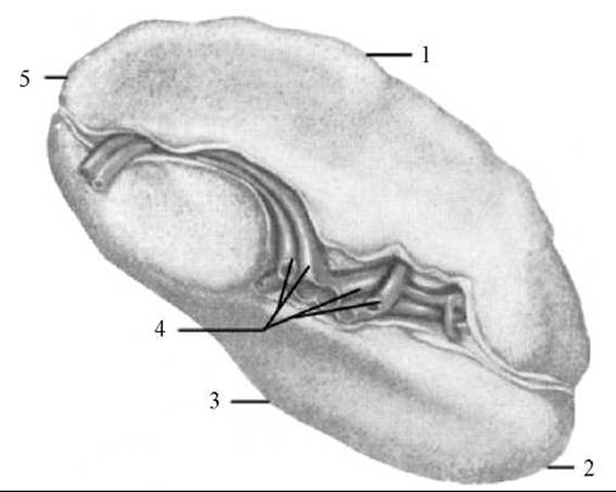
Fig. 222. Spleen (visceral surface)
1 — superior border; 2 — anterior extremity; 3 — inferior border; 4 — hilum of the spleen (Blood Vessels visible); 5 — posterior extremity
The diaphragmatic surface (facies diaphragmatica) is smooth, convex, and faces laterally and upward toward the Diaphragm. The visceral surface (facies visceralis) is uneven, concave, and features the hilum of the spleen (hilum splenicum), through which blood Vessels and nerves enter the organ. The visceral surface is further subdivided into areas adjacent to neighboring Organs. The gastric surface (facies gastrica) contacts the Cytology/practical/108.html">Fundus of the Stomach and is located anterior to the splenic hilum. The renal surface (facies renalis) lies posterior to the hilum, in contact with the upper pole of the left Kidney and the left suprarenal gland. The colic surface (facies colica) is situated inferior to the hilum, near the anterior border, where the spleen touches the left colic flexure. Just superior to the colic surface and directly posterior to the hilum is a small area approached by the tail of the Pancreas. The superior border (margo superior) of the spleen is sharp, separates the gastric surface from the diaphragmatic surface, and features 2–3 shallow notches. The inferior border (margo inferior) is more blunt, dividing the diaphragmatic and renal surfaces. The extremities (poles) of the spleen include the posterior extremity (extremitas posterior), which is rounded and directed upward and backward, and the anterior extremity (extremitas anterior), which is sharper.
Except for the area in contact with the tail of the pancreas, the spleen is entirely enclosed by Peritoneum, which forms its serous coat (tunica serosa). The serous coat is tightly fused with the fibrous coat (tunica fibrosa), composed of Collagen and elastic fibers along with smooth Muscle Cells. Where the peritoneum reflects from The surface of the spleen onto adjacent organs, it forms ligaments: the gastrosplenic ligament (lig. gastrosplenicum) and the phrenicosplenic ligament (lig. phrenicosplenicum), which anchor the organ to The Stomach and diaphragm. Because of these ligaments, the Movements of the diaphragm during Respiration, as well as the filling and emptying of the stomach, alter the Topography of the spleen.
From the fibrous capsule at the hilum, Connective Tissue trabeculae (trabeculae splenicae) extend into the substance of the spleen. The space between the trabeculae is filled with the splenic parenchyma, or pulp (pulpa splenicae), which is differentiated into red and white pulp.
The red pulp (pulpa rubra) constitutes approximately 80% of the total splenic parenchyma. It is situated between the venous sinuses of the spleen and consists of a reticular tissue meshwork filled with erythrocytes, leukocytes, lymphocytes, and macrophages.
The white pulp (pulpa alba) accounts for up to 20% of the total splenic parenchyma. It is formed by splenic lymphoid nodules and periarterial lymphatic sheaths, which consist of lymphocytes and other lymphoid cells embedded within a reticular stroma. The lymphoid nodules are roughly spherical. Each nodule is traversed by a central artery, which typically runs eccentrically. The periarterial lymphatic sheaths form sleeve-like structures around the pulp Arteries or the initial segments of the central splenic arteries. Distally, the periarterial lymphatic sheath extends toward the lymphoid nodule and even merges into it (Fig. 223).
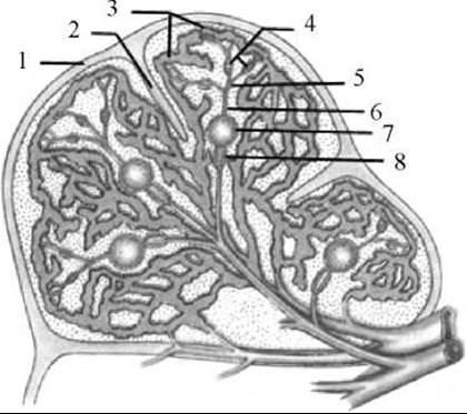
Fig. 223. Microscopic Structure OF THE spleen (diagram)
1 — fibrous capsule; 2 — splenic trabecula; 3 — splenic venous sinuses; 4 — ellipsoid macrophage sheath; 5 — penicillar arteriole; 6 — central artery; 7 — lymphoid nodule; 8 — periarterial lymphatic sheath
The function of the spleen involves supporting the development and activation of lymphocytes and their transformation into antibody-producing cells or cells mediating cellular Immunity. Key Functions of the spleen also include filtering microbes and foreign particles from the blood, destroying altered and aged erythrocytes (hence known as the "erythrocyte graveyard"), and acting as a blood reservoir.
Development and Age-related Features of the spleen. The spleen originates during the 5th to 6th week of Embryogenesis as a small mesenchymal Cell cluster within the dorsal mesentery. Soon, clefts—the future splenic vessels—appear within this mesenchymal primordium, around which the tissue Differentiation of the organ takes place. During the second month of intrauterine development, blood vessels form and hemopoietic foci appear in the spleen. By the end of the fourth and into the fifth month, clusters of lymphocytes emerge, representing the future lymphoid nodules and periarterial lymphatic sheaths. The number of lymphoid nodules gradually increases, developing germinal centers that displace the central artery. By the eighth month of fetal life, hemopoiesis in the spleen declines and eventually ceases altogether, whereas lymphopoiesis progressively increases.
In newborns, the spleen is rounded and lobulated, with a mass of 8–9 g. At this stage, the white pulp makes up 5% to 10% of the organ's mass. In a one-year-old child, the splenic mass reaches 20–28 g, and the proportion of white pulp increases to 20%. Following Puberty, the relative mass of the white pulp gradually declines, dropping to no more than 6% in elderly individuals. Small clusters of lymphoreticular tissue that separated from the main splenic primordium during embryonic development may occasionally persist in adults within the greater omentum or the gastrosplenic ligament, and are referred to as accessory spleens (splen accessorius).
The spleen receives arterial blood from the splenic artery, which divides into several branches entering the organ through the hilum. These splenic branches form 4–5 segmental arteries, which further branch into trabecular arteries. Pulp arteries with a diameter of 0.2 mm penetrate the splenic parenchyma, surrounded by periarterial lymphatic sheaths and splenic lymphoid nodules. Arteries passing through the nodules are termed central arteries. Upon exiting a nodule, each central artery divides into penicillar arterioles (about 50 µm in diameter). Each of these branches into 2–3 smaller arterioles, which then terminate in capillaries. These capillaries empty into wide splenic sinuses located in the red pulp. Venous blood from the splenic parenchyma drains via pulp Veins and subsequently trabecular veins. The splenic vein, formed at the hilum, empties into the portal vein.
Innervation of the spleen is mediated by sympathetic fibers that reach the organ as part of the splenic plexus. Afferent fibers represent the processes of sensory Neurons located in spinal ganglia.
Main Latin terms
Bone Marrow — medulla ossium
Red bone marrow — medulla ossium rubra
Yellow bone marrow — medulla ossium flava
Tonsils — tonsillae
Palatine tonsil — tonsilla palatina
Tonsillar fossa — fossa tonsillaris
Tonsillar capsule — capsula tonsillaris
Tonsillar fossulae — fossula tonsillae
Tonsillar crypts — cryptae tonsillares
Tubal tonsil — tonsilla tubaria
Lingual tonsil — tonsilla lingualis
Pharyngeal (adenoid) tonsil — tonsilla pharyngealis (adenoidea)
Aggregated lymphoid nodules of the vermiform Appendix — noduli lymphatici aggregati appendicis vermiformis
Aggregated lymphoid nodules (Peyer's patches) — noduli lymphatici aggregati (Peyer)
Solitary lymphoid nodules — noduli lymphatici solitarii
Spleen — splen, lien
Diaphragmatic surface — facies diaphragmatica
Visceral surface — facies visceralis
Hilum of spleen — hilus splenicum
Gastric surface — facies gastrica
Renal surface — facies renalis
Colic surface — facies colica
Superior border — margo superior
Inferior border — margo inferior
Posterior extremity — extremitas posterior
Anterior extremity — extremitas anterior
Serous coat — tunica serosa
Fibrous coat — tunica fibrosa
Trabeculae of spleen — trabeculae splenicae
Splenic pulp — pulpa splenicae
Red pulp — pulpa rubra
White pulp — pulpa alba
Accessory Spleen — splen accessorius
Last update: 08/08/2026
Editorial and Educational Adaptation: This material has been compiled based on the primary/original source text. The project team performed an editorial review, corrected technical inaccuracies, structured sections, and adapted the content for an educational format.
What was processed:
- elimination of formatting defects (OCR errors, structural breaks, corrupted characters);
- editorial organization of content;
- standardization of terminology in accordance with academic sources;
- verification of factual statements against the original source text.
All mentions of the author, publication year, and origin of the primary text have been preserved in accordance with the source.