Human Anatomy and Physiology - N. I. Fedyukovich 2003
Endocrine Glands
Pituitary and Pineal Glands
The Pituitary Gland (hypophysis). This is a small, oval-shaped gland located in the hypophyseal fossa of the Sphenoid bone, separated from the cranial cavity by a fold of the dura mater that forms the diaphragma sellae. The mass of the pituitary gland is about 0.5 g in men, 0.6 g in women, and may increase up to 1 g during Pregnancy. Its transverse diameter is 10–17 mm, anteroposterior diameter is 5–15 mm, and vertical diameter is 5–10 mm. Externally, the pituitary gland is covered by a capsule. It consists of the anterior, intermediate, and posterior lobes.
Class="center">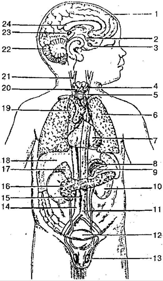
Fig. 86. Location of human Endocrine glands (diagram):
1 — cerebral hemisphere; 2 — hypothalamic nuclei; 3 — pituitary gland; 4 — Thyroid Gland; 5 — Trachea; 6 — lung; 7 — Pericardium; 8 — adrenal medulla; 9 — adrenal cortex; 10 — Kidney; 11 — aorta; 12 — Urinary Bladder; 13 — Testis; 14 — INFERIOR VENA CAVA; 15 — aortic paraganglion; 16 — Pancreas; 17 — Adrenal gland; 18 — Liver; 19 — Thymus; 20 — parathyroid gland; 21 — carotid glomus; 22 — Cerebellum; 23 — Pineal Gland (epiphysis); 24 — corpus callosum
The anterior lobe (adenohypophysis) is larger and denser, making up 70–80% of the total mass of the pituitary gland. It is subdivided into the pars distalis, pars intermedia, and pars tuberalis. The parenchyma of the intermediate lobe is represented by several types of glandular Cells interspersed with Blood Vessels. The posterior lobe (neurohypophysis) is located in the posterior part of the hypophyseal fossa. It consists of the infundibulum and the pars nervosa; it is formed by neuroglial cells (pituicytes), nerve fibers extending from the Hypothalamus to the neurohypophysis, and neurosecretory bodies.
Via nerve fibers and blood vessels, the pituitary gland is functionally connected to the hypothalamus of the Diencephalon, which regulates its activity.
The pituitary gland produces seven Hormones. Four of them affect peripheral endocrine glands and are referred to as tropic hormones (follicle-stimulating, luteinizing, thyroid-stimulating, and adrenocorticotropic), while three are effector hormones—Growth Hormone (somatotropic), prolactin (luteotropic hormone, melanocyte-stimulating hormone)—which directly affect target Organs and Tissues.
The anterior pituitary produces somatotropic hormone (growth hormone, GH). Its secretion is regulated by Hypothalamic hormones: releasing hormones and the inhibitory hormone Somatostatin. GH plays an active role in regulating GROWTH AND DEVELOPMENT in the young Organism. Following Puberty, the epiphyseal cartilages ossify, and GH ceases to affect longitudinal bone growth. Excessive oversecretion of this hormone in adults leads to the overgrowth of soft tissues as well as bone deformation and thickening, a condition known as acromegaly. Overproduction in youth, when bones are still capable of longitudinal growth, results in gigantism, whereas growth hormone deficiency leads to dwarfism (nanism). This condition is characterized by delayed growth (under 120 cm in women and 130 cm in men at age 20), proportional reduction of all body parts and Internal Organs, and sexual infantilism.
Another hormone of this lobe is adrenocorticotropic hormone (ACTH). It is essential for the normal development and function of the adrenal cortex, stimulating the production and secretion of glucocorticoids. ACTH synthesis is stimulated by hypothalamic corticotropin-releasing hormone; Adrenal Cortical Hormones also participate in the feedback regulation of ACTH secretion.
The anterior lobe also produces thyroid-stimulating hormone (TSH). Regulated by hypothalamic thyrotropin-releasing hormone, TSH stimulates the growth and Development of the thyroid gland, as well as the production and release of thyroxine (T4) and triiodothyronine (T3).
Gonadotropic hormones—follicle-stimulating hormone (FSH), luteinizing hormone (LH), and prolactin (PRL)—influence sexual maturation, regulate and stimulate ovarian follicle development, ovulation, mammary gland growth, milk production, and Spermatogenesis in men. The release of these hormones is stimulated by hypothalamic releasing factors.
The intermediate lobe produces melanocyte-stimulating hormone (MSH). Its primary function is to stimulate The Biosynthesis of the Skin pigment melanin, as well as to increase the size and number of pigment cells. The Cells of the intermediate lobe are regulated by hypothalamic releasing factors and inhibitory hormones. Vasopressin and Oxytocin are produced by neurosecretory cells of the hypothalamus and transported via axons of the hypothalamo-hypophyseal tract to the posterior pituitary, from which they enter the bloodstream.
Vasopressin exerts antidiuretic and vasoconstrictive effects, hence its name antidiuretic hormone (ADH). Insufficient secretion of this hormone leads to diabetes insipidus. This condition is characterized by The excretion of large volumes of urine (from 4 to 20 liters per day or more) with a low specific gravity. Renal Water loss and elevated plasma osmotic pressure cause insatiable thirst, leading patients to consume large amounts of water.
Oxytocin stimulates the contractile activity of the uterine musculature, enhances milk ejection by the Mammary Glands, affects the smooth Muscle tone of the gastrointestinal tract, and inhibits the function of the corpus luteum.
The pineal body (corpus pineale), or epiphysis. This is a small, oval, glandular Structure belonging to the diencephalon, situated in the shallow groove between the superior colliculi of the Midbrain.
The mass of the gland in adults is about 0.2 g, its length is 8–15 mm, width is 6–10 mm, and thickness is 4–6 mm.
Externally, the pineal body is covered by the pia mater, a delicate Connective Tissue membrane containing numerous anastomosing blood vessels. The cellular elements of the parenchyma comprise specialized glandular cells called pinealocytes and glial cells called gliocytes.
The endocrine role of the pineal body lies in the secretion of substances (serotonin, melatonin), Other Hormones, and Polypeptides by its cells. Melatonin acts as an antagonist to melanocyte-stimulating hormone, exhibits antigonadotropic activity, and inhibits gonad development. The pineal body participates in The regulation of electrolyte METABOLISM and influences, during early life, the complex of endocrine organs (pituitary, thyroid, adrenal cortex) involved in somatic and sexual development.
Last update: 08/08/2026
Editorial and Educational Adaptation: This material has been compiled based on the primary/original source text. The project team performed an editorial review, corrected technical inaccuracies, structured sections, and adapted the content for an educational format.
What was processed:
- elimination of formatting defects (OCR errors, structural breaks, corrupted characters);
- editorial organization of content;
- standardization of terminology in accordance with academic sources;
- verification of factual statements against the original source text.
All mentions of the author, publication year, and origin of the primary text have been preserved in accordance with the source.