MODERN BOTANY - P. RAVEN - 1990
SECTION IV. DIVERSITY
CHAPTER 18. SEED PLANTS
Angiosperms
The division of angiosperms, or flowering plants (Anthophyta), includes about 235,000 species—far more than any other plant group. Their vegetative Structure is extraordinarily diverse. It ranges from giant eucalypts, trees over 100 m tall with trunks nearly 20 m in circumference (Fig. 18-36), to tiny duckweeds, simply structured floating monocots that often do not exceed 1 mm in length (Fig. 18-37). Some angiosperms are lianas climbing high into the tropical rainforest canopy, while others are epiphytes inhabiting that canopy. Many flowering plants, such as cacti, are adapted for growth in extremely arid conditions. Representatives of this division have dominated the land for over 100 million years.
Class="center">Fig. 18-25. Pine life cycle. The gametophytes are greatly reduced and depend on the sporophyte for Nutrition. The immature male gametophytes, consisting of four Cells, are pollen grains, which are transported by wind to the female gametophyte (megagametophyte) located within the ovule. Incapable of active locomotion, the sperm cells produced by germinating pollen grains are delivered to the archegonial egg cells via pollen tubes (Water is not required as a medium for their transport). Following Fertilization, the ovule containing the megagametophyte matures into a seed. The well-developed suspender characteristic of pines degenerates by the time the embryo is fully developed. A pine seed consists of an embryo, a seed coat, and the megagametophyte, which serves as a nutrient reserve

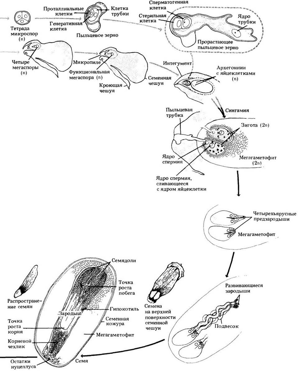
Fig. 18-26. Seeds of conifers of the yew family (Taxaceae) are surrounded by a fleshy cup called an aril. This structure attracts birds and other animals, which eat it and thereby disperse the seeds. A. In the yew (Taxus), native to the Northern Hemisphere, the arils are red. B. Sporophylls and microsporangia of yew microstrobils. Mega- and microstrobils are located on separate trees. Yew arils are poisonous and are the most frequent cause of Plant poisoning in children in the US, although fatalities are extremely rare
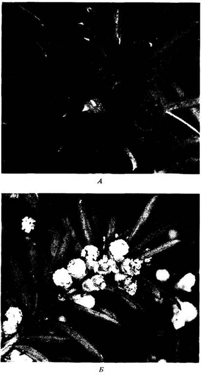
Fig. 18-27. Bald cypress (Taxodium distichum) is a deciduous representative of the taxodium family growing in swamps of the southeastern US. In this autumn photograph, the leaves of the tree have already begun to change color. Hanging abundantly from its branches is the so-called Spanish moss—actually a flowering plant, Tillandsia usneoides, related to the pineapple
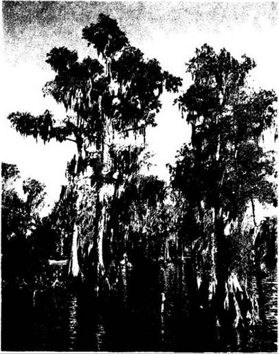
Fig. 18-28. Dawn redwood (Metasequoia glyptostroboides). This tree, growing in Hubei Province in central China, is over 400 years old
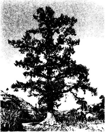
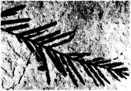
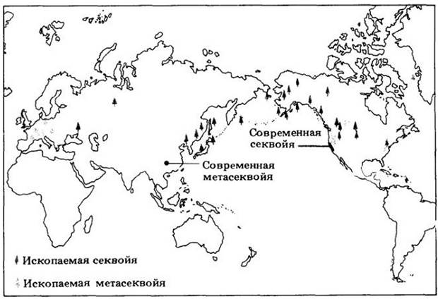
Fig. 18-29. A fossil twig of dawn redwood, approximately 50 million years old. The map shows the geographic distribution of some living and fossil Representatives of the taxodium family
Two classes of angiosperms are distinguished: Monocotyledones (monocots), with approximately 65,000 species (Fig. 18-38), and Dicotyledones (dicots) (Fig. 18-39). The similarities between them far outweigh the differences, yet they are easily recognizable natural groups. Among monocots are such well-known plants as grasses, lilies, irises, orchids, cattails, and palms. Dicots include almost all familiar trees and shrubs (except conifers) and many herbaceous plants. The main differences between these classes are summarized in Table 18-1.
Table 18-1. Main differences between monocots and dicots
Features |
Dicots |
Monocots |
Floral parts |
Usually in whorls of 4 or 5 |
Usually in whorls of three |
Pollen |
Tricolpate basically (with three furrows or pores) |
Monocolpate basically (with one furrow or pore) |
Cotyledons |
Two |
One |
Leaf venation |
Usually reticulate |
Usually parallel |
Primary vascular bundles in the stem |
Arranged in a ring |
Complexly arranged |
True Secondary Growth (involving cambium) |
Usually present |
Absent |
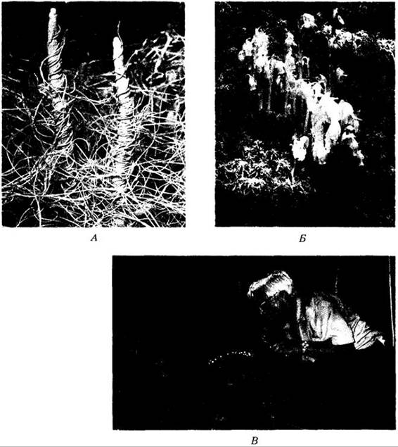
With few exceptions, angiosperms lead an independent lifestyle, but parasites and Saprophytes are also known among them (Fig. 18-40), largely or entirely devoid of chlorophyll. It has recently been shown that many, if not all, flowering "saprophytes" are obligately associated with mycorrhizal Fungi, which simultaneously interact with a green photosynthetic angiosperm. The fungus forms a kind of "bridge," actively transferring CARBOHYDRATES from the second plant to the colorless "saprophyte." There are about 2,800 species of parasitic dicots, including mistletoe and dodder (Fig. 18-40, A), and one parasitic gymnosperm (Parasitaxus from New Caledonia). Such flowering plants form specialized absorbing Organs, haustoria, which penetrate the Tissues of their hosts.
Fig. 18-30. Female and male plants of coontie (Zamia pumila)—the only wild-growing cycad species in the US. Its entirely or partially underground stems, along with the main roots, were used for food by the Seminole Indians. The two large gray "cones" in the foreground are female, while the smaller brown ones are male
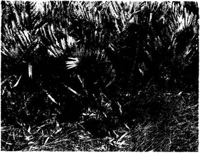
Fig. 18-31. A. The South African cycad Encephalartos altensteinii. A male specimen with strobili is shown. B. A female specimen of Cycas siamensis. The top of the female "cone" has been removed to expose the seeds on the upper surfaces of the megasporophylls
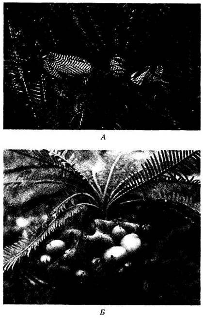
Fig. 18-32. A. Ginkgo biloba. B. Leaves and fleshy seeds of ginkgo on short shoots
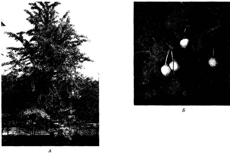
Fig. 18-33. The large, leathery leaves of the tropical Gnetum resemble those of certain dicots. Species of this genus are shrubs or woody lianas found in tropical and subtropical forests. A. Megasrobilus clusters. B. Microstrobilar clusters and leaves. C. Fleshy seeds and leaves. Photographs B and C were taken in the southern Venezuelan Amazon basin
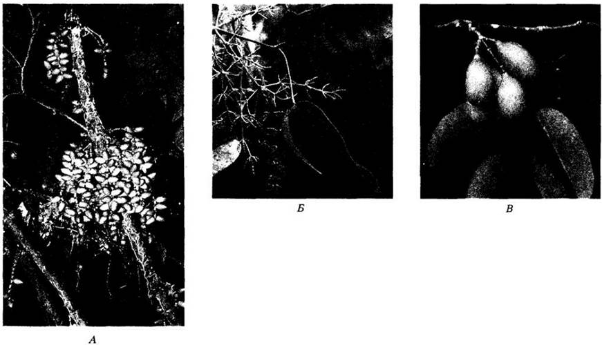
Fig. 18-34. Ephedra is the only one of the three gnetophyte genera found in the United States. A. Male plant of Ephedra viridis (California), a heavily branched shrub with scale-like leaves, typical of all species in the genus. B. Microsporangiate (male) strobili of E. viridis. Scale-like leaves are visible on the stem. C. Male strobili of Ephedra trifurca (Arizona). D. Female plant of Ephedra viridis with seeds
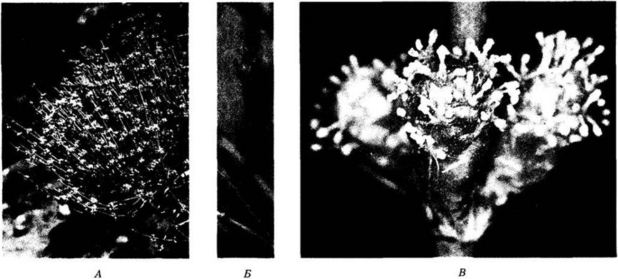
Fig. 18-35. Welwitschia mirabilis, a gnetophyte found exclusively in the Namib Desert and adjacent areas of southwestern Africa. Welwitschia produces only two leaves that continue to grow throughout the plant's lifetime. Because they fray at the tips and split longitudinally, older plants appear to have numerous leaves. A. Large plant with seeds. B. Microsporangiate strobili. C. Strobili with ovules; an insect (bug) is feeding on sap from the strobilus. Welwitschia is a dioecious plant
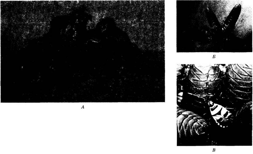
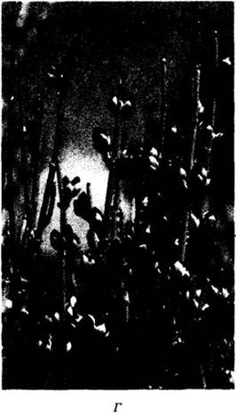
Fig. 18-36. Giant eucalyptus (Eucalyptus jacksonii) in the Valley of the Giants, southwestern Australia. The massive scale of this angiosperm is apparent when compared with the person standing near its fire-scarred base
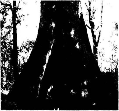
Fig. 18-37. Duckweeds (family Lemnaceae), the smallest of all flowering plants. Their structural features indicate that they are extremely reduced descendants of the Araceae (aroids), a family that includes calla lilies and philodendrons. A. A bee resting on a dense floating mat formed by three duckweed species. The largest plant here is Lemna gibba, 2–3 mm in length; the smaller ones are two Wolffia species, up to 1 mm long. B. A flowering plant of Wolffia borealis showing a circular, concave stigma (resembling a tiny bagel) and a small anther protruding from the central cavity just above it. The entire plant is less than 1 mm long. C. A flowering plant of Lemna gibba; two stamens and a style emerge from a pocket on the upper surface of the plant
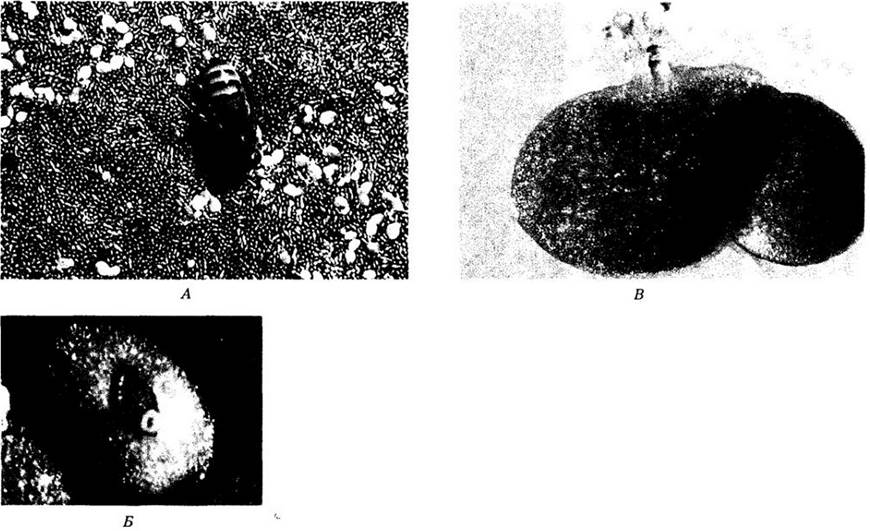
Fig. 18-38. Monocots. A. Coconut palm (Cocos nucifera) in Tehuantepec (Oaxaca, Mexico). Its fruit (the coconut) is actually a drupe rather than a nut (see Chapter 29). B. Flowers and fruits of the banana (Musa × paradisiaca). The Ovary in this plant is inferior, leaving a prominent scar at the apex of the fruit from the fallen floral parts. C. Rice (Oryza sativa) of the grass family (Poaceae)
Fig. 18-39. Dicots. A. Fragrant water lily (Nymphaea odorata). The highly aromatic flower of this species features numerous stamens and petals and is radially symmetrical. The genus Nymphaea is widely distributed in tropical and temperate regions worldwide. B. Saguaro cactus (Carnegiea gigantea). Cacti, comprising about 2,000 species, are found almost exclusively in the New World. Their thick, fleshy, water-storing stems contain METABOLISM/14.html">Chloroplasts and carry out Photosynthesis in place of leaves. C. Hepatica (Hepatica americana) blooming in deciduous forests in early spring. Its perianth segments (6–10 in number) are undifferentiated into sepals and petals. Numerous stamens and carpels are arranged spirally
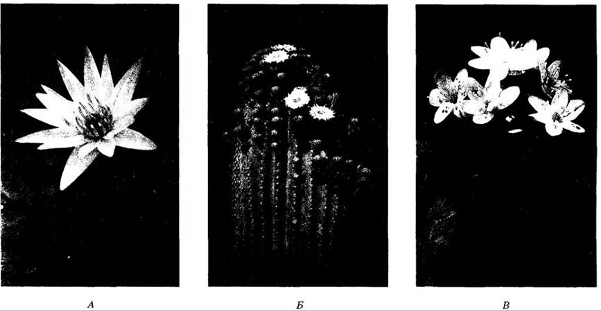
Fig. 18-40. Parasitic and saprophytic angiosperms. These plants are entirely or nearly devoid of chlorophyll and obtain nourishment from the photosynthetic products of other plants. A. Dodder (Cuscuta salina), a parasitic, bright orange or yellow plant of the morning glory family (Convolvulaceae). B. Indian pipe (Monotropa uniflora), a "saprophyte" that obtains nutrients from the roots of other plants via mycorrhizal fungal hyphae. C. The world's largest flower, Rafflesia arnoldii, on Mount Sago, Sumatra. Plants of this genus parasitize the roots of Vitaceae (grape family). All plants shown in the photographs are dicots
In Section V, we will examine in detail the structure and Development of the plant body, i.e., the sporophyte of flowering plants, while dedicating the remainder of this chapter to the most characteristic feature of angiosperms—the flower—and their mode of reproduction. Chapter 29 discusses the evolution of this division in depth.
The Flower
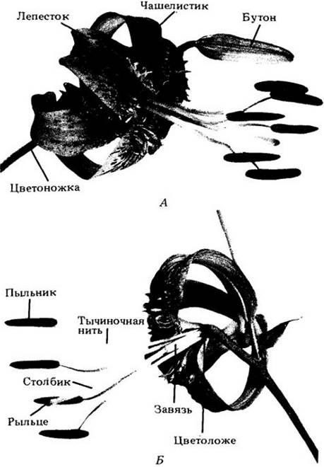
A flower is a determinate SHOOT bearing sporophylls (Fig. 18-41). The name Angiospermae translates as "enclosed seeds" and is derived from the Greek words angeion (vessel) and sperma (seed), because the defining floral structure—the carpel—is a kind of "vessel" enclosing the ovules, which develop into seeds after fertilization.
Fig. 18-41. Floral PARTS OF THE lily (Lilium henryi). A. Intact flower. Sometimes, as illustrated here, sepals and petals are similar in appearance and are collectively referred to as tepals. The sepals are attached to the receptacle below the petals. B. A flower with two tepals and two stamens removed to reveal the ovary. The gynoecium consists of the ovary, style, and stigma, while a stamen comprises a filament and an anther. Here, the sepals, petals, and stamens are attached to the receptacle below the ovary, which is consequently termed superior, making the flower hypogynous
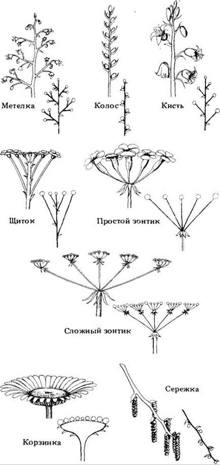
Flowers may be grouped in various ways into inflorescences (Figs. 18-42 and 18-43). The stalk of an inflorescence or a solitary flower is called a peduncle, whereas the stalk of an individual flower within an inflorescence is a pedicel. The receptacle is the region of the pedicel (or peduncle) to which the floral organs (whorls) are attached. Like any shoot apex, it consists of nodes and internodes, except that the internodes are extremely short, bringing the nodes into close proximity.
Fig. 18-42. Some common inflorescence types in angiosperms, shown with simplified diagrams.
Many flowers possess two sets of sterile appendages, the sepals and petals, attached to the receptacle below the fertile elements, the stamens and carpels (with the sepals originating below the petals, and the stamens below the carpels). Together, the former constitute the calyx and the latter the corolla, which collectively form the perianth. Sepals and petals are largely leaf-like in structure. The sepals are typically green, whereas the petals are brightly colored, although in many flowers both whorls are similar in hue (Fig. 18-41).
The stamens, which collectively form the androecium (Greek for "house of man"), are microsporophylls. In modern angiosperms, with few exceptions, they consist of a slender stalk (the filament) and an anther divided into two halves containing four microsporangia, or pollen sacs.
The carpels, which together make up the gynoecium (Greek for "house of woman"), are megasporophylls folded lengthwise and enclosing one or more ovules. A flower may contain one or several carpels, which are sometimes completely or partially fused. An individual carpel, or a structure formed by fused carpels, is frequently called a pistil because of its shape resemblance to a pestle used for grinding substances in a mortar.
In most flowers, the pistils are differentiated into a lower portion—the ovary, containing the ovules—and an upper portion, the stigma, which receives the pollen. The stigma and ovary are often connected by a more or less elongated structure known as the style. When carpels fuse, the individual styles or stigmas either merge or remain distinct. A compound ovary formed by fused carpels is usually (though not always) partitioned into two or more locules that house the ovules. The number of locules generally corresponds to the number of carpels in the gynoecium.
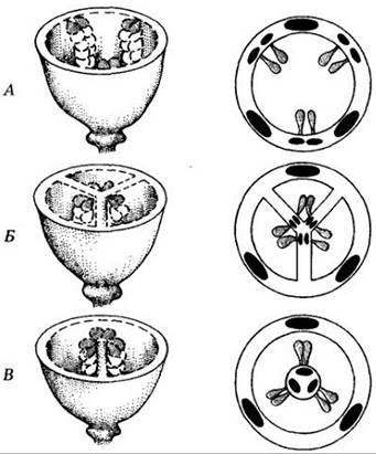
The regions within the ovary where ovules originate and remain attached until maturity are called placentae. Their arrangement (placentation), and consequently the positioning of the ovules, varies among different groups of flowering plants (Fig. 18-44). It may be parietal, meaning the ovules are located on the inner wall of the ovary or on its inward projections. In other cases, the ovules are borne on a central Column within a multi-locular ovary (axile placentation). In free-central placentation, ovules develop on a central column that is not connected by partitions to the ovary wall. Finally, in some flowers, a single ovule is located at the very base of a unilocular ovary (basal placentation). These differences are of great importance in the Classification of angiosperms.
Fig. 18-44. Types of placentation. A. Parietal. B. Axile. C. Free-central. Basal placentation is not shown here.
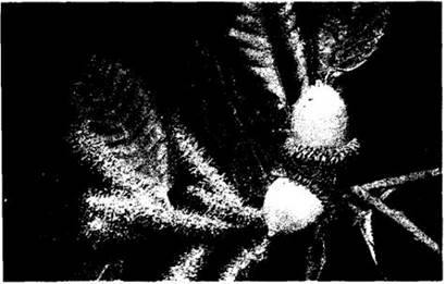
Despite a shared general underlying structure, substantial variations exist among the flowers of different plants. Most flowers bear both stamens and carpels, and are referred to as perfect (bisexual). If either stamens or carpels are absent, the flower is termed imperfect—either pistillate or staminate, respectively (Fig. 18-45). If staminate and pistillate flowers occur on the same plant (as in maize or oak), the species is said to be monoecious, whereas if they occur on different plants (as in willow, American holly, etc.), it is dioecious. Other seed plants with pollen- and seed-producing organs borne on separate individuals (such as ginkgo, cycads, and junipers) are also considered dioecious.
Fig. 18-45. Staminate and pistillate flowers of the giant chinquapin (Lithocarpus densiflora). Most members of the beech family (Fagaceae), which includes oaks (Quercus), are monoecious plants; their staminate and pistillate flowers are separate yet located on the same tree.
Any floral whorl—sepals, petals, stamens, or carpels—may be absent in a given group of angiosperms. Flowers possessing all four whorls are termed complete, whereas those lacking at least one are incomplete. Thus, an imperfect flower is invariably incomplete, but not all incomplete flowers are imperfect.
Floral parts are arranged either spirally on a more or less elongated receptacle, or equivalent elements (such as petals) are attached in whorls at a node. Within a single whorl, these parts may be fused to one another or adnate to elements of other whorls (for instance, stamens are frequently fused to the corolla). When the parts are free, the prefix "apo-" or "dialy-" is used in their description; when they are fused, the prefix "syn-" or "sym-" applies. Examples include a free-petalled (dialypetalous) versus a gamopetalous corolla, or an apocarpous versus a syncarpous gynoecium.
Flowers differ not only in the spiral or whorled arrangement of their parts, but also in the level of attachment of these parts to the floral axis relative to the gynoecium (Fig. 18-46). If the other floral elements originate from the receptacle below the ovary, the ovary is said to be superior, and the flower hypogynous (see Fig. 18-41). In some such flowers, the sepals, petals, and stamens fuse to form a cup-shaped extension of the receptacle known as a hypanthium (perigynous flowers; Fig. 18-47). In this case, the petals and stamens appear to arise from the rim of the cup. In other instances, they appear to emerge from the top of a so-called inferior ovary (epigynous flowers; Fig. 18-48).
Fig. 18-46. Flower types in three common dicot families, illustrating variations in ovary position. In many Rosaceae, the Ovaries are superior, and the bases of the floral parts fuse into a cup-shaped expansion (the hypanthium); such flowers are termed perigynous. In Apiaceae and Onagraceae, the ovaries are inferior, meaning the floral parts are attached above them; flowers of these two families are termed epigynous. When a hypanthium is absent and the floral parts are attached below the ovary, the flower is termed hypogynous.
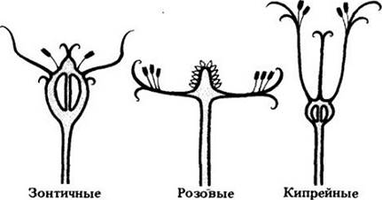
Fig. 18-47. A. Flowers of sweet cherry (Prunus) are perigynous—their sepals, petals, and stamens are attached to the hypanthium. In Cytology/practical/54.html">Longitudinal section of a bud (B), the stamens are congested within the hypanthium.
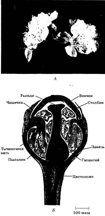
Fig. 18-48. A. Flowers of the crabapple (Malus sylvestris) are epigynous—their sepals, petals, and stamens appear to arise from the top of the ovary. B. Section of a nearly opened flower with stamens not yet fully straightened.
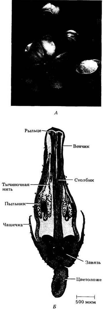
Finally, when discussing The Diversity of floral architecture, Symmetry must be considered. In some instances, the corolla consists of petals of identical shape radiating at equal distances from the center of the flower, resulting in radial symmetry. Such flowers are termed actinomorphic (from the Greek ROOT *actinos*, meaning "ray" or "star"). In other cases, one or more elements of at least one whorl differ from the others and exhibit bilateral symmetry. Such flowers are called zygomorphic (see Fig. 18-43, B, C).
Angiosperm Life Cycle
The gametophytes of flowering plants are drastically reduced in size—more so than in any other heterosporous plants, including gymnosperms. The mature male gametophyte consists of only three cells, whereas the female gametophyte, embedded within the sporophyte tissues throughout its life, typically consists of only seven cells in most species. Antheridia and archegonia are absent. Pollination is indirect, meaning pollen is deposited on the stigma, after which a pollen tube delivers two non-motile sperm cells to the female gametophyte. Following fertilization, the ovule develops into a seed enclosed within the ovary, which simultaneously (sometimes accompanied by accessory structures) matures into a fruit.
Microsporogenesis and Microgametogenesis
Microsporogenesis is The formation of microspores within the microsporangia, or pollen sacs, of an anther. Microgametogenesis is The Development of a microspore into a male gametophyte, or pollen grain.
In the Early stages of differentiation, an anther consists of a homogeneous mass of cells and a partially distinct epidermis. Four groups of fertile, or sporogenous, cells then differentiate within it. Each such group is surrounded by several layers of sterile cells that develop into the wall of the pollen sac. This wall includes nutritive cells that supply nutrients to the developing microspores and form the tapetum, the innermost layer of the wall (Fig. 18-49). The sporogenous cells become meiotically dividing diploid microsporocytes, which give rise to tetrads of haploid microspores. Microsporogenesis concludes with the formation of single-celled microspores.
Fig. 18-49. Two transverse sections of lily (Lilium) anthers. A. An immature anther with four pollen sacs containing microsporocytes surrounded by the tapetum. B. A mature anther with pollen grains. The partitions between adjacent pollen sacs break down during dehiscence.

During Meiosis, Cell wall formation either follows each nuclear division or divides the protoplasts of the four microspores only after the second meiotic division. The former is typical of monocots, the latter of dicots. The next stage involves the Formation of the principal Features of the pollen grains (Fig. 18-50). They become surrounded by a tough outer coat, the exine, and a cellulosic-pectic inner coat, the intine. The exine consists of a highly resistant substance, sporopollenin, apparently produced in part by the tapetum and in part by the microspore. The intine is deposited by the microspore protoplast.
Fig. 18-50. The pollen grain wall serves to protect the male gametophyte during its arduous journey from the anther to the stigma. Its outer layer, or exine, is composed largely of sporopollenin (a polymer consisting primarily of carotenoids). The rigid and durable exine is often intricately sculptured. This sculpturing is distinctive and varies among different species, as seen in these scanning electron micrographs. A. Horse chestnut (Aesculus hippocastanum). B. Lily (Lilium longiflorum). C. Detail of The surface of an L. longiflorum pollen grain. D. Ragweed (Ambrosia psilostachya). Ragweed pollen is a major cause of allergies in the United States. Spiny pollen grains similar to the one shown here are common in the aster family (Asteraceae), to which this genus belongs.
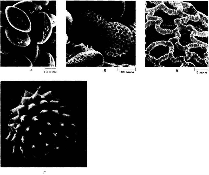
The size and shape of pollen grains, much like spores, vary widely. Their diameter ranges from less than 20 to more than 250 µm. They also differ in the number and structure of apertures through which the pollen tubes eventually emerge. Almost all families, many genera, and a significant number of species of flowering plants can be identified by The structure of their pollen grains, particularly their size, aperture number, and surface sculpture. Unlike larger plant fragments—such as leaves, flowers, and fruits—pollen grains are exceptionally well-represented in the fossil record thanks to The chemical composition of their exine. Consequently, pollen is a valuable source of information about past vegetation and climates.
Both pollen grains and spores have a sporopollenin wall and are products of meiosis. However, by the time they are dispersed, the former have undergone mitosis to produce two or three nuclei, whereas the latter contain only one. Spores germinate through a centrally located aperture or scar, whereas pollen grains germinate through pores. As a result, they are often easily distinguishable in the fossil state.
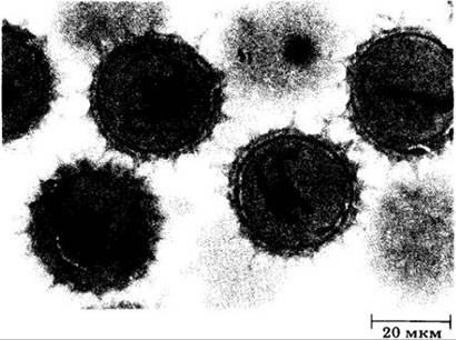
Microgametogenesis in angiosperms is uniform and begins with the mitotic division of the uninucleate microspore, yielding two cells within its original wall. One of these is called the tube cell, and the other is the generative cell (Fig. 18-51). In many species, the male gametophyte is at this two-celled stage when the pollen is shed during anther dehiscence. In other species, the generative cell divides slightly earlier to produce two male Gametes, or sperm cells (Fig. 18-52).
Fig. 18-51. A mature lily pollen grain containing a two-celled male gametophyte. The spindle-shaped generative cell will divide by mitosis to form two sperm cells; the larger tube cell will later produce the pollen tube.
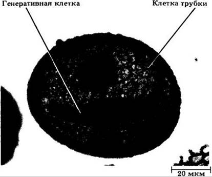
Fig. 18-52. Mature pollen grains with three-celled male gametophytes of Silphium (Asteraceae). Prior to pollination, each pollen grain contains two filamentous sperm cells suspended in the Cytoplasm of the larger tube cell. In other words, the pollen of this plant is dispersed at the three-celled stage, rather than the two-celled stage seen in the lily in the previous figure.
Megasporogenesis and Megagametogenesis
Megasporogenesis is The process of megaspore formation within the nucellus (megasporangium). Megagametogenesis is the development of the megaspore into the female gametophyte.
An ovule is a relatively complex structure consisting of a stalk (the funiculus) that Supports the nucellus, which is enclosed by one or two integuments. Depending on the species, anywhere from one to numerous ovules develop on the placentas. A developing ovule initially consists entirely of the nucellus, but one or two protective layers (integuments) soon emerge, leaving a small opening, the micropyle, at one end (Fig. 18-53).
At an early stage of ovule development, a single diploid megasporocyte differentiates within the nucellus. It undergoes meiosis (Fig. 18-53, 15) to produce four haploid megaspores, typically arranged in a linear tetrad. This marks the completion of megasporogenesis. Three of the megaspores usually degenerate, while the fourth, located furthest from the micropyle, develops into the female gametophyte.
The functional megaspore soon begins to enlarge at the expense of the nucellus, and its Nucleus undergoes three successive mitotic divisions. By the end of the third mitosis, the resulting eight daughter nuclei are arranged in two groups of four—one group near the micropylar end of the megagametophyte and the other at the opposite, chalazal, end. One nucleus from each group migrates to the center of the eight-nucleate cell; these are known as the polar nuclei. The three nuclei remaining at the micropylar end form the egg apparatus, which consists of the egg cell and two synergid cells. Cell walls also form around the nuclei at the chalazal end, giving rise to the so-called antipodal cells. The polar nuclei remain in the binucleate central cell. This eight-nucleate, seven-celled structure constitutes the mature female gametophyte, referred to as the embryo sac (Fig. 18-53, 19).
Fig. 18-53. Lily. Selected stages of ovule and embryo sac development. A. Two young ovules, each with a single large megasporocyte. The integuments have not yet begun to form. B. An ovule with developing integuments. The megasporocyte is in prophase I of meiosis. C. An ovule containing an eight-nucleate embryo sac (only six nuclei are visible here). The polar nuclei have not yet migrated to the center.
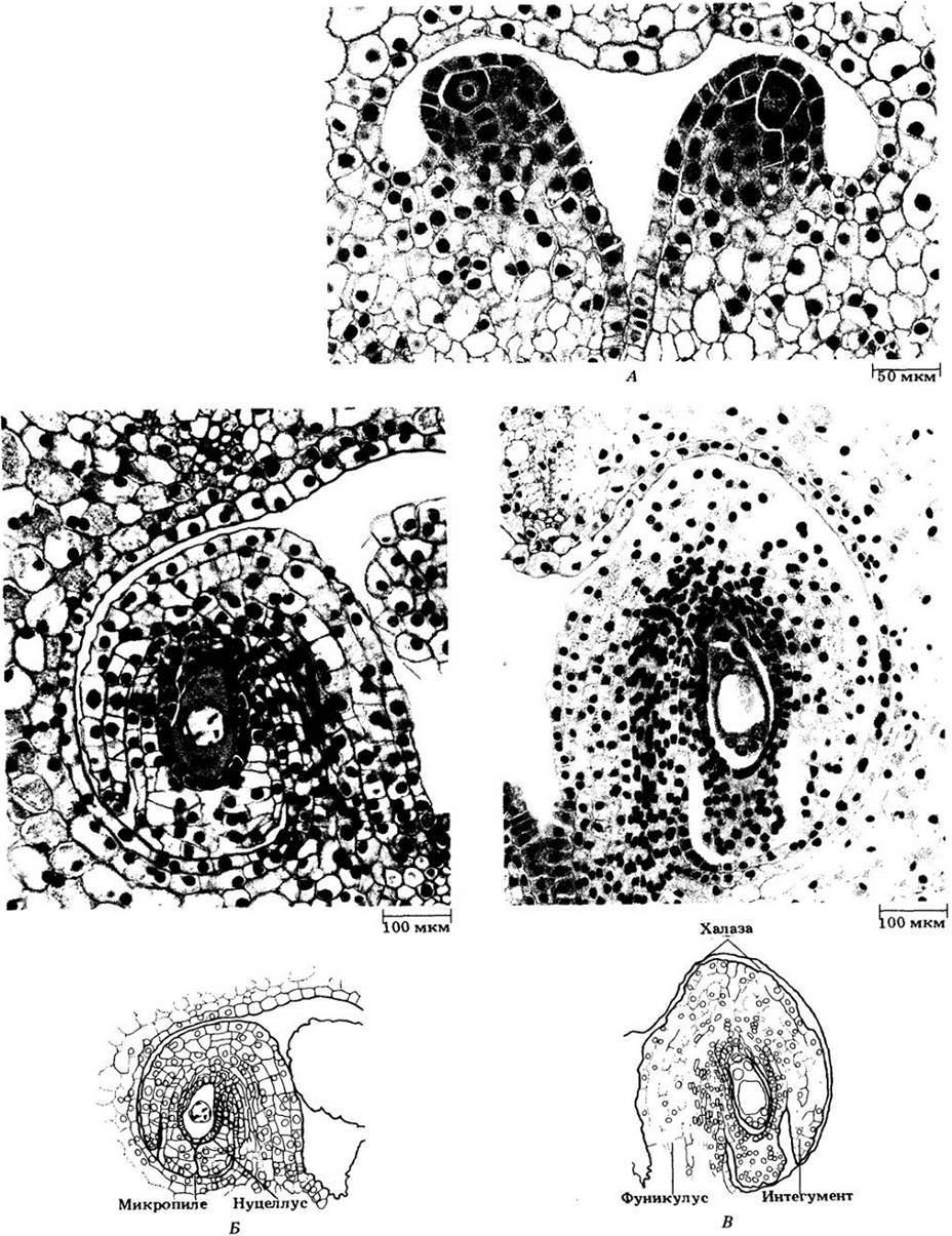
Although the type of development described above is the most common, it varies in approximately one-third of the angiosperm species studied in this regard.
Upon anther dehiscence (the release of their contents), pollen grains are transferred to stigmas in numerous ways (see Chapter 29); this transfer process is called pollination. Upon contacting the stigma, the pollen grains absorb additional water from the surface cells (along a water potential gradient). Once hydrated, they germinate to produce a pollen tube. The generative cell, if it has not already divided, soon divides to form two sperm cells. The germinated pollen grain, complete with a tube nucleus and two sperm cells, represents the mature male gametophyte (Fig. 18-55).
The stigma and style are structurally and physiologically adapted to facilitate pollen germination and pollen tube growth. The surface of many stigmas is formed largely of glandular tissue (stigmatic tissue) that secretes a sugary solution. This tissue is continuous with the transmitting tissue of the style, which serves as a pathway for the growing pollen tubes. In some styles, the transmitting tissue lines open canals, and the pollen tubes grow either along this lining or among its cells. However, in most angiosperms, the styles are sol-
tissues (solid) with one or more strands of conducting tissue between the stigma and the ovules. Depending on the plant species, pollen tubes here grow either among its cells or within their thick walls.
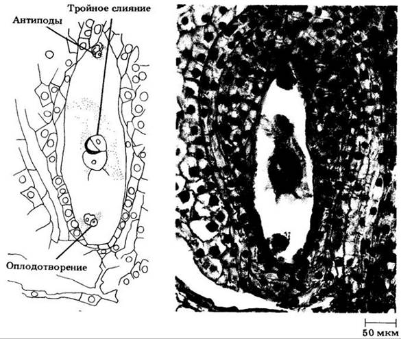
Typically, the pollen tube enters the ovule through the micropyle and penetrates one of the synergids, which begins to degenerate shortly after pollination, yet before the pollen tube reaches the embryo sac. Two sperm cells and a tube nucleus are discharged into this synergid through a subterminal pore that opens in the pollen tube. Subsequently, The Nucleus of one sperm enters the egg cell, while the other enters the central cell, where it fuses with two polar nuclei (Fig. 18-56). This differs from gymnosperms, in which only one of the two sperm is functional and fuses with the egg cell, whereas the second degenerates. The fusion of one sperm with the egg cell and the other with the polar nuclei—double fertilization—represents a unique feature of angiosperms. As noted above, "true" fertilization, or syngamy, implies solely the fusion of gametes, in this case the egg cell and the sperm, resulting in a diploid zygote. The union of the other sperm with two polar nuclei, i.e., triple fusion, yields a triploid primary endosperm nucleus. Meanwhile, the tube Cell Nucleus degenerates, and the remaining synergid and antipodals degenerate along with it or during the early stages of embryo sac differentiation.
Seed and Fruit Development
Following double fertilization, several processes commence: the primary endosperm nucleus divides to form the endosperm; the zygote develops into the embryo; the integuments transform into the seed coat, and the ovary wall along with associated structures gives rise to the fruit.
In contrast to the Embryogenesis of most gymnosperms, which begins with a free-nuclear stage, in angiosperms this process proceeds much like that in vascular spore-bearing plants, meaning that the first nuclear division of the zygote is accompanied by the formation of a cell wall. During early developmental stages, The sequence of cell divisions in dicot and monocot embryos is similar; in both cases, spherical bodies are formed. Later on, a distinction emerges: the dicot embryo develops two cotyledons, whereas the monocot develops only one. The details of angiosperm embryogenesis are examined in Section V.
Endosperm formation begins with the mitotic division of its primary nucleus, which typically occurs earlier than the First Division of the zygote. In some angiosperms, cell wall formation is preceded by a varying number of free nuclear divisions (nuclear type of endosperm development); in others, both the first and subsequent mitoses are accompanied by cytokinesis (cellular type of endosperm development). Although endosperm formation occurs through different pathways, the function of the resulting tissue is always the same: to supply the developing embryo, and in many cases the young seedling, with essential nutrients. In the seeds of certain angiosperm groups, the nucellus proliferates into a storage tissue called perisperm. Some seeds contain both endosperm and perisperm, as in beets (Beta). However, in many dicots and some monocots, all or most of the storage tissue is absorbed by the developing embryo even before the seed enters dormancy (in peas, beans, etc.). Embryos in such seeds typically develop fleshy, nutrient-laden cotyledons. The primary reserve nutrients stored in seeds are carbohydrates, Proteins, and Lipids.
The seeds of gymnosperms and angiosperms differ in THE ORIGIN OF these substances. In the former, they are provided by the female gametophyte; in the latter—at least initially—by the endosperm, which is neither gametophytic nor sporophytic tissue.
The development of the ovule into a seed is accompanied by the transformation of the ovary (and sometimes other parts of the flower or inflorescence) into a fruit. In the course of this process, the ovary wall (pericarp) often thickens and differentiates into distinct layers—the outer exocarp, the middle mesocarp, and the inner endocarp (sometimes only into exocarp and endocarp)—which are usually more conspicuous in fleshy fruits than in dry ones. Fruits are discussed in greater detail in Chapter 29.
The life cycle of angiosperms is illustrated in Fig. 18-54.
Fig. 18-54. Life Cycle of soybean (Glycine max), a representative flowering plant. Following germination, the seed develops into a mature sporophyte which, like other angiosperms, eventually produces flowers. Microspore mother cells, or microsporocytes, form within the anthers of the flower. Meiosis of each microsporocyte gives rise to four haploid microspores, which divide once to form a tube cell and a generative cell. This two-celled structure represents an immature microgametophyte, or pollen grain. During germination or even earlier, the generative cell divides into two sperm cells. They are delivered to the egg apparatus by the pollen tube. A germinated pollen grain with a tube nucleus and two sperm cells is a mature male gametophyte. Within the ovule, a megaspore mother cell, or megasporocyte, develops and ultimately undergoes meiosis to produce four megaspores, three of which degenerate. The fourth develops into the female gametophyte, which in its mature state is a seven-celled, eight-nucleate embryo sac. The pollen grain germinates on the stigma, producing a pollen tube that grows down toward the ovary through the style and enters the ovule via the micropyle. The nucleus of one sperm from the pollen tube fuses with the egg cell to form a zygote, while the Nucleus of the second sperm fuses with the two polar nuclei of the embryo sac to form the triploid primary endosperm nucleus. Such double fertilization is known exclusively in angiosperms. An embryo develops within the embryo sac, and the ovule integuments transform into the seed coat. Eventually, the seeds are dispersed


Fig. 18-55. Mature male gametophyte of Solomon's seal (Polygonatum). Sperm cells and the tube nucleus are visible within the pollen tube
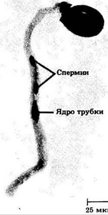
Fig. 18-56. Double fertilization in lily. The fusion of the sperm and egg cell nuclei—"true" fertilization—is visible in the lower part of the photomicrograph. Above is the triple fusion of the second sperm nucleus and the two polar nuclei
Last update: 07/08/2026
Editorial and Educational Adaptation: This material has been compiled based on the primary/original source text. The project team performed an editorial review, corrected technical inaccuracies, structured sections, and adapted the content for an educational format.
What was processed:
- elimination of formatting defects (OCR errors, structural breaks, corrupted characters);
- editorial organization of content;
- standardization of terminology in accordance with academic sources;
- verification of factual statements against the original source text.
All mentions of the author, publication year, and origin of the primary text have been preserved in accordance with the source.