BIOCHEMISTRY - L. Stryer - 1984
VOLUME 2
PART II GENERATION AND STORAGE OF METABOLIC ENERGY
CHAPTER 16. GLYCOGEN AND DISACCHARIDE METABOLISM
16.18. Liver Glycogen Metabolism Regulates Blood Glucose Levels
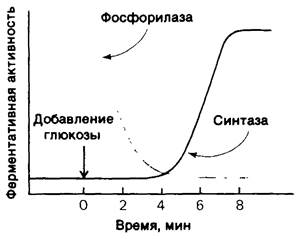
The control of Glycogen Synthesis and degradation in the Liver plays a central role in regulating Blood glucose levels. Normally, this concentration fluctuates between 80 and 120 mg per 100 mL. The liver is sensitive to blood glucose concentrations: if the Blood Glucose Level exceeds a threshold value, the liver takes up glucose; conversely, if the level drops below this threshold, the liver releases glucose. The amount of phosphorylase a in the liver rapidly decreases upon glucose infusion (Fig. 16.10). After a lag period, the amount of glycogen synthase a increases, leading to glycogen synthesis. It has recently been established that in liver Cells, phosphorylase acts as a glucose sensor. The binding of glucose to phosphorylase a shifts the allosteric equilibrium from the R state to the T state (see Fig. 16.5). As a result, the phosphoryl group at Serine-14 becomes accessible to Hydrolysis by phosphatase. A significant role in this process is played by the fact that phosphatase, being tightly bound to phosphorylase a, exerts its catalytic action only after the latter transitions to the T state induced by glucose.
How is glycogen synthase activated by glucose? Recall that the same phosphatase acts on both phosphorylase and glycogen synthase. Unlike the a form, phosphorylase b does not bind phosphatase. Consequently, The conversion of phosphorylase a to phosphorylase b is accompanied by the release of phosphatase, which can now be utilized to activate glycogen synthase. The removal of the phosphoryl group from inactive synthase b converts it into the active a form. Initially, there are about ten molecules of phosphorylase a per molecule of phosphatase. Therefore, an increase in synthase activity can begin only after the majority of phosphorylase a has transitioned to the b form (Fig. 16.10). This remarkable, glucose-sensitive system relies on key elements: (1) the coupling between the allosteric site for glucose and phosphoserine, (2) The Use of the same phosphatase for the inactivation of phosphorylase and the activation of glycogen synthase, and (3) the binding of phosphatase to phosphorylase a to prevent premature activation of glycogen synthase.
Class="center">Fig. 16.10. Glucose infusion leads to the inactivation of phosphorylase, which is accompanied by the activation of glycogen synthase
16.19. A Number of Genetically Determined Glycogen Storage Diseases Are Known
The first glycogen storage disease was described by Edgar von Gierke in 1929. The patient presented with a massive abdomen due to marked hepatomegaly. Pronounced hypoglycemia was observed between meals. Furthermore, administration of epinephrine and Glucagon failed to raise blood sugar levels. Children with this condition may experience seizures associated with low blood glucose.
The Nature of the enzymatic defect in von Gierke's disease was elucidated by the Coris in 1952. They established that the liver of such patients lacks glucose-6-phosphatase. This was the first discovered case of congenital hepatic enzyme deficiency. Liver glycogen in these patients had a normal Structure, but was present in unusually large amounts. The absence of glucose-6-phosphatase in the liver causes hypoglycemia because glucose cannot be formed from glucose-6-phosphate. Furthermore, the phosphorylated sugar cannot leave the liver because it is unable to cross Cell/30.html">The Plasma Membrane. A compensatory enhancement of hepatic Glycolysis occurs, resulting in elevated blood lactate and Pyruvate levels. Individuals with von Gierke's disease are also characterized by an increased reliance on fat METABOLISM.
To date, A number of Glycogen Storage Diseases have been characterized (Table 16.1). Cori and coworkers elucidated the biochemical defect underlying another glycogen storage disease (Type III), which cannot be distinguished from von Gierke's disease (Type I) by physical examination alone. Type III disease is characterized by an abnormal structure of Muscle and liver glycogen and a significant increase in its amount. The most striking deviation from normal is the very short length of the outer Branches of the glycogen molecule. Patients with this condition lack the debranching enzyme (α-1,6-glucosidase); consequently, they can effectively utilize only the outermost glucose residues of the glycogen branches. Therefore, only a small fraction of this abnormal glycogen is functionally active as an available glucose reserve.
Table 16.1. Glycogen storage diseases

Defects in the metabolism of muscle glycogen exclusively occur in McArdle's disease (Type V). In this condition, phosphorylase activity is absent in Skeletal Muscle, and patients exhibit a limited capacity for strenuous Physical Exercise due to painful muscle cramps. No other abnormalities are detected, and patients develop normally. Thus, the efficient utilization of muscle glycogen is not essential for life.
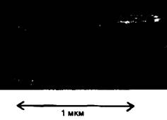
Fig. 16.11. Electron micrograph of skeletal muscle from an infant with Type II glycogen storage disease (Pompe disease). Lysosomes are engorged with glycogen due to a deficiency of α-1,4-glucosidase. This glycogen storage defect is restricted to lysosomes, whereas cytosolic glycogen levels remain normal.
16.20. Starch as a Reserve Polysaccharide in Plants
We now turn to other widespread Polysaccharides. The reserve polysaccharide in plants is starch, which exists in two forms. Amylose, the unbranched form of starch, consists of glucose residues linked by α-1,4 glycosidic bonds. In amylopectin, the branched form, there is approximately one α-1,6 linkage for every thirty α-1,4 linkages. Thus, it is similar to glycogen, differing from the latter in its lower degree of branching.
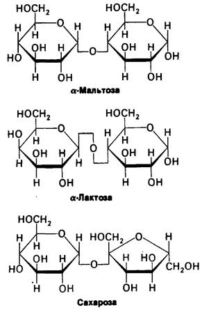
Starch accounts for more than half of the CARBOHYDRATES consumed by humans. Both amylopectin and amylose are rapidly hydrolyzed by α-amylase, which is secreted by the Salivary Glands and the Pancreas. Alpha-amylase cleaves internal α-1,4 bonds, yielding maltose, maltotriose, and α-dextrin. Maltose consists of two glucose residues joined by an α-1,4 linkage (Fig. 16.12); maltotriose consists of three such residues. Alpha-dextrin is composed of several glucose residues joined by α-1,6 linkages In addition to α-1,4 linkages. Maltose and maltotriose are hydrolyzed to glucose by maltase, whereas α-dextrin is hydrolyzed to glucose by α-dextrinase. Another type of amylase, called β-amylase, is present in malt; it hydrolyzes starch to maltose. Beta-amylase acts exclusively on the non-reducing ends of the chains.
Fig. 16.12. STRUCTURE OF THE most common Disaccharides: α-maltose, α-lactose, and sucrose. The prefix α refers to the configuration of the anomeric carbon atom at the reducing end of the disaccharide, rather than to the configuration of the glycosidic bond.
Another major plant polysaccharide is Cellulose, which serves a structural rather than a nutritive function. In fact, Cellulose is the most abundant organic compound in the biosphere, accounting for more than half of all organic carbon. It is an unbranched polymer of glucose residues linked by β-1,4 bonds. Mammals lack cellulases and are therefore incapable of digesting wood and plant fibers. However, the digestive tract of certain ruminants harbors Bacteria that produce cellulase, enabling these animals to digest cellulose.
Dextran is another polysaccharide composed exclusively of glucose residues joined predominantly by α-1,6 linkages, with branch points formed via α-1,2, α-1,3, or α-1,4 bonds depending on the Organism. Dextran serves as a reserve polysaccharide in Yeast and bacteria. The exoskeleton of insects and crustaceans contains Chitin, which is constructed of N-acetylglucosamine residues linked by β-1,4 bonds. Thus, chitin is similar to cellulose, with the sole difference that the C-2 substituent is an acetylated amino group rather than a hydroxyl group.
16.21. Maltose, sucrose, and lactose: widespread disaccharides
The structures of these three common disaccharides are shown in Fig. 16.12. As mentioned above, maltose is produced by the Hydrolysis of Starch and is subsequently hydrolyzed by maltase to glucose. Sucrose, common table sugar, is obtained commercially from sugar cane or sugar beets. The anomeric carbon atoms of the glucose and fructose residues in sucrose are linked by an α-glycosidic bond. Consequently, unlike many other sugars, sucrose lacks a reducing end group. The hydrolysis of sucrose to glucose and fructose is catalyzed by sucrase. Lactose is a disaccharide found
in milk and is not found in significant amounts anywhere else. It is hydrolyzed by lactase to yield galactose and glucose. Lactase, sucrase, maltase, and α-dextrinase are associated with the mucosal Cells of the Small Intestine.
UDP-sugars are activated intermediates in the synthesis of sucrose and lactose, just as UDP-Glucose serves as a glucosyl donor in glycogen synthesis. In fact, nucleoside diphosphate sugars serve as activated intermediates in almost all processes involving the synthesis of glycosidic bonds. For example, depending on the plant species, cellulose is synthesized from adenosine diphosphate glucose (ADP-glucose), cytidine diphosphate glucose (CDP-glucose), or guanosine diphosphate glucose (GDP-glucose). Sucrose is synthesized by transferring glucose from UDP-glucose to fructose-6-phosphate to form sucrose-6-phosphate, which is then hydrolyzed to sucrose.
Last update: 06/08/2026
Editorial and Educational Adaptation: This material has been compiled based on the primary/original source text. The project team performed an editorial review, corrected technical inaccuracies, structured sections, and adapted the content for an educational format.
What was processed:
- elimination of formatting defects (OCR errors, structural breaks, corrupted characters);
- editorial organization of content;
- standardization of terminology in accordance with academic sources;
- verification of factual statements against the original source text.
All mentions of the author, publication year, and origin of the primary text have been preserved in accordance with the source.