Human Biochemistry, Volume 2 - Murray R. 1993
Structure, Function, and Replication of Information Macromolecules
Recombinant DNA Technology
RNA
Ribonucleic acid is a copolymer of purine and pyrimidine ribonucleotides linked together by 3'—5' phosphodiester bridges, similar to DNA (Fig. 37.6). Although these two Types of Nucleic acids share many common features, they differ in several key respects.
1. In RNA, the sugar moiety to which the purine or pyrimidine bases and phosphate groups are attached is ribose rather than 2'-deoxyribose (as found in DNA).
2. The pyrimidine components of RNA differ from those of DNA. Both RNA and DNA contain adenine, guanine, and cytosine NUCLEOTIDES. However, RNA (with a few special exceptions discussed below) lacks thymine, which is replaced in the RNA molecule by uracil.
3. RNA is a single-stranded molecule (unlike the double-stranded Structure of DNA); however, when regions of complementary sequences (of opposite polarity) exist within the RNA chain, the single strand can fold back on itself to form so-called “hairpins,” which possess double-helical characteristics (Fig. 37.7).
Class="center">
Fig. 37.6. Fragment of a ribonucleic acid (RNA) molecule in which the purine and pyrimidine bases—adenine (A), uracil (U), cytosine (C), and guanine (G)—are held together by a phosphodiester backbone connecting the ribosyl residues, which are linked via N-glycosidic bonds to their respective nucleic acid bases. Note that the RNA chain exhibits a definite directionality indicated by the 5'- and 3'-terminal phosphate residues.
4. Because the RNA molecule is a single strand complementary to only one of the DNA strands, the guanine content does not necessarily equal the cytosine content, nor does the adenine content necessarily equal the uracil content.
5. RNA can be hydrolyzed by alkali into 2',3'-cyclic diesters of mononucleotides; a 2',3',5'-triester acts as a reaction intermediate, which is not formed during the alkaline Hydrolysis of DNA due to the absence of 2'-hydroxyl groups in the latter. The alkaline lability of RNA (relative to DNA) is a useful property for both diagnostic and analytical purposes.
The information contained in single-stranded RNA is expressed as a specific sequence of purine and pyrimidine bases (i.e., the Primary Structure) of the polymer chain. This sequence is complementary to the coding strand of the Gene from which the RNA is “transcribed.” Owing to this complementarity, the RNA molecule can specifically bind (hybridize) with the coding strand, but does not hybridize with the noncoding strand of DNA. The RNA sequence (except for the substitution of T for U) is identical to The sequence of the noncoding strand of the gene (Fig. 37.8).
BIOLOGICAL Functions OF RNA
Several types of RNA are known. Almost all of them are directly involved in The process of METABOLISM/35.html">Protein Biosynthesis. Cytoplasmic RNA molecules that function as templates for Protein Synthesis are called messenger RNAs (mRNAs). Another type of cytoplasmic RNA, ribosomal RNA (rRNA), serves as a structural component of Ribosomes (Organelles that play a crucial role in protein synthesis). The adaptor molecules of Transfer RNAs (tRNA) participate in translating mRNA information into the Amino Acid Sequence of Proteins.
A significant portion of primary RNA transcripts formed in Eukaryotic Cells, including mammalian cells, undergoes degradation in The Nucleus and does not play any structural or informational role in the Cytoplasm. A class of Small nuclear RNAs (snRNAs) has been discovered in cultured human cells; these do not directly participate in protein synthesis, but can influence RNA Processing and overall cellular “architecture.” The sizes of these relatively small molecules vary, containing anywhere from 90 to 300 nucleotides (Table 37.3).
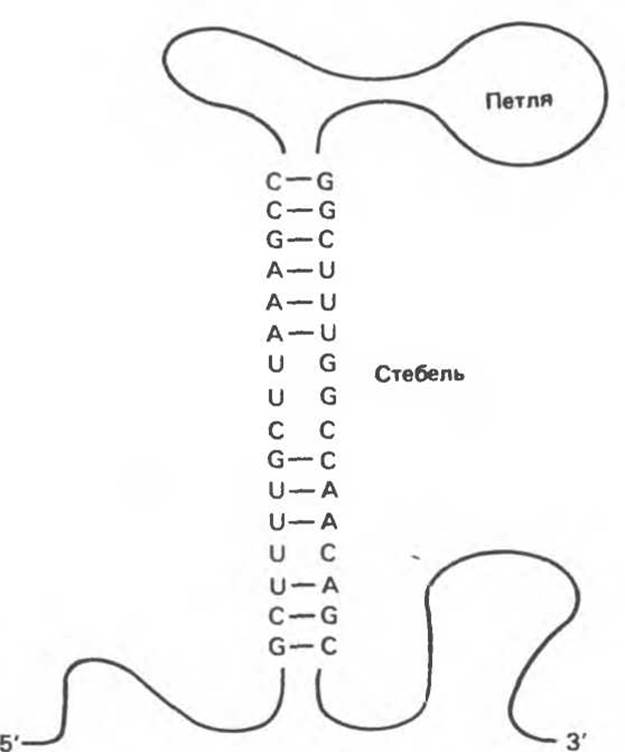
Fig. 37.7. Introduction/11.html">Secondary structure of an RNA molecule featuring a “stem-loop” (“hairpin”) motif, formed via intramolecular hydrogen bonding between complementary pairs of nucleic acid bases.
RNA serves as the primary genetic material in certain animal and plant Viruses. Some RNA-containing Viruses never undergo the stage of reverse Transcription of RNA into DNA. However, most known animal viruses, such as Retroviruses, are characterized by the reverse transcription of their RNA genome, catalyzed by an RNA-dependent DNA polymerase (Reverse Transcriptase) to form a double-stranded DNA copy. In many cases, the resulting double-stranded DNA transcript integrates into the host genome and subsequently drives the expression of viral genes as well as The production of new copies of viral RNA genomes.
Structural Organization of RNA
In all eukaryotic and prokaryotic organisms, there are three main classes of RNA molecules: Messenger RNA (mRNA), Transfer RNA (tRNA), and ribosomal RNA (rRNA). Representatives of these classes differ in size, function, and stability.
Messenger RNA (mRNA) is the most heterogeneous class with respect to size and stability. All members of this class serve as carriers of information from the gene to the protein-synthesizing machinery of The Cell. They act as templates for the synthesized polypeptide, thereby determining The amino acid sequence of the protein (Fig. 37.9).
Messenger RNAs, particularly eukaryotic ones, possess several unique structural features. The 5' end of mRNA is “capped” with a 7-methylguanosine triphosphate attached to the 5'-hydroxyl of the adjacent 2'-O-methylribonucleoside via a triphosphate bridge (Fig. 37.10). mRNA molecules frequently contain internal 6-methyladenine residues and 2'-O-methylated ribonucleotides. Although the exact significance of “capping” is not yet fully understood, it is presumed that the resulting 5'-terminal structure of mRNA is used for specific recognition by the translational machinery. Protein synthesis initiates at the 5'-capped end of the mRNA. The other end of most mRNA molecules (the 3' end) contains a polyadenylate tract of 20—250 nucleotides. The specific functions of this 3'-poly(A) tail are not yet definitively established, though it is hypothesized that this structure is responsible for maintaining the intracellular stability of mRNA. Some mRNAs, including histone mRNAs, lack a poly(A) tail. The presence of poly(A) in the mRNA structure is exploited to separate mRNA from Other types of RNA by fractionating total RNA on oligo(dT) columns immobilized on a solid support such as Cellulose. The binding of mRNA to the Column occurs through complementary interactions between the poly(A) tail and the immobilized oligo(dT).

Fig. 37.8. Sequence of a gene and its RNA transcript. The coding and noncoding strands are shown, and their polarity is indicated. The RNA transcript, possessing a 5'→3' polarity, is complementary to the coding strand (with 3'→5' polarity) and identical in sequence (except for the T to U substitution) and polarity to the noncoding strand of the DNA.
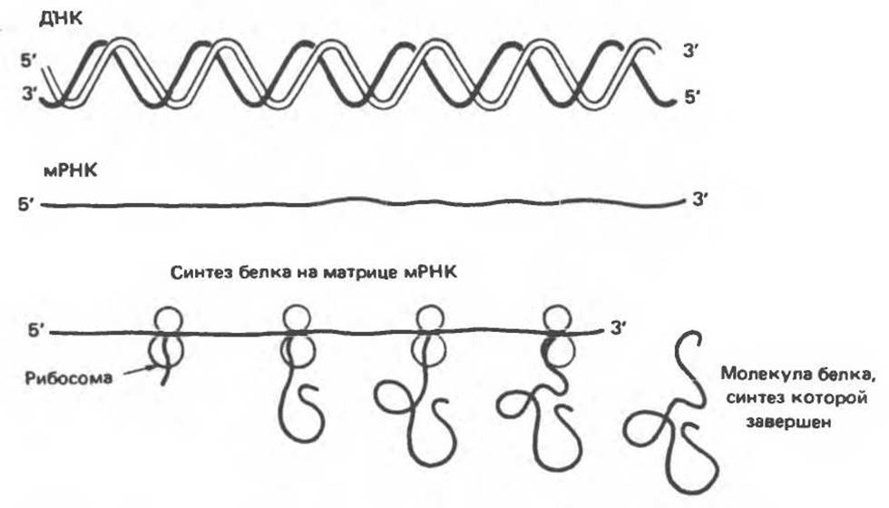
Fig. 37.9. Expression of genetic DNA information in the form of an mRNA transcript and subsequent Translation involving ribosomes to yield a specific protein molecule.

Fig. 37.10. STRUCTURE OF THE "cap" located at the 5'-end of most eukaryotic messenger RNAs. 7-methylguanosine triphosphate is attached to the 5'-end of the mRNA, which usually contains a 2'-O-methylpurine nucleotide.
In mammalian cells, including human cells, mature mRNA molecules in the cytoplasm are not exact copies of the transcribed gene region. The polyribonucleotide produced by transcription is a precursor to cytoplasmic mRNA and undergoes specific processing before leaving the nucleus. Unprocessed transcription products found in mammalian cell nuclei form a fourth class of RNA molecules. Such nuclear RNAs are highly heterogeneous and reach considerable sizes. Heterogeneous nuclear RNA (hnRNA) molecules can have a molecular weight exceeding 107, whereas the Molecular Weight of mRNA typically does not exceed 2∙106. hnRNAs undergo processing in the nucleus, and the resulting mature mRNAs enter the cytoplasm, where they serve as templates for protein biosynthesis.
Transfer RNA (tRNA) molecules typically contain about 75 nucleotides, with a molecular weight of approximately 25,000. tRNAs are also formed through the specific processing of corresponding precursor molecules (see Chapter 39). Transfer RNAs act as intermediates during mRNA Translation. Every cell contains at least 20 Different types of tRNA molecules. Each type (and sometimes multiple types) of tRNA corresponds to one of the 20 Amino Acids required for protein synthesis. Although each specific tRNA differs from the others in its nucleotide sequence, they all share common features. Due to several intrachain complementary regions, all tRNAs possess a secondary structure known as the "cloverleaf" (Fig. 37.11).
Molecules of all tRNA types feature four main arms. The acceptor arm consists of a "stem" of paired nucleotides and ends with the CCA sequence (5'→3'). It is via the 3'-hydroxyl group of the adenosine residue that binding to the carboxyl group of the amino acid occurs. The remaining arms also consist of "stems" formed by complementary Base Pairs and loops of unpaired bases (Fig. 37.7). The anticodon arm recognizes the nucleotide triplet, or codon (see Chapter 40), in the mRNA. The D-arm is named for the presence of dihydrouridine, and the TψC-arm is named after the T-pseudouridine-C sequence. The extra arm is the most variable structure and serves as the basis for tRNA Classification. Class 1 tRNAs (75% of the total) have an extra arm 3–5 base pairs long. The extra arm in Class 2 tRNA molecules consists of 13–21 base pairs and frequently includes an unpaired loop.
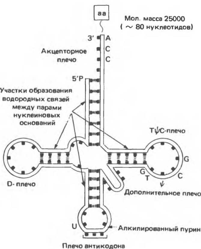
Fig. 37.11. Structure of an aminoacyl-tRNA molecule with an amino acid (aa) attached to its 3'-CCA end. Intramolecular Hydrogen Bonds and the locations of the anticodon, TTC, and dihydrouracil (D-) arms are indicated. (From J. D. Watson, Molecular Biology of the Gene, 3rd ed., Copyright 1976, 1970, 1965 by W. A. Benjamin, Inc., Menlo Park, Calif.)
The secondary structure, determined by a system of complementary base interactions within the respective arms, is characteristic of all tRNA types. The acceptor arm contains seven base pairs, the TψC-arm contains five base pairs, and the D-arm contains three (or four) base pairs.
tRNA molecules are quite stable in prokaryotes and somewhat less stable in eukaryotes. The reverse is true for mRNA, which is relatively unstable in prokaryotes but exhibits considerable stability in eukaryotic organisms.
Ribosomal RNA. A ribosome is a cytoplasmic nucleoprotein structure designed for protein synthesis using an mRNA template. The ribosome provides the specific contact between mRNA and tRNA through which The nucleotide sequence read from a particular gene is translated into the amino acid sequence of the corresponding protein.
Table 37.2 presents the components of mammalian ribosomes, which have a molecular weight of 4.2×10⁶ and a sedimentation coefficient of 80S (Svedberg units). Mammalian ribosomes consist of two nucleoprotein subunits: a large subunit with a molecular weight of 2.8∙106 (60S) and a small subunit with a molecular weight of 1.4∙106 (40S). The 60S subunit contains 5S ribosomal RNA (rRNA), 5.8S rRNA, and 28S rRNA, along with more than 50 different Polypeptides. The small 40S subunit includes a single 18S rRNA and approximately 30 polypeptide chains. All Ribosomal RNAs, except for 5S RNA, share a common precursor: 45S RNA located in the nucleolus (see Chapter 40). The 5S RNA molecule has its own unique precursor. In the nucleolus, highly methylated ribosomal RNAs are packaged with ribosomal proteins. In the cytoplasm, ribosomes are quite stable and capable of undergoing numerous rounds of translation.
Table 37.2. Components of Mammalian Ribosomes 1)
|
Component |
Molecular weight |
Protein components |
RNA components |
|||
|
Number |
Molecular weight |
Size |
Molecular weight |
Number of bases |
||
|
40S subunit |
1.4∙106 |
~35 |
7∙105 |
18S |
7∙105 |
1900 |
|
60S subunit |
2.8∙106 |
~ 50 |
1∙106 |
5S |
35000 |
120 |
|
5,8S |
45000 |
160 |
||||
|
28S |
1.6∙106 |
4700 |
||||
Ribosomal subunits are classified by their sedimentation velocity in Svedberg units (40S and 60S); the table lists the mass of both subunits, the number of individual proteins and their mass, and for the RNA components of each subunit, provides their size (in Svedberg units), molecular weight, and number of bases.
Small Stable RNAs. Eukaryotic cells contain A large number of discrete, highly conserved, small, and stable RNA molecules. Most RNAs of this type are found as components of ribonucleoproteins and are localized in the nucleus, the cytoplasm, or both compartments simultaneously. The sizes of these molecules range from 90 to 300 nucleotides, and their Abundance is 100,000 to 1,000,000 copies per cell.
Small nuclear ribonucleoprotein particles (often called snurps) likely play a crucial role in the Regulation of Gene Expression. U7-type ribonucleoprotein particles appear to be involved in The formation of 3'-ends of histone mRNAs. Particles U4 and U6 are probably essential for polyadenylation, whereas U1 is required for intron removal and mRNA Processing (see Chapter 39). Table 37.3 summarizes some characteristics of small stable RNAs.
Table 37.3. Certain Types of Small Stable RNAs Found in Mammalian Cells
|
Name |
Length (number of nucleotides) |
Number of molecules per cell |
Localization |
|
U1 |
165 |
1∙106 |
Nucleoplasm (hnRNA) |
|
U2 |
188 |
5∙105 |
Nucleoplasm |
|
U3 |
216 |
3∙105 |
Nucleolus |
|
U4 |
139 |
1∙105 |
Nucleoplasm |
|
U5 |
118 |
2∙105 |
Nucleoplasm |
|
U6 |
106 |
3∙105 |
Perichromatin granules |
|
4,5S |
91—95 |
3∙105 |
Nucleus and cytoplasm |
|
7S |
280 |
5∙105 |
Nucleus and cytoplasm |
|
7-2 |
290 |
1∙105 |
Nucleus and cytoplasm |
|
7-3 |
300 |
2∙105 |
Nucleus |
Darnell J. et al. Molecular Cell Biology, Scientific American Books, 1986.
Hunt T. DNA Makes RNA Makes Protein, Elsevier, 1983. Lewin B. Genes, 2nd ed., Wiley, 1985.
Rich A. et al. The chemistry and biology of left-handed Z-DNA, Annu. Rev. Biochem., 1984, 53, 847.
Turner P. Controlling roles for snurps, Nature, 1985, 316, 105. Watson J. D. The Double Helix, Atheneum, 1968.
Watson J. D., Crick F.H.C. Molecular Structure of Nucleic Acids. Nature, 1953, 171, 737.
Zieve G. W. Two groups of small stable RNAs, Cell, 1981, 25, 296.
Last update: 06/08/2026
Editorial and Educational Adaptation: This material has been compiled based on the primary/original source text. The project team performed an editorial review, corrected technical inaccuracies, structured sections, and adapted the content for an educational format.
What was processed:
- elimination of formatting defects (OCR errors, structural breaks, corrupted characters);
- editorial organization of content;
- standardization of terminology in accordance with academic sources;
- verification of factual statements against the original source text.
All mentions of the author, publication year, and origin of the primary text have been preserved in accordance with the source.