Glycoproteins - Hughes R. 1985
Biosynthesis
Assembly of N-glycans
Carrier lipids
As we have already seen, the Structure of N-glycans in Glycoproteins is remarkably diverse. The architecture of hybrid and complex types of mannose-rich oligosaccharide chains was described in Chapter 2. Despite this structural variety, The Biosynthesis of N-glycans in most (and possibly all) glycoproteins proceeds via a common polyisoprenoid lipid intermediate known as dolichol [23, 24]. This pathway is strikingly conserved throughout evolution [24].
The Role of polyisoprenols in glycosylation was first elucidated during studies on the Biosynthesis of certain Introduction/37.html">Bacterial Cell wall polymers: Polysaccharides bearing antigenically active, species-specific carbohydrate sequences were investigated by Phillips Robbins and his colleagues, whereas The structure of The Cell wall peptidoglycan was studied by Jacques Strominger and his research group. Later, the distinguished Argentine biochemist Luis Leloir provided compelling evidence that similar isoprenoid carriers participate in protein glycosylation.
The general structure of an active polyisoprenoid is shown in Fig. 3.4. The value of n varies, but in animal Tissues it typically ranges from 17 to 22. The terminal isoprene unit is saturated and bears a primary hydroxyl group, which in its active form is phosphorylated. This reaction is carried out by a specific CTP-dependent kinase. When extracts from various animal Cells, as well as from insects, plants, and certain lower eukaryotes (such as Yeast, specific Fungi, and Protozoa), were incubated with dolichyl phosphate and GDP-mannose or UDP-glucose, UDP or GDP was released, and organic-solvent-soluble sugar derivatives were formed. The sugar residues are linked to the polyisoprenoids via a phosphodiester bond with a ß-configuration (Fig. 3.4). Because nucleotide sugars possess an a-linked configuration, The formation of dolichyl phosphate sugars proceeds with an inversion of configuration at C-1. In contrast, N-acetylglucosamine-1-phosphate is transferred from UDP-N-acetylglucosamine to dolichyl phosphate with the release of UMP (Fig. 3.4). Consequently, the a-configuration of the sugar moiety is retained in dolichyl pyrophosphate-N-acetylglucosamine.
Class="center">
Fig. 3.4. Structure of dolichols and phosphodolichyl Monosaccharide Derivatives active in glycoprotein biosynthesis.
The simple dolichol derivatives shown in Fig. 3.4 are by no means the only lipophilic compounds synthesized by cell extracts. Leloir made a fundamental discovery by detecting dolichyl phosphate derivatives with unusual solubility properties, and he demonstrated that these derivatives contain Oligosaccharides attached via a pyrophosphate bond to the primary hydroxyl group of dolichol. These findings were confirmed across many cell types and tissue extracts, and subsequently, the complete reaction sequence involving dolichyl phosphates and nucleotide sugars was established (Fig. 3.5). Furthermore, certain regulatory aspects of this unique metabolic pathway have now been elucidated.

Fig. 3.5. The lipid cycle and STRUCTURE OF THE end product.
The content of dolichyl phosphates in Cells and Tissues is regulated by at least two Enzymes: the aforementioned kinase and a phosphatase. The reaction of UDP-N-acetylglucosamine with dolichyl phosphate is catalyzed by a specific enzyme that yields a pyrophosphate derivative containing an N-acetylglucosamine residue linked to dolichol. This residue serves as an attachment site for the enzyme that transfers a second N-acetylglucosamine residue from UDP-N-acetylglucosamine, forming a (ß1→4)-linkage. This is followed by The addition of a ß-mannosyl residue to the C-4 position of this new unit. GDP-mannose serves as the mannose donor in this reaction. Subsequent steps of biosynthesis involve The transfer of eight mannose residues, forming a-linkages with dolichol, and three glucose residues, leading to the formation of a uniquely branched structure [23, 24]. At present, these mannosyltransferases have not yet been isolated in purified form. However, kinetic data obtained from experiments with whole cells clearly indicate that the addition of a-mannosyl units is a highly ordered process (Fig. 3.6), likely fixed during evolution. First, an (a1→3)-mannosyl residue is attached to the central ß-mannosyl unit, followed by an (a1→6)-mannosyl residue. The branch containing the a-mannosyl residue added first is then elongated by two (a1→2)-mannosyl units before the polymannose chain is assembled on the second branch. Once again, the (a1→3)-linked branch is assembled prior to the (a1→6)-linked branch, and glucose is added at the Conclusion of this process [25]. It is highly probable that various a-mannosyltransferases and three glucosyltransferases are required for these stages of biosynthesis.
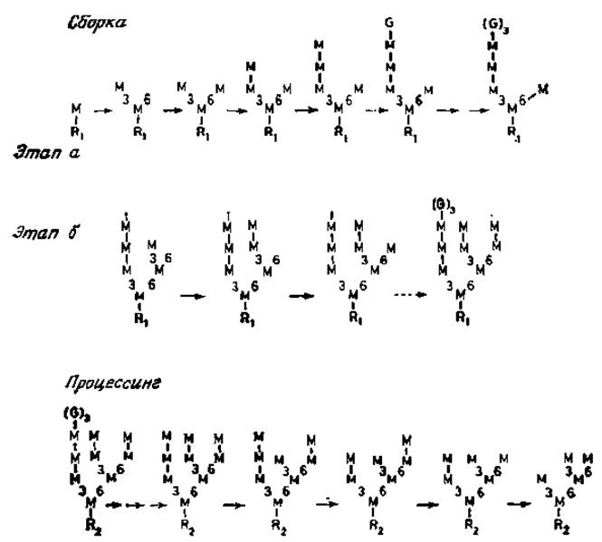
Fig. 3.6. Assembly of oligomannosyl Lipids and Processing stages in CHO (Chinese hamster Ovary) cells. R1 — glcNAcß1→4glcNAc pyrophosphoryldolichol. R2 — glcNAcß1→glcNAc Asn. The positions of mannose (M) and glucose (G) residues in the chain are shown in Fig. 3.5. Pathway a represents the minor pathway, and pathway b represents the major pathway.

Fig. 3.7. Final stages of asparagine residue glycosylation in Proteins.
In cells with specifically blocked dolichol phosphomannose synthesis, only truncated lipid intermediates containing four mannose residues are formed (Fig. 3.5). In normal cells, such truncated dolichol phosphomannoses are merely minor components. This implies that the initial a-mannosyl residues are transferred from GDP-mannose, whereas the subsequent four residues originate from dolichol phosphomannose [26, 27]. The remaining assembly stages involve the attachment of three glucose residues—most likely from dolichyl phoshoglucose—to one of the Branches of the mannose-rich structure. The major end product (Fig. 3.5) serves as a direct precursor for N-glycans in glycoproteins: the oligosaccharide moiety of the lipid intermediate is transferred intact (as a single block) to an asparagine residue within the polypeptide acceptor. Dolichol pyrophosphates are released and presumably recycled via dephosphorylation back into the general dolichol pool (Fig. 3.7).
The enzyme capable of transferring such a block has been identified in numerous tissues actively synthesizing glycoproteins. This enzyme exhibits remarkable versatility. Enzyme preparations from a single source display activity toward various acceptors derived from the same tissue or from entirely different sources. Even more remarkably, short Peptides containing asparagine can also function as acceptors in this reaction [28, 29]. When comparing the acceptor activity of unfolded Ribonuclease and a-lactalbumin with that of synthetic peptides whose sequences matched the region surrounding the glycosylated asparagine of the respective glycoprotein Polypeptides, the acceptor activity of these synthetic peptides was found to be high (Table 3.1). However, not all peptides were active; activity was restricted to those containing the Asn-x-Ser or Thr sequence, where x can be virtually any amino acid except Proline and possibly aspartic acid (Table 3.1). To function as an acceptor, both the asparagine and hydroxyamino acid residues must have blocked amino and carboxyl terminal groups. The essential role of the asparagine residue in acceptor activity is unsurprising, given that it is this specific residue that undergoes glycosylation. Consequently, analogous peptides containing glutamine or aspartic acid residues instead of asparagine are completely devoid of activity. The acceptor requirements for oligosaccharide transfer to a polypeptide, established in these studies, depend entirely on the peptide sequence surrounding the glycosylated asparagine residue within the given glycoprotein [30]. Many such sequences are now known, and in all cases, Asn-x-Ser or Thr is localized within the glycosylated polypeptide chain. The inhibitory effect of proline was also predicted from the analysis of glycosylated sites in glycoproteins. Although the exact mechanism of this inhibition remains unknown, one hypothesis is that a Hydrogen bond can form between the asparagine amide and the hydroxyl group of Serine or Threonine, whereas a proline residue prevents the formation of such a bond. It is possible that this bond is required to induce the proper conformation for replacing the amide group with an appropriate oligosaccharide group, for instance, through the Modification of the amide group's ionization.
Table 3.1. Acceptor requirements for oligosaccharide transfer [25, 29]
|
Synthetic peptides |
Relative activity, % |
|
RNase (Asn34-Leu-35-Thr) |
100 |
|
Na-acetyl Asn-Leu-ThrNHCH3 |
473 |
|
Na-acetyl Asn-Leu-Thr Lys |
293 |
|
Ser-Arg-Asn-Leu-Thr-Lys |
120 |
|
Na-acetyl Asn-Leu-Thr |
5 |
|
Asn-Leu-Thr-Lys |
1 |
|
Asn-Leu-Thr |
1 |
|
a-Lactalbumin (Asn15-Gln16-Ser17) |
100 |
|
Na-acetyl Asn-Leu-Ser-Leu |
18 |
|
Asn-Leu-Ser-Leu |
<1 |
|
Tyr-Asn-Leu-Thr-Ser-Val |
100 |
|
Tyr-Asp-Leu-Thr-Ser-Val |
0 |
|
Tyr-Gln-Leu-Thr-Ser-Val |
0 |
|
Tyr-Asn-Leu-Val-Ser-Val |
0 |
|
Tyr-Asn-Pro-Thr-Ser-Val |
0 |
Although it is evident that oligosaccharide transfer to asparagine requires a tripeptide with a specific sequence, this requirement alone is insufficient. Many proteins contain the requisite sequences, yet they remain either entirely unglycosylated or only partially glycosylated during normal cellular biosynthesis. Pancreatic ribonuclease is a prime example of this phenomenon: only 20% of its protein molecules are glycosylated at asparagine-34 (Table 3.1), while the remaining molecules stay unglycosylated and serve as excellent acceptors only if their polypeptide moiety is first denatured (unfolded) by appropriate chemical treatments (such as disulfide bond Cleavage and carboxymethylation under denaturing conditions). Similarly, a-lactalbumin contains two recognizable tripeptides, Asn-Gln-Ser and Asn-Thr-Ser. However, the majority of these sequences are not glycosylated, and only a small fraction is modified at the first glycosylation site. Denatured proteins are readily glycosylated at any of these tripeptides, and synthetic peptides designed from their sequences likewise act as excellent acceptors. These findings lead to two Conclusions: (1) the tripeptide must be appropriately exposed on the protein surface for glycosylation to occur during glycoprotein assembly within the cell, and (2) existing control mechanisms ensure the glycosylation of only those asparagine residues located within sequences that become exposed on the protein surface post-biosynthesis, as the fully folded polypeptide takes its final conformation. It is clear that bulky carbohydrate groups consistently reside On the surface of protein globules and are typically located within peptide regions exhibiting a beta-sheet structure. Such Conformations are particularly favorable for accommodating glycosylation sequences.
As noted previously, under special conditions, the complete assembly of glucose-containing lipid intermediates can be prevented in intact cells. Nevertheless, N-glycosylated proteins continue to be formed in such cells, raising questions regarding the Specificity of the Glycosyltransferases that catalyze oligosaccharide transfer to the polypeptide. It appears that certain dolichol pyrophosphate oligosaccharides can function as intermediates in this reaction. Even dolichol pyrophospho-N-acetylglucosaminyl-ß1→4-N-acetylglucosamine is utilized by the enzyme to transfer a chitobiose unit to a synthetic peptide, albeit with reduced transfer efficiency [28]. This observation, combined with the fact that normal cells contain large glucose-containing carrier lipids (predominantly dolichol-linked oligosaccharides), strongly indicates that cellular N-glycosylation proceeds primarily via the transfer of such glucose-containing oligosaccharides to the polypeptide. Direct proof supporting this hypothesis has come from pulse-labeling experiments conducted in normal or virus-infected cells. These studies demonstrated that the oligosaccharide initially transferred to the polypeptide in cells or viral glycoproteins is identical in both Size and Structure to the large glucose-containing dolichol intermediate. The glucose moiety of the fully assembled intermediate carrier appears to be an essential factor governing the efficiency of transfer to proteins, at least in animal cells [31].
Last update: 06/08/2026
Editorial and Educational Adaptation: This material has been compiled based on the primary/original source text. The project team performed an editorial review, corrected technical inaccuracies, structured sections, and adapted the content for an educational format.
What was processed:
- elimination of formatting defects (OCR errors, structural breaks, corrupted characters);
- editorial organization of content;
- standardization of terminology in accordance with academic sources;
- verification of factual statements against the original source text.
All mentions of the author, publication year, and origin of the primary text have been preserved in accordance with the source.