Biochemistry and Molecular Biology - Belyasova N.A. 2002
Metabolism. Processes Requiring Energy Input
Biological Role and Regularities of Antibiotic Biosynthesis
Mechanisms of Antibiotic Action on Cellular Targets
Cell wall synthesis inhibitors. This action is characteristic of ß-lactam Antibiotics, which include Penicillins, Cephalosporins, nocardicin A, sulfazecin, thienamycin, and others. All of these compounds contain a ß-lactam ring in their Structure (Fig. 18.1). The target of these antibiotics is the bacterial enzyme glycopeptide transpeptidase, which catalyzes The formation of cross-links in the murein molecule. ß-Lactam antibiotics share a structural similarity with a substrate fragment of this enzyme (-D-Ala-D-Ala-OH) and bind to the Active Site of the transpeptidase in such a way that the ß-lactam ring ends up in close proximity to the Serine residue. The hydroxyl group of serine participates in forming a stable covalent bond with the carbon atom of the unstable ß-lactam ring (Fig. 18.1). As a result, an inactive acylated form of transpeptidase is produced, which is incapable of catalyzing the transpeptidation reaction (Chapter 14). This Enzyme Inhibition is irreversible, and the action of the antibiotics is bactericidal: a defective cell wall lacks the necessary mechanical strength and ruptures as The Cell expands during growth or due to the osmotic influx of Water.
In addition to ß-lactam antibiotics, the polypeptide antibiotic bacitracin A disrupts the synthesis of bacterial cell walls. It has been shown that the bacitracin molecule can bind a phosphorylated lipid carrier molecule into a ternary complex with divalent Metal Ions. Consequently, the dephosphorylation of the lipid carrier does not occur, meaning its free form cannot be regenerated. Under these conditions, all stages of murein synthesis that involve the lipid carrier are halted, preventing the Formation of the cell wall (bactericidal effect).
Class="center">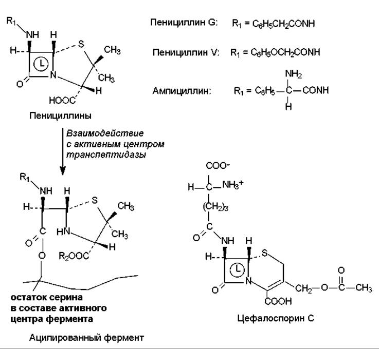
Fig. 18.1. Structure and MECHANISM OF ACTION of certain ß-lactam antibiotics: L — ß-lactam ring
It should be added that these antibiotics are most active against Gram-positive Bacteria, as their Cell walls contain a high concentration of murein. They exhibit low toxicity toward eukaryotes, which is explained by the absence in Eukaryotic Cells of the primary target — the Enzymes and other components involved in murein synthesis.
The antibiotic cycloserine, an amino acid derivative of serine, inhibits The activity of two enzymes involved in murein synthesis: Alanine racemase and D-alanyl-D-alanine synthetase. It is active against a wide range of bacteria and has low toxicity, although it can cause Nervous system disorders in humans.
Membrane-active antibiotics. The primary target of these antibiotics is the bacterial Plasma Membrane. Polymyxins (a group of peptide antibiotics with a cyclolinear structure) are capable of binding to the phosphate groups of cardiolipin, phosphatidylethanolamine, and other acidic Lipids that make up the membrane lipid bilayer. This binding disrupts membrane permeability—a very severe form of damage that affects overall cellular METABOLISM (most notably causing the "leakage" of ions that establish the membrane gradient). Furthermore, polymyxins activate phospholipases in the outer membrane of Gram-negative bacteria, leading to the destruction of The Lipid Bilayer. These disruptions are bactericidal, and polymyxins act primarily against Gram-negative bacteria, which possess an additional (outer) membrane.
Polyene antibiotics (amphotericin B, nystatin, levorin, trichomycin) affect membranes in a similar manner. Their molecules contain a conjugated system of double bonds, with the membrane serving as their target. Polyene antibiotics bind to specific sterols that predominate in fungal membranes. As a result, large pores form in the membranes, leading to permeability disruption.
Ionophore antibiotics, described in Chapter 4, also disrupt membrane permeability.
Translation inhibitors. This group includes a significant number of antibiotics (Fig. 18.2), among which Tetracyclines, aminoglycoside antibiotics (streptomycin, kanamycins, neomycins, gentamicins), chloramphenicol, and macrolide antibiotics (erythromycins, oleandomycin, leucomycins, tylosin) are the most widely used. A defining feature of these substances is their ability to bind to Ribosomes, thereby blocking Protein Synthesis. It is worth noting that most of these antibiotics are characterized by selective binding specifically to the subunits (or their constituent parts) of 70S ribosomes found in Prokaryotic Cells. Consequently, these antibiotics are specific to prokaryotes. Their action is largely reversible, which usually results in a bacteriostatic effect.
Upon binding to ribosomes, translation inhibitors either prevent aminoacyl-Transfer RNAs from interacting with the ribosomes (tetracyclines), inhibit Translation initiation (streptomycin), inhibit the activity of peptidyl transferase — a structural component of the 50S ribosomal subunit (chloramphenicol), or disrupt ribosomal translocation (erythromycin), among other mechanisms.
The antibiotic puromycin bears a structural resemblance to the terminal fragment of the aminoacyl-tRNA acceptor arm, though it is significantly smaller in size. Due to this, puromycin can rapidly bind to the A-site of ribosomes (both 70S and 80S); however, once a peptide residue is attached to puromycin's amino group (via the peptidyl transferase reaction), the entire complex dissociates from the ribosome due to the lack of codon-anticodon interaction. Puromycin exhibits high toxicity toward eukaryotic cells because it lacks selective binding to 70S ribosomes. This antibiotic is active against bacteria, Protozoa, helminths, and certain malignant tumors.

Fig. 18.2. Structure of tetracyclines, actinomycin D, streptomycin, and chloramphenicol
Intercalating antibiotics. The action of these antibiotics is based on their ability to insert themselves (intercalate) into DNA molecules. Examples of such agents include actinomycin D (Fig. 18.2), adriamycin, and daunomycin. Specifically, the actinomycin molecule inserts its flat heterocyclic moiety between the parallel planes of DNA Base Pairs. Daunomycin behaves similarly, with two molecules intercalating their heterocyclic cores between the planes of G/C base pairs. This interaction causes localized distortion of the Introduction/20.html">DNA Structure, resulting in the inhibition of Replication and Transcription processes.
Because The structure of DNA is highly conserved, intercalating antibiotics are also active against eukaryotic DNA, making them highly toxic. These antibiotics are used in the Chemotherapy of malignant tumors, whose cells are known to divide faster than normal cells, meaning that the template processes targeted by these antibiotics are much more active in them.
DNA-tropic antibiotics. These antibiotics interact directly with DNA molecules, leading to their degradation. Classical examples include mitomycins (A, B, C) and porfiromycin. The action of these antibiotics occurs via two pathways: first, through the modification of purine bases, and second, by forming cross-links between antiparallel DNA strands. As a result, replication is suppressed, replication errors arise, and breaks occur within the DNA molecule. Another type of antibiotic, bleomycins, induces numerous single- and double-strand breaks in DNA molecules.
Most DNA-tropic antibiotics are highly toxic and are used as antitumor drugs.
Transcription inhibitors. In addition to the aforementioned intercalating antibiotics, Rifamycins also suppress transcription. These antibiotics interact with prokaryotic DNA-dependent RNA polymerases, specifically with the ß-subunit of the core enzyme. This non-covalent binding is nonetheless quite strong, thereby disrupting the synthesis of RNA chains.
Inhibitors of enzymatic processes. These antibiotics include the previously discussed penicillins, cephalosporins, cycloserine, chloramphenicol, and others. Additionally, noteworthy examples include antimycin, which suppresses electron transport between Cytochromes in the Respiratory Chain; oligomycin, which binds to enzymes responsible for coupling substrate oxidation with phosphorylation; novobiocin, an inhibitor of the DNA gyrase enzyme (which is involved in replication); and actinomycin A, which inhibits cytochrome c reductase activity.
Last update: 06/08/2026
Editorial and Educational Adaptation: This material has been compiled based on the primary/original source text. The project team performed an editorial review, corrected technical inaccuracies, structured sections, and adapted the content for an educational format.
What was processed:
- elimination of formatting defects (OCR errors, structural breaks, corrupted characters);
- editorial organization of content;
- standardization of terminology in accordance with academic sources;
- verification of factual statements against the original source text.
All mentions of the author, publication year, and origin of the primary text have been preserved in accordance with the source.