Biological Chemistry - Berezov, T. T., Korovkin, B. F. 1998
Protein Chemistry
Structural Organization of Proteins
Tertiary Structure of Protein
The Tertiary Structure of a protein refers to the spatial orientation of the polypeptide helix or the manner in which the polypeptide chain is folded within a specific volume. Since neither the Primary Structure nor the types of helices or combinations of helical and linear segments of the polypeptide chain provide an indication of the volume and shape of the polypeptide chain, researchers are consistently faced with the necessity of determining the three-dimensional or spatial configuration of the protein. X-ray crystallography with high resolution has played a fundamental role in solving these problems. As noted, this method successfully resolves two main challenges of Protein Chemistry: the regularity of amino acid residue sequences in the polypeptide and the regularity of the protein molecule's configuration. Interatomic distances in organic molecules range from 0.1 to 0.2 nm, while the maximum resolving power of modern apparatus is 0.2 nm. This does not allow for determining the exact position of every individual atom, although distinct combinations of atoms can be clearly distinguished, especially upon introducing heavy metal atoms into the protein molecule (the latter are used as reference points in the mathematical Processing of X-Ray Diffraction patterns due to their high electron density).
Class="center">
Fig. 1.19. Domain Introduction/12.html">Structure of Globular Proteins (after A. A. Boldyrev).
a — ß-subunit of Hemoglobin; b — constant domain of an immunoglobulin; c — flavodoxin; d — chicken egg white Lysozyme.

Fig. 1.20. Model of the tertiary structure of a Myoglobin molecule (after J. Kendrew). Structural domains are designated by Latin letters, and the heme group is shown in red.
Sperm whale myoglobin was the first protein whose tertiary structure was elucidated by J. Kendrew using X-ray Diffraction Analysis. It is a relatively small protein with a Molecular Weight of 16,700, containing 153 amino acid residues (its primary structure is completely known), and is represented by a single polypeptide chain. The primary function of myoglobin is Oxygen transport in Muscles. The polypeptide chain of myoglobin (Fig. 1.20) is depicted as a curved tube folded compactly around the heme (a non-protein, iron-containing component; see Chapter 2).
Over recent decades, driven by improvements in the resolution of X-ray crystallography, the tertiary structure of more than 1,000 proteins has been deciphered, including hemoglobin, Pepsin, Chymotrypsin, Ribonuclease, lysozyme, Trypsin and its inhibitor, A number of human immunoglobulin fragments, cytochrome c, human Carbonic anhydrase, aspartate aminotransferase, Insulin, and others. Examples of the three-dimensional structure of some of these proteins are shown in Fig. 1.21.
X-ray crystallography makes it possible to determine the conformation and the path of the polypeptide chain in space; therefore, a three-dimensional model can be constructed for each protein, reflecting the positions of its linear and helical regions. Studies on Globular proteins have demonstrated that their Spatial Structure heavily depends on several factors, particularly the Ionic strength and pH of the solution, Temperature, etc. State-of-the-art X-ray diffraction techniques have enabled the deciphering of the crystal structures of over 100 Enzymes. Recently, low-temperature computational techniques, as well as mathematical and computer-based Methods for determining volumetric structures from Amino Acid Sequence data, have also been successfully applied to elucidate the three-dimensional structure of proteins.
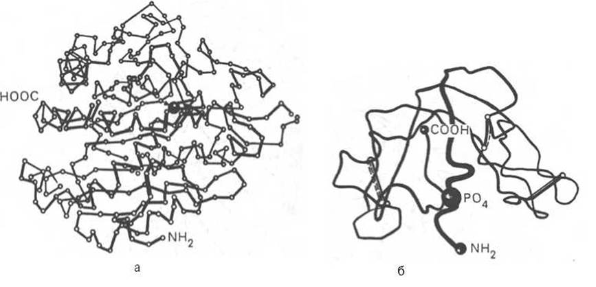
Fig. 1.21. Spatial configuration of carboxypeptidase (a) and ribonuclease (b).
Currently, undeniable evidence has been obtained that, alongside covalent bonds (peptide and Disulfide Bonds), so-called non-covalent bonds play the primary role in stabilizing the Spatial structure of Proteins (Fig. 1.22). These bonds include Hydrogen Bonds, Electrostatic Interactions between charged groups, intermolecular Van der Waals forces, interactions between non-polar amino acid side chains (the so-called hydrophobic interactions), etc.
According to modern concepts, once its synthesis in Ribosomes is complete (see Chapter 14), the tertiary structure of a protein forms entirely automatically, spontaneously, and is fully predetermined by its primary structure. The main driving force behind The Emergence of a three-dimensional structure is the interaction between amino acid side chains and Water molecules. In this process, non-polar hydrophobic amino acid residues are, as it were, buried within the protein molecule, forming dry interiors, while polar residues become oriented toward the water. At a certain point, the thermodynamically most favorable and stable conformation* of the molecule is achieved. In this form, the protein molecule is characterized by a minimal Free energy. Protein molecules In aqueous solutions typically adopt a range of stable Conformations induced not only by changes in pH and temperature, but also by low-molecular-weight compounds. Two main conformational forms are distinguished: the T-form (from the English "tensed") and the R-form (from the English "relaxed"). Transitions occur between these forms, which are correspondingly reflected in their biological properties.
* Conformation is conventionally defined as the spatial arrangement of atoms within a protein molecule. This term denotes a structural state of a protein molecule that can transition into another structural state without the Cleavage of covalent bonds, caused, for example, by rotation around a single bond.
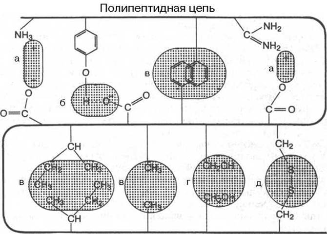
Fig. 1.22. Types of non-covalent bonds stabilizing the tertiary structure of a protein.
a — electrostatic interaction; b — Hydrogen bond; c — hydrophobic interactions of non-polar groups; d — dipole-dipole interactions; e — disulfide (covalent) bond.
During the folding of the synthesized polypeptide chain—a process known as folding, which establishes the native spatial structure—Cells select a single stable and biologically active conformation out of a multitude of sterically possible states, most likely determined by the primary structure. A number of inherited human diseases have been described whose development is associated with disruptions in the folding process due to Mutations (pigmentoses, fibroses, etc.). Consequently, researchers currently focus intense attention on clarifying the relationship between The amino acid sequence of The Cell-synthesized polypeptide chain (primary structure) and The formation of the three-dimensional spatial structure that endows the protein molecule with its native properties. There is considerable experimental evidence indicating that this process is not entirely automatic, as previously assumed, but is most likely regulated and controlled by intracellular molecular mechanisms whose details are not yet fully understood. Several classes of proteins, termed chaperones or heat Shock proteins, have been isolated from cells; they are located between the N-terminal signal peptide and the matrix protein. It is hypothesized that the primary Functions of chaperones include preventing the polypeptide chain from forming non-specific (chaotic) random coils or protein aggregates, and ensuring their delivery (transport) to subcellular targets, thereby creating favorable conditions for the completion of protein folding. These findings suggest the potential existence of a "second half of METABOLISM/28.html">The Genetic Code," thereby heightening researchers' interest in The problem of polypeptide chain folding and the formation of its native spatial conformation.
Thus, the linear, one-dimensional structure of a polypeptide chain (i.e., The sequence of amino acid residues dictated by the genetic code of Protein Synthesis) carries another type of information—conformational information, which represents the formation of a protein molecule with a strictly prescribed shape and a defined spatial arrangement of its individual parts. In other words, the tertiary (volumetric) structure of a protein molecule is determined by the amino acid sequence of the polypeptide chain, or more specifically, by the size, shape, and polarity of the amino acid residues' side chains. These concepts can serve as a basis for predicting the conformation of a protein molecule based on its amino acid sequence. It should be noted, however, that the mechanism underlying this close and subtle link between the amino acid sequence and the three-dimensional structure of a protein molecule remains an intriguing puzzle. Interestingly, Polypeptides with nearly identical sequences sometimes form entirely different structures, whereas polypeptides with distinct sequences can form the same three-dimensional structure.
In turn, the three-dimensional structure of a protein molecule also contains information, but of an entirely novel type—namely, functional information, which Academician V. A. Engelhardt termed intramolecular information. As will be demonstrated hereafter, all biological Properties of Proteins (catalytic, hormonal, antigenic, etc.) are tied to the preservation of their tertiary structure, commonly referred to as the native conformation. Any influences (thermal, physical, chemical) that disrupt this molecular conformation (the cleavage of Hydrogen bonds and other non-covalent bonds) are accompanied by a partial or complete loss of the protein's biological properties.
Last update: 06/08/2026
Editorial and Educational Adaptation: This material has been compiled based on the primary/original source text. The project team performed an editorial review, corrected technical inaccuracies, structured sections, and adapted the content for an educational format.
What was processed:
- elimination of formatting defects (OCR errors, structural breaks, corrupted characters);
- editorial organization of content;
- standardization of terminology in accordance with academic sources;
- verification of factual statements against the original source text.
All mentions of the author, publication year, and origin of the primary text have been preserved in accordance with the source.