Biochemical Foundations of Human Vital Activity - Volkov N.I., Nesen E.N. 2000
Biochemical Foundations of Human Vital Activity
Biochemistry of Carbohydrates
Characteristics of Carbohydrate Classes
Depending on The complexity of their molecular Structure, CARBOHYDRATES are classified into three main groups: Monosaccharides, Oligosaccharides (mainly Disaccharides), and Polysaccharides. Their General characteristics are presented in Table 14.
Monosaccharides
Monosaccharides are simple carbohydrates that cannot be hydrolyzed into simpler molecules. Based on the number of carbon atoms in the molecule, monosaccharides are divided into trioses (С3Н6O3), tetroses (С4Н8O4), pentoses (С5Н10О5), hexoses (С6Н12O6), and heptoses (С7Н14O7). Other monosaccharides do not occur naturally, but can be produced synthetically.
The most important roles in The Human Body are played by hexoses—glucose and fructose; pentoses—ribose and deoxyribose; and trioses—glyceraldehyde and dihydroxyacetone.
Class="center">TABLE 14. Classes of carbohydrates and their main characteristics

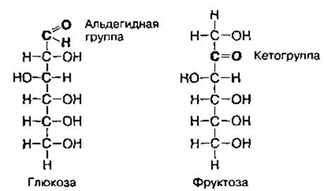
Glucose and fructose. These are the main energy substrates of the human body. They have the same molecular formula (С6Н12O6), but different molecular structures due to having different functional groups. Glucose contains an aldehyde group, while fructose contains a keto group, meaning they are positional isomers of the carbonyl group 
Monosaccharides are also characterized by spatial isomerism, or stereoisomerism, because they contain asymmetric carbon atoms (marked with *), which are bonded to four different atoms or groups of atoms. D- and L-forms of isomers are distinguished for glucose and other monosaccharides. In these forms, the hydroxyl group at the fourth carbon atom occupies different spatial positions:
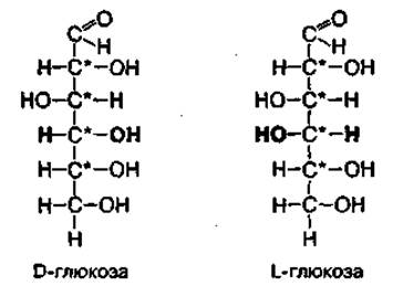
The human body can assimilate only the D-form of monosaccharides, whereas Amino Acids are utilized exclusively as L-isomers. Intracellular Enzymes are capable of distinguishing between optical isomers of substances. Galactose and mannose are stereoisomers of glucose:

Galactose is a component of lactose, the primary disaccharide in milk. In the Liver, it can be converted into glucose through enzymatic action.
In an aqueous environment, glucose and fructose exist predominantly in cyclic form. Molecule cyclization occurs due to the intramolecular interaction of the aldehyde group in glucose or the keto group in fructose with one of the hydroxyl groups of the same monosaccharide:
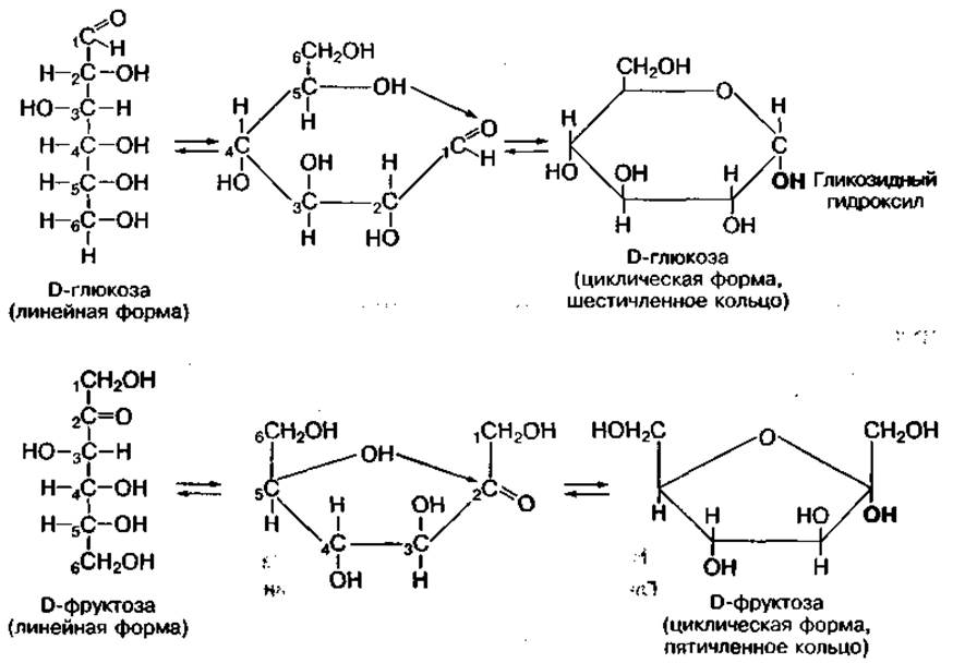
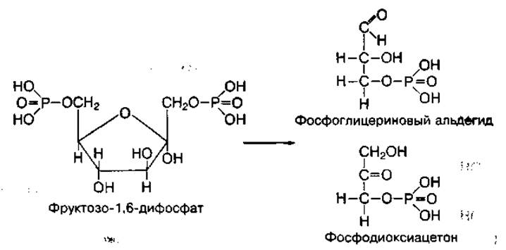
The cyclic forms of monosaccharides acquire a biologically reactive hydroxyl group at the C1 or C2 carbon atom, known as the glycosidic hydroxyl. It plays a crucial role in the Chemical transformations of these monosaccharides, specifically participating in The formation of di- and polysaccharides and phosphoric esters. For instance, glucose participates in METABOLISM and Energy turnover in the form of the phosphoric ester glucose-1-phosphate, which initiates glucose breakdown and Polysaccharide synthesis. Monosaccharides also readily form other phosphoric esters: glucose-6-phosphate, fructose-6-phosphate, and fructose-1,6-diphosphate:
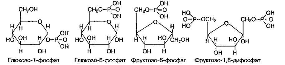
During metabolism, phosphorylated forms of glucose and fructose can interconvert and also break down into trioses—phosphoglyceraldehyde and phosphodihydroxyacetone:
Amino sugars are formed from monosaccharides when hydroxyl groups are replaced by an amino group (-NH2). In the human body, the most important amino sugars are glucosamine and galactosamine:
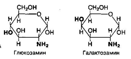
They are components of complex carbohydrates known as mucopolysaccharides, which perform protective and specific Functions characteristic of mucous secretions, the vitreous humor of the eyes, synovial fluid of joints, the Blood clotting system, etc.
Through reduction or oxidation processes, glucose yields many functionally important substances: ascorbic acid (Vitamin C), sorbitol, gluconic, glucuronic, sialic, and other acids.
Ribose and deoxyribose. These carbohydrates rarely occur in a free state. More often, they are part of complex substances, meaning they are utilized in plastic processes within the body. Ribose participates in the Biosynthesis of NUCLEOTIDES (ATP, ADP, AMP, etc.) and RNA, as well as many Coenzymes (NAD, NADP, FAD, FMN, CoA). Deoxyribose participates in The biosynthesis of deoxynucleotides, which serve as Structural components of DNA. Ribitol, a derivative of ribose alcohol, is a component of vitamin B12 and certain respiratory enzymes.
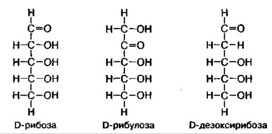
Ribose and deoxyribose are aldoses. The deoxyribose molecule lacks an oxygen atom at the second carbon atom. Ribulose is a positional isomer of the functional carbonyl group in ribose:
In the body, ribose and other pentoses also exist in the cyclic D-form:
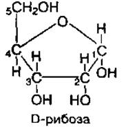
Ribose and ribulose are synthesized in body Tissues during The oxidation of glucose via the Pentose Phosphate Pathway. Deoxyribose is formed from ribose through its deoxygenation.
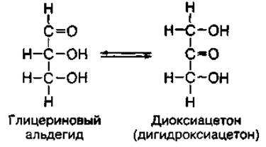
Glyceraldehyde and dihydroxyacetone. These are produced in body tissues during the Catabolism of glucose and fructose. As isomers, these trioses are capable of interconversion:
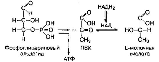
During carbohydrate and Lipid Metabolism IN body tissues, phosphoric esters of glyceraldehyde and phosphodihydroxyacetone are formed. Phosphoglyceraldehyde serves as a high-energy substrate for Biological Oxidation. Its oxidation yields an ATP molecule along with oxidation products such as pyruvic acid (PA) and lactic acid:
Disaccharides
Disaccharides are the primary group of oligosaccharides, consisting of a small number (from 2 to 10) of monosaccharides. In disaccharides, two monosaccharide residues are linked together by 1,4- or 1,2-glycosidic bonds. The principal disaccharides are sucrose, maltose, and lactose. Their molecular formula is С12Н22О11
Sucrose consists of a glucose residue and a fructose residue linked together by a 1,2-glycosidic bond, which is formed through the interaction between the hydroxyl group at the first carbon atom of glucose and the hydroxyl group at the second carbon atom of fructose:
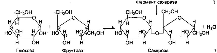
Sucrose is the main component of table sugar. During Digestion, under the action of the highly specific enzyme sucrase, it breaks down into glucose and fructose.
Maltose consists of two glucose residues linked together by a 1,4-glycosidic bond:
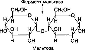
Maltose is formed in the gastrointestinal tract during the Hydrolysis of dietary starch or Glycogen. During digestion, it is broken down into glucose molecules by the enzyme maltase. Maltose is abundant in cereal malt extracts and sprouted grains.
Lactose (milk sugar) consists of glucose and galactose residues linked together by a 1,4-glycosidic bond:
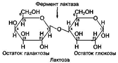
Lactose is synthesized in the Mammary Glands during Lactation. Its content is about 5% in cow's milk and approximately 6% in human breast milk. In the human Digestive System, lactose is broken down into glucose and galactose by the enzyme lactase. The Dietary intake of lactose promotes the growth of lactic acid Bacteria, which inhibit putrefactive processes in the gut. However, individuals with low activity of the lactase enzyme, also known as galactosidase, develop milk intolerance.
The disaccharides discussed above, especially sucrose (table sugar), have a sweet taste and high nutritional value. Therefore, they are not recommended for the diet of individuals suffering from obesity and diabetes. They are replaced by artificial substances, such as saccharin, which taste sweet but are not metabolized by the body.
Polysaccharides
Polysaccharides are complex carbohydrates consisting of many hundreds or thousands of linked monosaccharide residues, predominantly glucose residues. A distinction is made between Homopolysaccharides, which consist of identical monosaccharide residues (e.g., glucose), and Heteropolysaccharides, which consist of residues of different MONOSACCHARIDES AND THEIR derivatives.
The main homopolysaccharides that perform vital biological functions and consist of glucose molecules are starch and Cellulose in plants, and glycogen in humans and animals. These polysaccharides are non-sweet, poorly soluble in Water, and form colloids. They share a general molecular formula of (С6Н10О5)n, yet differ in quantitative composition and molecular structure.
Starch is a plant reserve polysaccharide consisting of A large number of D-glucose residues (up to 300). It serves as the primary dietary polysaccharide and a source of glucose for the human body. Starch has a high molecular weight, ranging from 50,000 to 300,000. Structurally, it is heterogeneous, consisting of a mixture of helical amylose chains (10-20%) and branched amylopectin chains (80–90%). The glucose residues in amylose are linked by 1,4-glycosidic bonds, whereas at the branching points of amylopectin they are linked by 1,6-glycosidic bonds (Fig. 57, a, b).
Amylose is readily soluble in water, whereas amylopectin is insoluble and forms a colloidal solution known as starch paste. Partial degradation of the starch structure yields lower-molecular-weight compounds (dextrins), which are also readily soluble in water. The main enzymes that break down dietary starch are amylases found in saliva and pancreatic juice.
Glycogen is the primary reserve polysaccharide in Human and Animal tissues. It consists of a large number of glucose molecules (up to 30,000) linked together by glycosidic bonds (Fig. 57, c). Its molecular weight reaches 1–10 million. The glycogen molecule has a structure similar to plant starch amylopectin, but with a higher degree of branching. Thanks to this structure, glycogen is readily soluble in water.
Glycogen is stored (deposited) primarily in the liver (about 100 g) and skeletal Muscles (about 400 g), providing a vital glucose reserve for the body. Its tissue concentration depends on diet, physical activity, and environmental factors (such as heat or Hypoxia). An insufficient carbohydrate intake or intense physical exertion depletes glycogen reserves, whereas an abundant glucose intake restores them. Liver glycogen is utilized to maintain blood glucose levels between meals or during intensive glucose oxidation, while Skeletal Muscle glycogen provides the energy required by the muscles themselves (Fig. 58).
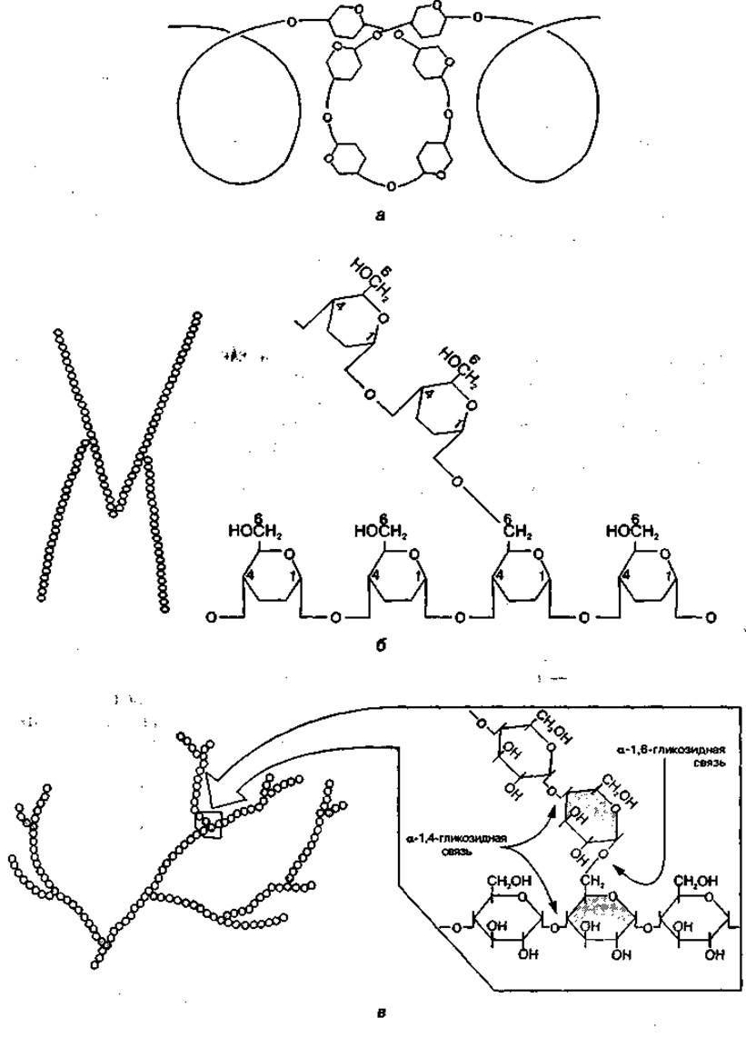
Fig. 57 Cytology/cytology/92.html">SCHEMATIC STRUCTURE OF starch chains — amylose (a), amylopectin (b), and a section of a glycogen molecule (c)

Fig. 58 Utilization of liver and skeletal muscle glycogen
Dietary fiber (cellulose) is a structural plant polysaccharide that provides strength and elasticity. It is an unbranched polymer composed of numerous glucose residues. Although human body cannot digest cellulose, it is essential for regulating gut motility and The activity of Small Intestine enzymes.
Pectic substances belong to homopolysaccharides composed of galacturonic acid derivatives. They are synthesized in plants and fall into two main categories: protopectins and Pectins. Protopectins are water-insoluble because they exist as a complex of pectin and cellulose. Pectins dissolve in water, forming a gelatinous colloidal mass. Due to their unique structure, they can adsorb various toxic compounds, including heavy metals such as lead. In the body, they act as a natural sorbent, clearing metabolic waste and toxins from the gastrointestinal tract. Consequently, dietary pectin fibers are vital for human health.
Heteropolysaccharides in the body are represented mainly by mucopolysaccharides. Mucopolysaccharides comprise a large group of polysaccharides with diverse chemical compositions and structures, found in the Skin, tendons, Cartilage, Cell membranes, intercellular matrix, and synovial fluid. The most clinically significant ones for the body include hyaluronic acid, chondroitin sulfate, and heparin.
Hyaluronic acid is an unbranched polymer of glucuronic acid and glucosamine:
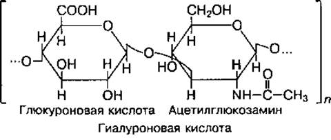
It plays a key role in binding water within the body, provides lubricating properties to synovial fluid to reduce friction during joint movement, and regulates cell membrane permeability by acting as a biological filter that traps microbes and prevents them from entering Cells.
Chondroitin sulfate is a polymer composed of glucuronic acid and acetylgalactosamine sulfate:
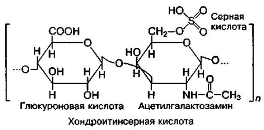
Combined with the protein Collagen, it is a key component of bones, cartilage, Heart Valves, blood vessel walls, and skin, serving a vital supportive function in the body.
Heparin is a polymer of glucuronic acid and glucosamine:
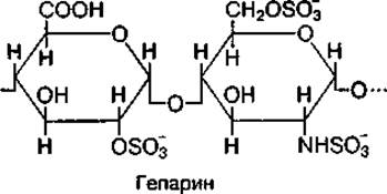
It was first isolated from the liver, which gave it its name. Heparin is also found in the Lungs, Connective Tissue, and other Organs, from which it is released into the blood and intercellular fluid. As a natural anticoagulant (preventing blood clotting), heparin is widely used clinically during blood transfusions, as well as for the Prevention and Treatment of thrombosis in the Circulatory system. In addition, heparin exhibits anti-inflammatory properties, regulates potassium and sodium metabolism, and performs an antihypoxic function.
Last update: 06/08/2026
Editorial and Educational Adaptation: This material has been compiled based on the primary/original source text. The project team performed an editorial review, corrected technical inaccuracies, structured sections, and adapted the content for an educational format.
What was processed:
- elimination of formatting defects (OCR errors, structural breaks, corrupted characters);
- editorial organization of content;
- standardization of terminology in accordance with academic sources;
- verification of factual statements against the original source text.
All mentions of the author, publication year, and origin of the primary text have been preserved in accordance with the source.