Amino Acids, Peptides and Proteins - Dévényi T., Gergely J. 1976
Quantitative determination of amino acids using automatic amino acid analyzers
Principle of the method (Fig. 35). The analyzed material is fractionated on Column K, packed with Dowex 50 x 8 resin. The Buffer solutions (Б1, Б2, Б3) are delivered by pump H1 and regulated by valve Кр. The effluent from the column enters mixer c containing a ninhydrin (Нг) solution, which is supplied by pump Н2. This reaction mixture passes through capillary Кn immersed in a Water bath at 100°C, where the color reaction characteristic of Amino Acids takes place. The colored eluate then passes through spectrophotometer Ф, the readings of which are recorded by chart recorder См.
Applications. Chemical and biochemical protein research, synthesis of Amino Acids and Peptides, hormone research, clinical Diagnostics, experimental and industrial enzymology, plant breeding, etc.
Class="center">
Fig. 35. Schematic diagram of an automatic amino acid analyzer (for designations, see text).
PROCEDURE
1. Selection of the analytical method. Amino acids can be determined using either a two-column or a single-column method. In the former case, basic Amino acids are analyzed on a separate column with a resin bed height of 6–8 cm, while acidic and neutral amino acids are analyzed on another, longer column with a height of 50 cm. In this case, the determination of basic amino acids takes about 1 h, and the determination of acidic and neutral amino acids takes about 3 h.
In the second method, only one column is used to determine all amino acids. The time required for analysis in this case depends on the number of buffer solutions used, which in turn is determined by the available instrumentation. If the instrument, such as the Unichrom analyzer from Beckman (USA), allows for a single automatic switch from one buffer solution to another, it is preferable to use a two-buffer system. In this case, the entire analysis takes 4 h. If the instrument can automatically change the flowing buffer solution two or more times, a three-buffer system is used, which reduces the analysis time to 3 h. With an appropriate combination of Methods, the analysis time of the two-column method can be further reduced (see below).
2. Instrumentation. Currently, several types of automatic amino acid analyzers are commercially available, each having its own Advantages and disadvantages. For routine purposes, simpler analyzers are more economical. This is determined not so much by price, but by reliability in ensuring continuous operation and by the method of programming the instrument. Apparently, an instrument equipped with an automatic sample loader that reliably ensures continuous operation can be considered highly cost-effective.
An amino acid analyzer is a complex instrument that requires constant maintenance, even with careful handling. The analyzer should not be shut down for extended periods (1–2 days). Buffer solutions are excellent media for the growth of Fungi and other microorganisms. Even a 1- to 2-day shutdown of the instrument can lead to bacterial contamination of the solutions, distorting the analytical results (see sources of error). If the instrument is not used continuously, it should be turned on daily for a few minutes to circulate the solutions, changing the positions of all Valves. When the instrument is shut down for a long period, the buffer solutions must be removed and the system flushed with distilled water. The resin should be stored in an alkaline solution within the column or in a separate vessel.
3. Resin. Modern analyzers use spherical bead resins, which allow for relatively high flow rates through the column at relatively low pressures. If the resin particles are uniform in size and the column pressure remains constant within certain limits, the resin does not compress significantly. However, even with perfectly uniform resin particles, after 30 analyses, the resin should be removed from the column and incubated in 2 N NaOH at 80°C for 30 min. After incubation, the liquid is decanted and the resin is suspended in 0.2 N NaOH. Once the resin particles settle, the supernatant is decanted again, and the ion exchanger is suspended in a buffer solution of a specific molarity depending on the method used. The resin is packed into the column in this same buffer solution, which is then used (three column volumes) to thoroughly wash the column before Sample application.
4. Buffer solutions. Sample application solution: 0.2 M NaCl in 0.01 N HCl. Preparation: Dissolve 11.6 g of NaCl and 0.8 mL of concentrated HCl in deionized distilled water to a final volume of 1 L.
Eluting solutions: the COMPOSITION OF THE eluting buffer solutions is given in Table 8.
Table 8 Buffer solutions for an automatic amino acid analyzer
A |
B |
C |
D |
E |
F |
|
Na+, M |
0,2 |
0,2 |
0,35 |
0,8 |
0,8 |
1,6 |
pH |
3,28 |
4,25 |
5,28 |
4,25 |
4,25 |
6,0 |
Final volume, ml |
5000 |
5000 |
5000 |
5000 |
5000 |
5000 |
Citric acid (monohydrate), g |
70,5 |
282,0 |
123,0 |
282,0 |
70,5 |
35,25 |
HCl (sp. gr. 1.19), ml |
61,6 |
167,5 |
32,5 |
167,0 |
41,88 |
— |
NaOH, g |
40,0 |
160,0 |
70,0 |
160,0 |
40,0 |
20,0 |
NaCl, g |
— |
— |
— |
— |
175,32 |
438,75 |
Ethanol, ml |
200 |
— |
— |
— |
— |
— |
Detergent (Brij 35), g |
10 |
10 |
10 |
10 |
10 |
10 |
Deionized distilled water is used to prepare all solutions; it can be obtained by passing distilled water through a column packed with cation and anion exchangers.
It is essential that solutions A and C have a strictly defined pH, as even a slight change in pH causes a significant deterioration in Separation. When eluting with solution A, cystine can serve as a marker on the chromatogram. Ideally, it elutes between Alanine and valine. If the pH is above 3,28, the cystine peak shifts closer to the alanine peak or even merges with it. On the other hand, if the pH of the solution is below 3,28, the cystine peak shifts toward the valine peak, overlapping it or even preceding it on the chromatogram. In the former case, the buffer is acidified by adding 0,5—1,0 ml of concentrated HCl per 1 l of solution, and in the latter, the pH of the solution is increased by adding the same amount of a 50% NaOH solution. It should be noted that alkalinization increases the concentration of sodium ions and, consequently, increases the molarity of solution A. When the Na+ concentration increases from 0,2 to 0,25 N, the peaks of aspartic acid, Threonine, Serine, glutamic acid, and Proline merge. When eluting with solution C, Histidine serves as a marker. If the pH of the solution is too high, the histidine peak overlaps with the ammonia peak, and if it is too low, the time required for elution increases. For a column 6—8 cm high at a flow rate of 68 ml/h, the maximum elution time is 1 h.
Even if the analyzer is used continuously, no more than 5 l of buffer solutions should be prepared due to the risk of bacterial growth. The first sign of Microbial growth (when the solution does not yet contain flakes) is peak Asymmetry, especially on the leading edge, and the merging of ammonia and Lysine peaks in a single-column procedure. In the case of heavy contamination, both peaks completely overlap. The contaminated solution must be discarded immediately, and the glassware thoroughly washed first with detergent and then with deionized water; the same procedure must be performed on the connecting tubing. Contaminated resin must not be washed inside the column; it should be removed and washed with a warm alkaline solution.
Slight fluctuations in the pH of solutions D, E, and F are not as critical during elution as changes in sodium ion concentration. If the Na+ concentration is decreased by 0,05 M, the distance between the ammonia, lysine, histidine, and Arginine peaks on the chromatogram increases. If the sodium ion concentration is increased by 0,05 M, ammonia elutes as a double peak with lysine. The sodium ion concentration in solution D depends on the quality of the NaOH used, and in solutions E and F, on the purity of NaCl. In our experience, most NaCl preparations contain significant amounts of water; therefore, we recommend using Reagents pre-dried at 100°C for 6 h.
It is very important that the solutions are ammonia-free. If the solutions, especially A, contain ammonia, a so-called "ammonia plateau" can be observed on the chromatogram in the single-column method, starting before the ammonia peak and ending after the histidine peak. The higher this plateau, the lower the sensitivity of the instrument in the region where the ammonia, lysine, and histidine peaks elute. To avoid a high Background, high-purity reagents and deionized water should be used, and smoking must be prohibited in the room where the instrument is located.
5. Column packing. Packing the short column for basic amino acid determination. About 15 ml of resin is mixed with 0,2 N NaOH. After the particles settle, the supernatant is decanted, and the resin is suspended in solution C, stirring at room Temperature for 30 min. After the particles settle, the supernatant is decanted again, and the entire procedure is repeated. Then, the resin is mixed with two volumes of solution C. A round filter is placed on the metal mesh at the bottom of the column, and about 2 ml of solution C is poured in. The resin is suspended in 2 volumes of solution C, poured into the column, and allowed to stand for 5—10 min. Then, solution C is connected to the column. The height of the settled resin in the column should be 6—8 cm. 0,2 N NaOH and solution C are alternately passed through the column 3 times.
Packing the long column for the separation of acidic and neutral amino acids in the two-column method. About 50 g of resin is required for one column. The long column is packed in the same way as the short one (described above), except that following the alkali Treatment, the resin is suspended in solution A, and subsequent treatment in the column is also carried out with alkali and solution A.
Column packing in the single-column method. In the single-column analysis method using a two-solution system, these solutions differ in Na+ concentration by a factor of 4. In a three-buffer system, the first and third solutions differ in Na+ concentration by a factor of 8. Such large differences can affect the resin volume in the column if packing is performed in the usual manner. The higher the molarity of the solution, the less the Swelling and, accordingly, the volume of the resin in the column. Therefore, if the first low-molarity buffer was used during column packing, and a buffer solution of higher molarity was used in subsequent treatment, the resin volume in the column may decrease significantly, which will lead to very tight compaction during the repeated cycle of passing solutions. To avoid this, column packing is carried out as follows.
The resin is mixed with 0,2 N NaOH, the supernatant is decanted, and the sediment is poured with solution D or E when working in a two-buffer system, and with solution F when working in a three-buffer system. After 30 min of equilibration, the liquid is decanted, and the resin is resuspended in the same solution. After another decantation, the resin is mixed with two volumes of the solution and poured into the column as described above. It is important to pack the column in a high-molarity solution. Once the resin bed in the column reaches 50 cm, a high-molarity solution is passed through the column for 20 min for final packing. Then, the passage of solutions is repeated, starting with the initial one, and this treatment cycle is performed 3 more times. It should be noted that due to differences in molarity, the duration of each cycle takes at least 20 min.
Due to the high concentration of sodium ions (0,8 and 1,5 N) in the solutions used, it is not necessary to regenerate the column with alkali every time. It is advisable to perform alkali treatment once after every 15—20 analyses, and after 30 analyses, it is recommended to remove the resin from the column and treat it with warm alkali.
6. Sample preparation. Hydrolysis of Peptides and Proteins. The analyzed material is dissolved or suspended in 6 N HCl, which is taken in approximately a 200-fold excess. Nitrogen is bubbled through the resulting acidic solution or suspension, and the ampoule is sealed. Hydrolysis continues for 48—72 h at 106°C, and upon completion, the acid is removed over KOH and P2O5 at 80°C in a vacuum desiccator.
It is important to remove Hydrochloric acid relatively quickly, as its slow removal may lead to partial degradation of amino acids.
During acid hydrolysis, Tryptophan is completely destroyed, while Serine and threonine are degraded by 5–10%; cystine, Cysteine, and Methionine are also destroyed. Methionine is primarily converted to methionine sulfoxide, which is partially converted back to methionine during hydrolysis. Sulfur-Containing Amino Acids can only be determined in oxidized samples (e.g., oxidized with performic acid).
The acid-free hydrolysate is dissolved in a mixture of 0.2 N NaCl and 0.01 N HCl (loading solution).
Preparation of Blood serum samples. The serum sample is mixed with sulfosalicylic acid (20:1) and centrifuged. The supernatant is decanted into a 5 ml flask. The precipitate is washed with two 1.5 ml portions of the loading solution, and the volume of the combined supernatant is adjusted to 5.0 ml with the same solution.
Preparation of urine samples. To 5 ml of urine, 2 N NaOH is added to adjust the pH to 11–12, and the solution is dried in a vacuum desiccator to remove ammonia. The dry residue is dissolved in 10 ml of the loading solution.
Notes. Physiological fluids—such as serum, urine, etc.—contain both Monoaminodicarboxylic Acids and their amides (asparagine and glutamine). Their separation requires Special Methods, which have been reviewed by Benson et al. [1].
When analyzing any material (protein hydrolysate, physiological fluid, synthetic material, etc.), the average amount of the sample should be 0.25–0.35 µmol (with a cuvette path length of 6.6 mm), with a lower limit of no less than 0.1 µmol and an upper limit of no more than 1.0 µmol. If the analyzer is equipped with a microspectrophotometer, 0.05–0.075 µmol can be used for analysis, and the lower and upper limits will be 0.001 and 0.3 µmol, respectively.
7. Sample Loading. The sample can be loaded onto the column either manually or automatically. Detailed instructions are usually provided with automatic sample loaders, so the automatic loading method will not be discussed here.
The analyte is dissolved in the aforementioned loading buffer, which has an optimal pH for binding. The sample volume can vary, but loading excessively large volumes is time-consuming. A volume of 0.1 to 0.5 ml is probably optimal. The solution above the resin is removed, and the sample is introduced using an appropriate pipette under compressed nitrogen. Any remaining sample residue is washed down from the column walls three times with the starting buffer using a circular motion of the pipette.
8. Programming. Programming the analysis, i.e., selecting the appropriate analytical method, depends on the specific task. If a specialized laboratory routinely performs routine studies, such as analyses of hydrolysates or physiological fluids, it is recommended to limit the work to a single optimal method. The literature on automated Amino acid analysis is extensive, and a significant number of different Procedures have been described to date. There is no need to list all of these methods; therefore, only those that we consider most convenient are presented below (Figs. 36 and 37).
General analysis of hydrolysates by the single-column, two-buffer method [2]
Resin bed height in the column |
55 cm |
Buffer solution 1 |
A |
Buffer solution 2 |
C or D |
Buffer flow rate |
100 ml/h |
Ninhydrin flow rate |
50 ml/h |
Buffer changeover time |
90 min |
Total analysis time |
240 min |
Buffer pressure at 55°C |
18—25 atm |
Notes. After the elution of arginine, the column can be regenerated without alkali treatment by passing solution A. Alkaline treatment is only required after every 15 analyses.
General Analysis of Protein hydrolysates by the single-column, three-buffer method [3]
Resin bed height in the column |
55 cm |
Buffer solution 1 |
A |
Buffer solution 2 |
D |
Buffer solution 3 |
E |
First buffer change |
90 min |
Second buffer change |
On the rising curve of the ammonia peak |

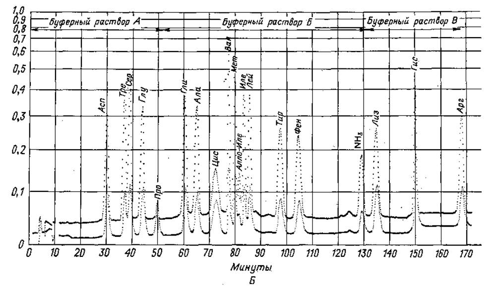
Fig. 36. Quantitative determination of amino acids on a single column. A. Two-buffer system. B. Three-buffer system.


Fig. 37. Chromatograms obtained during rapid programmed amino acid analysis.
A. Determination of Met and Lys. 1 — calibration mixture containing a 4-fold excess of Met and Lys; 2 — acid hydrolysate of plant seeds.
B. Determination of Trp. 1 — calibration mixture without Trp; 2 — calibration mixture containing 1 µmol of Trp.
Buffer flow rate |
100 ml/h |
Ninhydrin flow rate |
50 ml/h |
Analysis time |
180—190 min |
Buffer pressure at 55°C |
30—35 atm for solution A, 25—28 atm for solution D, 20—22 atm for solution E |
Notes. In this case, regeneration is optional. Alkaline treatment is performed as described above.
When determining acidic and neutral amino acids by the two-column method, the instrument is programmed in the same way as for the single-column method, but the analysis is stopped after the phenylalanine peak elutes.
With solutions C and D, such an analysis takes 2 hours. If the classical procedure using solution B is applied, this time increases to 3 hours; therefore, the latter option is hardly justified.
After the analysis, the column should be regenerated with 0.2 N NaOH and washed with the starting buffer solution (A).
Determination of basic amino acids on a short column [5]
Resin bed height in the column |
8—10 cm |
Buffer solution |
B |
Buffer flow rate |
68 ml/h |
Ninhydrin flow rate |
34 ml/h |
Buffer pressure at 55°C |
4—8 atm |
Analysis time |
about 80 min |
Notes. The duration of the analysis depends on the resin height in the column. Regeneration is required only after 10–15 analyses.
Rapid determination of methionine and lysine [4]
Resin height in the column |
14 cm |
Buffer solutions |
A and D |
Buffer flow rate |
100 ml/h |
Ninhydrin flow rate |
50 ml/h |
Buffer pressure at 55°C |
8—12 atm |
Analysis time, including regeneration |
75 min |
Programming |
0—25 min solution A, 25—62 min solution D, 62—63 min 0.2 N NaOH, 63—73 min solution A |
Notes. In many cases, especially in plant breeding and feed evaluation, it is sufficient to determine only the methionine and lysine content. The rapid analysis method allows for a whole series of screening experiments. On a short column in an analyzer using the program described above, methionine elutes as a separate peak between the valine and isoleucine peaks, and the lysine peak appears between the ammonia and histidine peaks.
9. Evaluation of results. The instrument manual typically describes how to evaluate the chromatogram, so we do not address this issue here.
10. Sources of error. An automatic amino acid analyzer is a rather complex instrument; when operating it, one should keep in mind that artifacts may occur due to the following reasons: a) mechanical malfunctions of the instrument, b) Chemical factors, c) operational inaccuracies.
Obviously, the Main sources of error may vary across different laboratories and types of instruments; therefore, below we will focus only on errors of a more general nature.
Observed malfunctions |
Possible cause |
Asp, Thr, Ser, Glu peaks overlap |
Column incorrectly equilibrated with a buffer of too high concentration). |
Glu and Pro peaks overlap or swap places |
Fault in the column temperature control system; the temperature is too low. Increased buffer solution pressure on the resin. |
Asymmetric peaks |
Resin contamination with heavy metals (the resin should be removed from the column and washed with EDTA and acid). Bacterial growth (hot alkali treatment required). Protein contamination (hot alkali treatment required). |
Unstable baseline, "spikes" on the chromatogram at each wavelength |
Air bubbles or floating particles in the flow Cell (e.g., resin beads). Flow cell holders are not secured in the spectrophotometer. Malfunction in the recorder electronic circuit (amplifier). |
Periodic baseline fluctuations (drift up and down) |
Pump malfunction, leak in the valves. |
Sudden drop in buffer solution pressure |
Faults in the buffer delivery system: a) air bubbles in the pump, b) puncture in the feed line, c) leak in the pump valve, d) rubber gaskets are worn or damaged. |
Sudden drop in ninhydrin solution pressure |
Air in the ninhydrin pump (ninhydrin depleted); puncture in the connecting line. |
Baselines at 570 and 440 nm swapped places |
Eluate-to-ninhydrin ratio has changed; one of the pumps is malfunctioning. |
Intermittent line appears on the recorder |
Poor contacts in the electronic tube system; oxidized contacts of the recorder microswitch. |
Appearance of an unusual peak on the chromatogram; most often, new components appear before ammonia and arginine |
Contaminated sample loading buffer. |
Baseline rise between ammonia and histidine peaks |
Ammonia in the buffer solution; contaminated distilled water. |
Peaks are too low |
Ninhydrin decolorized (reoxidation); "aged" lamps; heavily contaminated resin. |
Ammonia and lysine peaks overlap |
Heavily contaminated resin; damaged valve between the vessels with buffer solutions 2 and 3 |
No color reaction |
Malfunction in the system maintaining the required temperature of the reaction mixture; no ninhydrin. |
1. Benson J. V., Gordon М. J., Patterson J. A., Anal. Biochem., 18, 228.(1967).
2. D ev enyi T., Acta Biochim. Biophys. Acad. Sсi. Hung., 3, 429 (1968).
3. D ev enyi T., Acta Biochim. Biophys. Acad. Sci. Hung., 4, 297 (1969).
4. D ev enyi T., Acta Biochim. Biophys. Acad. Sci. Hung., 6, 129 (1971).
5. Spackman D. H., Stein W. H., Moore S., Anal. Chem., 30, 1181 (1958).
Last update: 06/08/2026
Editorial and Educational Adaptation: This material has been compiled based on the primary/original source text. The project team performed an editorial review, corrected technical inaccuracies, structured sections, and adapted the content for an educational format.
What was processed:
- elimination of formatting defects (OCR errors, structural breaks, corrupted characters);
- editorial organization of content;
- standardization of terminology in accordance with academic sources;
- verification of factual statements against the original source text.
All mentions of the author, publication year, and origin of the primary text have been preserved in accordance with the source.