Amino Acids, Peptides and Proteins - Dévényi T., Gergely J. 1976
Paper chromatography of amino acids and peptides
Principle of the method. Due to differences in partition coefficients, Amino Acids and Peptides can be separated on filter paper using Water-saturated organic Solvents in a closed chamber of appropriate shape and size.
Scope of application. Qualitative and semi-Quantitative determination of amino acids, peptide mapping (fingerprinting), micropreparative Separation and purification of peptides.
PROCEDURE
1. Chromatography chamber. For ascending chromatography, where the solvent front moves up the paper, a Glass cylinder of suitable size can be used. The maximum dimensions of the paper sheet are approximately 45 x 50 cm, so the height of such a cylinder should be slightly over 50 cm. The sheet of paper is rolled into a cylinder. To maintain this shape, the two ends of the sheet are sewn together with nylon thread. Typically, the roll diameter does not exceed 12–15 cm. With these dimensions, one can either perform fingerprinting of the material or obtain one-dimensional chromatograms of 8–10 different samples simultaneously in the same glass cylinder.
After applying a layer of vacuum grease to the rim of the cylinder, it is sealed with a glass plate, which is weighted down from above, for example, with a bottle filled with liquid. In such a hermetically sealed cylinder, an atmosphere uniformly saturated with solvent vapors can be created. This is maintained by the evaporation of liquid poured into a 50 ml vessel placed at the bottom of the cylinder inside the paper roll. Care should be taken to ensure that the paper does not Touch either the walls of the cylinder or the edges of the vessel containing the equilibrating liquid; otherwise, the solvent front will change its migration rate at the points of contact.
To obtain several chromatograms simultaneously under identical conditions, a rectangular chamber is more suitable, which can be used for both ascending and descending chromatography. For example, a wooden box with glass walls measuring 70 x 70 x 70 cm, with a strip of foam rubber attached along the upper edge, can be used as such a chamber. The chamber is covered from above with thick (heavy) glass to ensure the system is airtight. In ascending chromatography, the lower edge of the filter paper is clamped between two glass rods immersed in one of the glass troughs placed at the bottom of the chamber. For descending chromatography, plastic or glass troughs are required. The upper edge of the filter paper is immersed in such a trough located in the upper part of the chamber and secured there with a heavy glass rod.
Chromatography chambers of similar design are commercially available and can be used for A wide variety of purposes.
2. Selection of filter paper. For qualitative and semi-quantitative chromatographic analysis, Schleicher & Schuell 20436 and Whatman No. 1 papers are commonly used. Micropreparative Isolation of peptides is best performed on Whatman No. 3 and Whatman 3MM papers. The clearest peptide maps are obtained on Whatman 3MM paper.
In micropreparative isolation, special attention should be paid to the method of fraction elution. Whatman paper allows elution with distilled water, so the Background in the measured samples is quite low. At the same time, elution of fractions during chromatography on Schleicher & Schuell paper can only be carried out with a dilute solution of acetic acid, which leads to a very high background.
Class="center">Table 9 Composition of standard amino acid solutions
Solution A |
Concentration, mg/10 ml |
Solution B |
Concentration, mg/10 ml |
Cysteic acid |
70 |
59 |
|
62 |
69 |
||
Aspartic |
53 |
42 |
|
acid |
|||
30 |
Glutamic |
59 |
|
acid |
|||
47 |
35 |
||
46 |
Methionine sulfone |
59 |
|
72 |
Leucine |
54 |
|
Valine |
46 |
||
Phenylalanine |
55 |
3. Standard amino acid solutions. For qualitative and semi-quantitative chromatography of amino acids, two standard solutions are used, the compositions of which are given in Table 9. Due to the instability of Cysteine, cystine, and methionine, they are replaced in the standard solutions by their oxidized derivatives, i.e., cysteic acid and methionine sulfone.
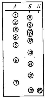
Fig. 38. Distribution of amino acids on paper during descending chromatography in a butanol system.
A and B — amino acid mixtures; H — neutral red; 1 — Cys SO3H; 2 — His; 3 — Asp; 4 — Gly; 5 — Thr; 6 — Pro; 7 — Phe; 8 — Lys; 9 — Arg; 10 — Met SO2; 11 — Ser; 12 — Glu; 13 — Ala; 14 — Tyr; 15 — Val; 16 — Leu.
The relative positions of various amino acids during chromatography of standard amino acid mixtures are shown in the chromatogram presented in Fig. 38. The spot labeled H contains neutral red. This indicator has an Rf value identical to that of leucine, and can therefore be used to monitor when leucine—the fastest-migrating component of the standard mixture—reaches the lower edge of the chromatogram during descending chromatography.
4. Sample application. The test sample can be applied to the chromatography paper using a micropipette, a graduated capillary, or by the so-called polyethylene film method.
For semi-quantitative analysis, a precisely known volume of the test solution must be applied to the chromatography paper. This is most easily and accurately done using a micropipette. Several types of commercially available micropipettes have fixed or adjustable volumes. Since commercially manufactured micropipettes are quite expensive, researchers often use homemade capillaries, which can be prepared as follows.
Thin capillaries about 12 cm long are drawn from glass tubes or test tubes softened in a flame and calibrated to 0.05 or 0.01 ml. Degreased and thoroughly dried capillaries are calibrated using a graduated 0.1 ml micropipette. To do this, exactly 0.05 or 0.01 ml of water is transferred from the micropipette into the capillary, and THE POSITION OF the meniscus is marked on the Capillary Wall with a glass-marking pencil or colored paste. With some experience, this calibration can be done quite accurately. It is important to ensure that the pipette tip is clean, the capillary itself is degreased, and both the capillary and the micropipette are held horizontally during the water transfer.
In a unique method of applying the test material to chromatography paper using a polyethylene film, several depressions are made in the film with the bottom of a flame-heated glass test tube. Approximately 0.05 ml of the test material, such as an acid protein hydrolysate, is introduced into each depression. Three to five samples can be placed on a single film (Fig. 39). Very carefully, avoiding smearing the drops, the film is transferred to a vacuum desiccator for drying. After about 10–15 min, when the drops have dried, the film is removed, and a small drop of water is applied to the edge of each dried spot using the pointed end of a glass rod, which is used to redissolve all the material in that drop. A sheet of chromatography paper is placed on clean glass, and the application spots are marked with a pencil. The polyethylene film with the test material is then applied to this area of the paper and pressed against the paper from the back with a finger. If the wetted paper area does not exceed 1 cm2, the chromatography will result in well-resolved, regularly shaped small spots. This application method has major advantages: 20–30 samples can be applied without loss within a few minutes. This eliminates the need to prepare and wash capillaries, use hot-air drying during application, etc.
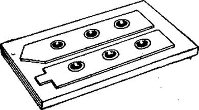
Fig. 39. Strips of polyethylene film with test samples.
Samples are applied to the starting area of the chromatogram using a capillary pipette or as a print.
5. Micropreparative chromatography. The material to be fractionated is applied to Whatman No. 3 or Whatman 3MM paper along the starting line, which should not exceed 20 cm in length. A maximum of 3–4 mg can be applied per 10 cm. Upon completion of chromatography, the chromatogram is dried, 0.5 cm wide strips of paper are cut from both sides and stained to locate the zones to be eluted (see Fig. 17); these zones are then cut out and the fractions are eluted.
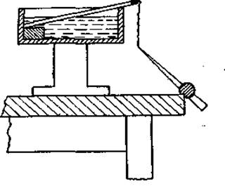
Fig. 40. Elution of components from filter paper with small volumes of solvent using a capillary (for a detailed explanation, see text).
For elution, one end of the cut-out section is clamped between two glass plates immersed in a trough containing distilled water. Once the entire paper is wet, a capillary tube, secured with plasticine, is brought to the lower pointed end. The eluate begins to flow slowly into the capillary (Fig. 40). If the capillary length is 30 cm, the volume of liquid filling it is quite sufficient to elute a chromatogram strip measuring 1x8 cm. The eluate is transferred from the capillary into a small 2–3 ml weighing bottle for drying in a vacuum desiccator.
Regardless of the chosen method of sample application (using a micropipette, capillary, or polyethylene film), the test material must be as concentrated as possible and distributed over a minimal area. If the starting spot diameter is too large or the application zone is too wide on a one-dimensional preparative chromatogram, it is quite difficult to achieve good separation, as the fractions will form large spots and zones, and adjacent components may overlap. Ideally, the sample should be applied as a single point. While this is hardly possible, quite sharp chromatograms can be obtained if the starting spot diameter does not exceed 5–6 mm and the starting zone is no wider than 2–3 cm. During application, it is often difficult to manage without special devices, such as a commercially available sample application table. To dry the samples during application, the simplest method is to use an ordinary Hair dryer. Hot air directed at the paper rapidly evaporates the water in the applied material. This allows for repeated Applications without increasing the starting spot diameter by more than a few millimeters or the starting zone width by more than a few centimeters.
6. Solvents. A great variety of solvents used for qualitative and semi-quantitative analysis, as well as for micropreparative purposes, have been described in the literature. Here we present only those that can be successfully applied in many cases.
Butanol — water — acetic acid (120:50:30) (by volume; homogeneous system, requiring no equilibrating solvent) |
This solvent can be used with equal success for qualitative and semi-quantitative analysis of amino acids, micropreparative separation, and peptide mapping. Freshly prepared solvent can be used immediately; it should not be stored for more than 10—12 days, as Esterification occurs after this period. |
Isoamyl alcohol — pyridine — water (35:35:30) (by volume; homogeneous system, requiring no equilibrating solvent) |
First, isoamyl alcohol is mixed with pyridine, and water is added dropwise to this mixture with constant stirring. If the resulting solution is opalescent, it should be filtered. Opalescence reappears after 8—10 days, at which point the solvent becomes unusable. This is one of the best mixtures for peptide mapping and micropreparative chromatography. |
The solvents listed below can be used for a combination of electrophoretic and chromatographic separation of the test material: butanol—pyridine—acetic acid—water (60 : 40 : 12 : 48); water saturated with phenol in an NH3 atmosphere; sec-butyl alcohol — 3% ammonia solution (3 : 1); m-cresol — phenol saturated with water (1 : 1); isobutanol—water—formic acid (695 : 295 : 10); isobutanol—methyl ethyl ketone — water (40 : 30 : 20); n-propanol—water (70 : 30); ethanol—water (77 : 23).
7. Identification of amino acids and peptides in fractions. A. Ninhydrin reaction. Ninhydrin reacts with all amino acids containing an a-NH2 group, yielding a purple color, except for proline or hydroxyproline, which react with ninhydrin to give a yellow color. Certain impurities (copper, cadmium, etc.) can affect the color of the spots. For developing chromatograms, the so-called cadmium-ninhydrin is highly convenient; its reaction with amino acids is quite sensitive and leads to The Development of a stable red color, and collidine-ninhydrin is also useful, reacting selectively with Certain amino acids and allowing them to be differentiated by spot color.
1) Preparation of cadmium-ninhydrin. 1.0 g of ninhydrin is dissolved in 100 ml of acetone. 100 mg of cadmium acetate is dissolved in 10 ml of distilled water, and 5 ml of acetic acid is added. Both resulting solutions are combined and stored in a dark glass bottle.
The dried chromatogram is drawn through the prepared dye solution poured into a flat tray and dried in the air in the dark. At room Temperature, maximum color development occurs in about 2 h.
2) Preparation of collidine-ninhydrin. 5.0 g of ninhydrin is dissolved in 955 ml of acetone, then 25 ml of acetic acid and 20 ml of collidine are added.
B. Detection of histidine by the Pauly reaction.
1) Preparation of Pauly's reagent. Solution I: 50.0 g of sulfanilic acid is dissolved in 250 ml of 10% KOH solution, and 200 ml of 10% NaNО2 solution is added. Solution II: to 80 ml of concentrated HCl, 40 ml of distilled water is added.
To Solution II, cooled to 0°C, Solution I is added dropwise with cooling and constant stirring. The resulting precipitate is filtered off and dried at room temperature.
2) 0.1 g of Pauly's reagent is dissolved in 100 ml of 20% Na2CО3 solution, and the chromatogram is sprayed with the freshly prepared solution. To prevent the aqueous alkaline solution from smearing the spots and zones on the chromatogram, fine nebulization of the reagent and uniform spraying must be achieved. Histidine-containing peptides and free histidine yield a red color on a yellow background. Tyrosine-containing peptides and free tyrosine yield a purple or gray-blue color, but unlike the histidine-containing spots, their spots fade rapidly.
C. Detection of arginine and arginine-containing peptides by the Sakaguchi reaction. Solution I: 0.01% a-naphthol in 95% ethanol containing 5% urea. Solution II: 2.0 g of bromine in 100 ml of 8% NaOH solution. Before use, a few pellets of NaOH are added to Solution I, and after they dissolve, the chromatogram is sprayed with the reagent. The paper is then dried and sprayed with Solution II. Arginine and arginine-containing peptides turn red.
D. Detection of proline and hydroxyproline. 100 mg of isatin is dissolved in 50 ml of butanol, and 5 ml of acetic acid is added. The chromatogram to be developed is sprayed with this solution and dried at 40°C for 5 min. Proline and peptides containing N-terminal proline yield an intense blue color. Phenylalanine and Tyrosine turn greenish-blue, which interferes with the detection of proline and hydroxyproline.
E. Detection of Tryptophan. 0.5 g of p-dimethylaminobenzaldehyde is dissolved in 100 ml of 95% ethanol containing 2 ml of concentrated HCl. Tryptophan yields a purple color with the resulting solution.
F. Detection of tyrosine. 0.1 g of a-nitroso-ß-naphthol is dissolved in 100 ml of 75% ethanol. After spraying with this solution, the chromatogram is dried and sprayed with a 10% nitric acid solution. It is then dried again and kept at 90°C for 3 min. Tyrosine and tyrosine-containing peptides yield a red color on a pale green background.
G. Detection of glycine. 1) 0.2 g of phthalaldehyde is dissolved in 100 ml of acetone; 2) 1.0 g of KOH is dissolved in 100 ml of 96% ethanol.
The chromatogram is drawn through Solution 1 and dried at 100°C, then drawn through Solution 2 and dried again at 100°C for 10 min. As a result of the reaction, glycine yields a green color.
3. Detection of N-acyl derivatives and large peptides by reaction with chlorine. The dry chromatogram or electrophoregram is kept in a glass cylinder in a chlorine atmosphere for 30 min, then removed from the cylinder and sprayed with a 1% starch solution containing 1% KI. Compounds with peptide bonds in the molecule appear as dark blue zones or spots.
Recommended Literature
Zweig G., Whitaker J. R., Paper Chromatography and Electrophoresis. Vol. II, Academic Press, New York, London, 1971.
Last update: 06/08/2026
Editorial and Educational Adaptation: This material has been compiled based on the primary/original source text. The project team performed an editorial review, corrected technical inaccuracies, structured sections, and adapted the content for an educational format.
What was processed:
- elimination of formatting defects (OCR errors, structural breaks, corrupted characters);
- editorial organization of content;
- standardization of terminology in accordance with academic sources;
- verification of factual statements against the original source text.
All mentions of the author, publication year, and origin of the primary text have been preserved in accordance with the source.