INVERTEBRATE ZOOLOGY IN THREE VOLUMES - BOOK 2 - H.Y. Shcherbak - 1996
PHYLUM ARTHROPODA
SUBPHYLUM TRACHEATA
CLASS CHILOPODA
Centipedes, like other myriapods, lead a secretive lifestyle, inhabiting soil, leaf litter, decaying wood, under stones, etc., though some species can spend prolonged periods in Water. All centipedes are predatory. Over three thousand species have been described, with no more than 50 known in Ukraine.
Centipede sizes range from a few millimeters to 26.5 cm (Scolopendra gigantea). As a rule, larger species inhabit tropical and subtropical regions. Centipedes are typically yellowish or brown, although some scolopendrids are greenish or, rarely, blue.
The body of centipedes is noticeably dorsoventrally flattened.
Like other myriapods, it is divided into a distinctly separated HEAD and a segmented trunk. The head cuticle forms a head capsule. The head bears a pair of bead-like antennae and simple or compound eyes (sometimes absent). On the ventral side of the head, There is a pair of mandibles and two pairs of complex maxillae. The mouthparts also include the first pair of trunk appendages, which have transformed into poison claws (Fig. 96). Each of them ends in a sickle-shaped claw, near the tip of which opens the duct of a venom gland. The poison claws participate in capturing and holding prey, and are also used for defense against enemies.
The trunk can consist of 15–180 more or less homonomous segments. All trunk segments, except the last two, bear a pair of walking legs, which vary in Structure among representatives of different orders.
Class="center">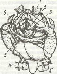
Fig. 96. Mouthparts of Lithobius forficatus (Chilopoda):
1 - antennae; 2 - Mandible; 3 - Maxilla; 4 - trunk leg; 5 - poison claw; 6 - maxilla
The sclerotization of the integument in different body regions of centipedes varies; however, apparently, all lack the waxy layer of the epicuticle, which causes centipedes to be highly dependent on the humidity of their habitats. The integument of the head, the posterior end of the body, and the appendages is well sclerotized, while the body segments are covered by separate sclerites, especially laterally (sometimes numbering more than 10), between which there are significant areas of non-sclerotized cuticle. Besides their protective function, the sclerites also act as an exoskeleton.
The structure of the trunk musculature in certain groups (Geophilomorpha, Scolopendromorpha) is characterized by the partial retention of a continuous layer of circular and longitudinal Muscles. Longitudinal muscles are often divided into bundles extending from one segment to the next or to the following one. The deepest positions are occupied by dorsoventral and pleurosternal bands (Fig. 97). The trunk muscles in these centipedes, together with appendage muscles, participate in locomotion.
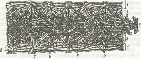
Fig. 97. Trunk musculature of Scolopendra sp.:
1, 2, 3 - ventral, lateral, and dorsal parts; 4 - longitudinal dorsal muscles; 5 - dorsoventral muscles; 6 - 8 - ventropleural, dorsopleural, and longitudinal ventral muscles, respectively
The Mouth opening is located on the ventral side of the head, flanked laterally by sickle-shaped mandibles and ventrally by the first maxillae. The second pair of maxillae does not directly participate in food fragmentation and is used solely for holding prey.
The digestive tract resembles a straight tube (Fig. 98, a), which in various centipedes differs only in the relative length of each of its three sections. For instance, in most Scolopendromorpha, the anterior ectodermal region accounts for nearly two-thirds of the total gut length. Salivary Glands also belong to the Digestive System, numbering from three to five pairs. The ducts of the salivary glands open into the Oral Cavity or on the second pair of mandibles.
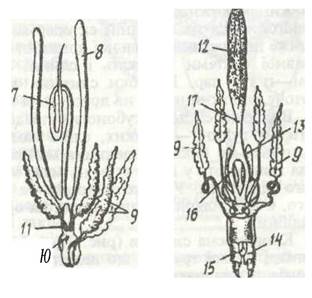
Fig. 98. Internal Organs of centipedes:
a - digestive system of Lithobius forficatus; b, c - male and female reproductive systems, respectively; 1 - Esophagus; 2 - salivary gland; 3 - Malpighian tubule; 4 - anus; 5 - hindgut; 6 - midgut; 7 - Testis; 8 - Seminal Vesicle; 9 - accessory gland; 10 - ejaculatory duct; 11 - paired part of the vas deferens; 12 - Ovary; 13 - seminal receptacle; 14 - gonopod; 15 - gonopore; 16, 17 - paired and unpaired PARTS OF THE oviduct
The excretory system of centipedes is represented by a pair of Malpighian tubules—thin, blindly ending tubes extending along the sides of the gut almost to the anterior end of the body and emptying into the gut at the boundary between the Midgut and Hindgut. In Lithobiomorpha and Scutigeromorpha, maxillary organs resembling the excretory organs of crustaceans are also present.
The Circulatory system (Fig. 99) is well developed. The Heart, in the form of a long tube lying above the gut along the trunk, is suspended from the body walls by specialized alary muscles. It is blindly closed at the posterior end. Corresponding to the segments, the heart is divided into chambers, each with two lateral ostia. Anteriorly, the heart continues into the main aorta, which reaches the Brain. On the sides of the aorta, an arterial ring branches off, encircling the gut and flowing into the ventral vessel. In addition to the ostia leading to the pericardial cavity, two lateral Arteries depart from each chamber.
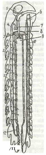
Fig. 99. CIRCULATORY SYSTEM OF Scutigera sp. (diagram of a lateral section):
1 - heart ostia; 2 — "tracheal Lungs"; 3 - arterial ring; 4 - aorta; 5 - brain; 6 - ventral nerve cord; 7 - esophagus; 8 - ventral vessel; 9 - midgut; 10 - lateral arteries; 11 - hindgut; 12 - hindgut artery
All vessels branching off from The Heart and the ventral vessel branch out to a greater or lesser extent. Blood flows from them into lacunae, from which it returns to the pericardial cavity, and then enters the heart through the ostia. Within the cardiac vessel, blood moves from the posterior end to the anterior, while in the ventral vessel it flows in the opposite direction.
The STRUCTURE OF THE tracheal system varies somewhat among representatives of different orders, but it is well developed in all of them. The tracheae are highly branched, with interconnections existing between the tracheal bundles of adjacent segments.
The Nervous system has a typical arthropod structure: it consists of the brain, circumesophageal connectives, subesophageal ganglion, and ventral nerve cord (Fig. 100).
Fig. 100. Nervous system of Lithobius forficatus (dorsal view):
1 - antennal nerve; 2 - supraesophageal ganglion; 3 - optic nerves; 4 - circumesophageal connective; 5 - subesophageal ganglion; 6 - ganglia of the ventral cord; 7 - connectives; 8 - appendage nerves
In chilopods, the sense of Touch and smell are associated with the antennae. Simple ocelli are present, located on the sides of the head in varying numbers (ranging from two to four up to large clusters resembling insect compound eyes).
Chilopods, like all myriapods, are dioecious animals. The Gonads are unpaired. In the male, a long testis lies above the gut, its posterior end transitioning into a small expansion whose walls form two sac-like Seminal Vesicles (see Fig. 98). Two Branches of the vas deferens extend from this expansion, loop around the gut, and join beneath it into an unpaired ejaculatory duct, into which the ducts of two pairs of accessory glands open. The genital opening is located on the penultimate segment. The FEMALE Reproductive System has a similar structure. The sac-like ovary transitions into an unpaired oviduct that bifurcates around the intestine. Beneath the gut, its branches reunite, and the ducts of two pairs of accessory glands and a pair of spermathecae empty into this unpaired section. The genital opening, as in the male, is situated on the penultimate segment. Fertilization is spermatophoric.
The eggs of chilopods are rich in yolk; therefore, Cleavage is meroblastic and superficial. Postembryonic development proceeds via two pathways: in some chilopods (Geophilus, Scolopendra), epimorphosis occurs, where a young individual hatches from the egg with all segments and legs already present. In the rest, development involves anamorphic stages: the animal hatches with an incomplete set of segments and 12 pairs of trunk legs, with the number of segments increasing during molts. Many representatives of this class exhibit parental care.
The class Chilopoda comprises five orders. One of them (Craterostigmomorpha) unites species found exclusively on the islands of Oceania and in Australia, while the other four are quite widespread, including in Ukraine, and it is these that we will examine.
Order Geophilomorpha. Representatives of this order can be found under fallen leaves, stones, tree bark, etc., but as their name implies, they most frequently inhabit the soil, where they live and hunt various invertebrates.
Their size is generally small (1–4 cm), but larger species exist, such as the thermophilic Orya barbarica, which reaches up to 22 cm in length; incidentally, this species is capable of intense Bioluminescence, much like certain others.
The body is more elongated and worm-like than in other chilopods, and in most species, it is ribbon-like and slender (Fig. 101).
Fig. 101. Geophilomorphs (Geophilus longicornis)
The head is relatively smaller than in other chilopods, heavily sclerotized, and always darker than the trunk. In some species, it tapers toward the anterior end. Eyes are absent.
The number of trunk segments ranges from 31 to 173. Unlike other chilopods, all segments, except for the modified first and the two posterior ones, share a uniform structure. Each of them bears a pair of spiracles located on the sides of the trunk near the Base of the legs. It should be noted that individuals of certain species possess a varying number of legs in adulthood; for instance, in Himantarium gabrielis, there may be from 133 to 173 pairs. The legs are relatively short and weak, allowing the animal to move very slowly. The last pair of legs resembles antennae in Structure and function: in narrow soil passages where geophilomorphs cannot turn around, they begin to move backward with the posterior end forward, probing their path with the hind legs.
The presence of a well-developed cutaneous-muscular sac enables geophilomorphs to move similarly to earthworms, allowing them not only to traverse existing passages and crevices in the soil but also to burrow their own. In this process, the circular muscles in the anterior third of the body contract, elongating it and pushing the head into the soil; then, the longitudinal muscles in this section contract, causing it to widen, while the narrowed posterior part of the body is simultaneously pulled forward. Backward slippage of the anterior body region is prevented by the legs bracing against the soil.
Geophilomorphs prey on earthworms—which are frequently much larger than themselves—as well as various insect larvae, while the species Scolioplanes acuminatus prefers heavily sclerotized millipedes. In doing so, they flip the prey onto its back, consuming the head first and the trunk afterward. Alongside animal matter, some species may occasionally feed on decaying succulent plant roots, such as sugar beet ROOT crops.
During reproduction, the male deposits a spermatophore or a drop of sperm onto silk threads woven through soil tunnels. Females pick them up using specialized modified appendages (gonopods) located near the genital opening. After laying 15–30 eggs in a microhabitat with near-saturation humidity, the female coils her body around the clutch and remains in this position until the young hatch. Without contact with the female's secretions, the eggs perish from putrefactive Bacteria.
Geophilomorphs are highly sensitive to moisture deficits. They use their antennae to locate environments with a humidity exceeding 85%.
Antennal amputation leads to a loss of behavioral response to desiccation. Despite lacking eyes, geophilomorphs exhibit a clear negative phototaxis.
Some species of geophilomorphs, such as Scolioplanes maritimus, have adapted to a truly marine lifestyle in the coastal zone. They inhabit rocky shores in the intertidal zone, where they safely remain under stones during high tide. At this time, air bubbles are clearly visible near their stigmata, mouth, and anal openings, apparently facilitating continued gas exchange. During low tide, geophilomorphs become highly active and prey on Mollusks, various crustaceans, and other small animals.
The most common species in Ukraine are Arctogeophilus macrocephalus and Pachymerium ferrugineum, which are predominantly found in deciduous and pine forests.
Order Scolopendromorpha. This group includes centipedes of medium and small size, such as Cryptops hortensis (15 mm long), as well as the largest chilopods, such as Scolopendra gigantea.
Their body is more robust than that of geophilomorphs, and the trunk typically consists of 25 segments bearing 21 pairs of legs (Fig. 102).
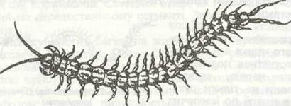
Fig. 102. Scolopendromorphs: Scolopendra cingulata
Their coloration is much more diverse than in other centipedes. Alongside yellowish and brown species, many are olive-green, green, or blue. Furthermore, the head and trunk may feature contrasting solid colors or distinct patterns.
The head is relatively small and dorsoventrally flattened. The antennae consist of 17 to 36 segments. Eyes are present and composed of several simple ocelli.
The trunk segments differ from one another primarily in the presence or absence of spiracles. The legs are well-developed, enabling these animals to run swiftly. In además to locomotion, the legs also assist in capturing and holding prey. The last pair of legs is structurally specialized: they are hook-shaped, heavily thickened, and often bear long, awl-like appendages. These serve as weapons for attacking prey and defending against predators; in addition, the centipede uses its rear legs to anchor firmly to the substrate when subduing large prey.
Scolopendromorphs most commonly inhabit areas beneath large, flat stones, where they tunnel horizontal passages and excavate rounded, fairly spacious living chambers. Like geophilomorphs, they achieve movement through the antagonistic contraction of circular and longitudinal muscles.
Scolopendras feed on earthworms, insect larvae, and other invertebrates. Larger species, such as Scolopendra gigantea, prey on small birds, lizards, and frogs. In addition to animal prey, they also consume succulent fruits.
Order Lithobiomorpha. Lithobiomorphs (Fig. 103) typically inhabit forest litter and herbaceous plant debris in meadows and steppes, and can also be found beneath tree bark and in moss; many species dwell near water or in caves.
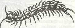
Fig. 103. Lithobiomorphs: Lithobius forficatus
Sizes range from 3 mm in the dwarf species Catanopsobius chilensis to 51 mm in Polybothrus fasciatus.
The coloration varies from yellowish to brown, featuring one to three longitudinal stripes along the dorsal side.
Externally, lithobiomorphs resemble scolopendras, but they differ in having a larger head, a shorter trunk consisting invariably of 18 segments, and relatively longer legs. Legs are present on all segments except the first and the final two.
The antennae have 13 to 100 segments; the eyes, when present, share the same structure as those of scolopendras.
The ventral structure of all segments is uniform; on the dorsal side, narrow tergites alternate with wide ones. The spiracles open on the ventral side, and their number ranges from two to seven pairs.
Lithobiomorphs are agile runners. At night, they hunt various small insects, spiders, and other invertebrates. They also ambush prey in shelters where victims likewise hide from sunlight.
The species most widely distributed in the fauna of Ukraine is Lithobius forficatus, which is frequently encountered near human dwellings, including urban areas. In forest litter, Monotarsobius curtipes is most commonly found.
Order Scutigeromorpha. This group comprises thermophilic centipedes inhabiting tropical and subtropical Regions of the world. Only about 150 species have been described, of which the fauna of Ukraine includes a single species, the house centipede (Scutigera coleoptrata), found on the southern coast of Crimea. It is currently listed in the Red Data Book of Ukraine.
Scutigeromorphs live in rock crevices, under stones, in conifer and deciduous leaf litter, and in various other shelters, including human buildings. They are small in size, ranging from 2.5 to 4.5 cm.
Unlike other chilopods, representatives of this order possess a rounded rather than flattened head (Fig. 104). Their long antennae consist of over 400 segments, and their large eyes strongly resemble the compound eyes of insects—a feature entirely absent in other centipedes.
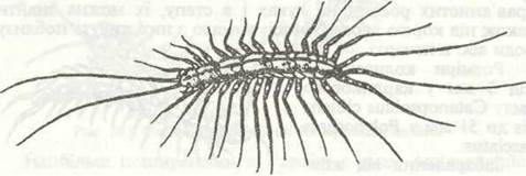
Fig. 104. Scutigeromorpha: Scutigera coleoptrata
The trunk consists of 18 segments whose external structure is similar to that of Lithobiomorpha, but the narrow tergites are nearly reduced, and the tergites of segments VII and VIII are fused into a single plate, leaving only eight tergites visible on the dorsal surface.
There are always 15 pairs of legs. They are long (hence Germans refer to the scutiger as a "spider-like" centipede) and terminate in a multi-jointed, flexible tarsus.
Unlike all other chilopods, the spiracles open on the broad tergites of the dorsal side. They lead into air chambers from which numerous tracheal trunks branch out, forming up to 600 tracheal tubules in each segment. The tracheae envelop The surface of the cardiac vessel, thereby oxygenating the blood and distributing it to every organ.
During reproduction, the male deposits a spermatophore directly onto the substrate, which is subsequently picked up by the female. Eggs are laid singly, eventually hatching into juveniles with an incomplete number of legs.
Scutigeromorpha move extremely rapidly, almost at lightning speed (up to 50 cm/s), across open surfaces (including building walls), preying on various Arthropods, especially insects and spiders—hence their common name, house centipedes.
Human attitudes toward scutigeromorphs are mixed: on the one hand, they are valued for eliminating large numbers of flies and various blood-sucking insects indoors; on the other hand, they often provoke revulsion due to the unfounded belief that they can bite humans.
Last update: 13/08/2026
Editorial and Educational Adaptation: This material has been compiled based on the primary/original source text. The project team performed an editorial review, corrected technical inaccuracies, structured sections, and adapted the content for an educational format.
What was processed:
- elimination of formatting defects (OCR errors, structural breaks, corrupted characters);
- editorial organization of content;
- standardization of terminology in accordance with academic sources;
- verification of factual statements against the original source text.
All mentions of the author, publication year, and origin of the primary text have been preserved in accordance with the source.