Molecular Biology of the Cell - Volume 1 - Alberts B., Bray D., Lewis J., Raff M., Roberts K., Watson J. 1994
Introduction to Cell Biology
How Cells Are Studied
Studying Cellular Macromolecules with Antibodies and Radioactive Isotopes
Virtually all properties of molecules—physical, chemical, and biological—can be used to study cellular macromolecules. In biological research, molecules inside Cells are usually detected by their optical properties (either in their pure form or complexed with Dyes), as well as by their biochemical activity. Here we will consider two Methods for identifying molecules inside cells: one involving The Use of radioactive isotopes, and the other, the use of Antibodies. Both methods are highly effective for detecting specific molecules in complex mixtures. Potentially, these methods are extremely sensitive and, under optimal conditions, make it possible to detect molecules in a sample whose total number is less than 1000.
4.5.1. Methods for detecting radioactive atoms are highly sensitive [31]
Most known natural elements are mixtures of isotopes that differ in the mass of their atomic Nucleus but nevertheless have the same set of electrons and, consequently, identical chemical properties. The nuclei of radioactive isotopes, or radioisotopes, are unstable and undergo spontaneous decay to form different atoms. When a nucleus decays, charged particles (such as electrons) or radiation (such as gamma rays) are emitted.
Because of their instability, radioisotopes are rare in nature, but they are produced in great Abundance in nuclear reactors, where stable atoms are bombarded with high-energy particles (Table 4-11). Today, many biological molecules are available in forms containing radioactive atoms. Various approaches are used to detect the radiation emitted by radioactive isotopes. Electrons (ß-particles) can be detected by the gas ionization they cause in a Geiger counter, or in a scintillation counter by the tiny flashes of light they produce in a scintillation fluid. These methods can reveal the presence of a specific radioactive isotope in a biological sample. The presence of radioactive isotopes in a sample can also be recorded by autoradiography (by their effect on silver grains in a photographic emulsion). This method is extremely sensitive, and under favorable conditions, it can record virtually every decay, meaning that almost every radioactive atom can be accounted for.
4.5.2. Radioactive isotopes are used to study the movement of molecules in cells and in the whole Organism [32]
One of the earliest Examples of using The phenomenon of radioactivity in biological research was The Study of carbon pathway in Photosynthesis. Unicellular green Algae were placed in an atmosphere containing radioactively labeled CO2 (14CO2) and exposed to sunlight for various periods of time. The radioactive Contents of the algae were then fractionated using paper Chromatography. Small molecules containing 14C atoms derived from CO2 molecules were detected on the chromatogram by placing a sheet of photographic film over the dried paper chromatogram. In this way, most of the major intermediates formed during the photosynthesis of sugars from CO2 were identified.
Radioactive molecules can be used to study virtually all intracellular processes. To do this, a precursor in radioactive form is typically added to the culture medium during the experiment; the radioactive molecules then mix with the non-radioactive ones present in the cells. The Cell uses both types of molecules because they differ only in the mass of their atomic nuclei. The change in the localization of radioactive molecules in the cell or their chemical transformations can be tracked over time. The sensitivity of such experiments is often increased by using the pulse-chase method. In this method, radioactive substances are added for a very short time (pulse), then removed and replaced with non-radioactive molecules (chase). Samples are taken at various time intervals, and at each point, the chemical nature and localization of the chemicals are determined (Fig. 4-54).
The value of the radioactive labeling method can hardly be overstated. It is this method that makes it possible to discriminate between chemically identical molecules with different histories—for example, those molecules that differ in their time of synthesis. Using radioactive methods, it was determined that almost all molecules in a living cell are constantly being broken down and replaced by other molecules. Such slow turnover processes might have gone unnoticed were it not for radioactive isotopes.
Today, industry produces virtually all common low-molecular-weight substances in radioactive form. Regardless of The complexity of biological molecules, almost any of them can be radioactively labeled. Radioactive molecules are often prepared with radioactive atoms introduced at specific positions in their Structure. This is done to make it possible to follow the independent fates of different parts of a single molecule during biological reactions (Fig. 4-55).
Class="center">
Fig. 4-54. Diagram illustrating The Essence of a typical pulse-chase experiment. The letters A, B, and C label reservoirs that correspond to different cell compartments (detected by autoradiography or in Cell Fractionation experiments) or to different chemical compounds (detected chromatographically or by other chemical methods).
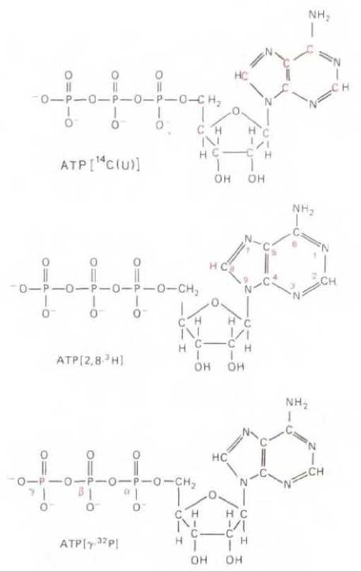
Fig. 4-55. Three commercially available radioactive forms of ATP. Radioactive atoms are highlighted in color. The notations used to indicate the position and type of radioactive atoms are shown.
One of the most important Applications of radioactive isotopes in cell biology is determining the localization of radioactive compounds in sections of cells or living Tissues by autoradiography. In this method, living cells are subjected to brief (pulse) labeling, followed by incubation for various periods of time in a non-radioactive medium. The cells are then fixed and processed for light or Electron Microscopy. Each prepared specimen is coated with a thin layer of photographic emulsion and left in the dark for several days—the time during which the radioactive isotope decays. The emulsion is then developed. The Location of radioactive molecules in each cell can be determined by THE POSITION OF dark silver grains. If cells are incubated with a radioactive DNA precursor (3H-thymidine), it can be seen that DNA is synthesized in The Nucleus and remains there. Conversely, labeling cells with a radioactive RNA precursor (3H-uridine) shows that RNA is initially synthesized in the nucleus and then rapidly accumulates in the Cytoplasm of the cells.
4.5.3. Antibodies can be used to detect and isolate specific molecules [33]
Antibodies are Proteins produced by vertebrates as a defense against infection (see Chapter 18). The number of different antibody forms reaches millions; this is what distinguishes antibodies from other proteins. Each antibody form has specific binding sites designed to recognize the specific molecules that stimulated the antibody synthesis. These molecules are called Antigens. The high Specificity of antibodies for their antigens makes them a powerful tool for cell biology research. After labeling antibodies with fluorescent dyes, they can be used to determine the Intracellular Localization of specific macromolecules using Fluorescence Microscopy (Fig. 4-56). Labeling with electron-dense microparticles, such as colloidal gold microspheres, allows antibodies to be used to localize cellular antigens by electron microscopy (Fig. 4-57). Antibodies can act as biochemical links to detect and quantify molecules in cell extracts and to identify specific proteins after their Separation by Polyacrylamide gel Electrophoresis. Coupling antibodies to an inert matrix yields affinity columns suitable for isolating and purifying specific molecules from crude cell extracts. The sensitivity of antibodies used as probes to detect specific macromolecules in Cells and Tissues is often increased by signal Amplification methods. For example, a marker molecule such as a fluorescent dye can be directly coupled to antibodies and used for direct antigen detection (primary antibodies). Even greater signal amplification can be achieved by using unlabeled primary antibodies and then detecting them with labeled secondary antibodies that bind to the primary antibodies (Fig. 4-58, A).
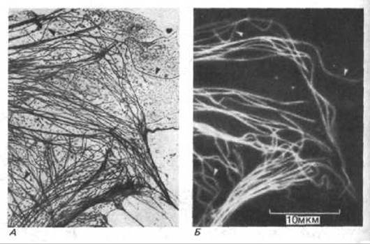
Fig. 4-56. A. In the electron micrograph of the peripheral region of a cultured epithelial cell, the arrangement of microtubules and other filaments can be distinguished. B. Using indirect immunocytochemistry, the same region is stained with fluorescent antibodies to tubulin, which is the monomer of microtubules (see Fig. 4-58). Arrows indicate individual microtubules that are clearly visible in both micrographs (Osborn M., Webster R., Weber K., J. Cell Biol., 77, R27-R34, 1978, reproduced with permission of Rockefeller University Press.)
Another signal amplification system is based on the exceptionally high affinity of biotin (a low-molecular-weight soluble vitamin) for streptavidin (a bacterial protein). When primary antibodies are covalently linked to biotin, streptavidin can be directly labeled with a marker and used instead of secondary antibodies. Streptavidin can also be used to link individual biotin-labeled antibody molecules to a branched network of biotin-labeled molecules (Fig. 4-58, B). Such networks are obtained by modifying the method (Fig. 4-58, A) through the use of a Third Layer of antibodies.
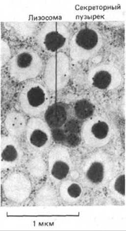
Fig. 4-57. Immunocytochemical localization of specific protein molecules in electron micrographs using labeling with antibodies coupled to colloidal gold particles. A thin section of an Insulin-secreting cell is shown, where insulin molecules are labeled with anti-insulin antibodies coupled to tiny gold microspheres (each appearing as a black dot). Most of the insulin accumulates in the dense core of secretory granules; in addition, the contents of some secretory granules degrade in Lysosomes. (L. Orci, Diabetologia, 28, 528-546, 1985.)
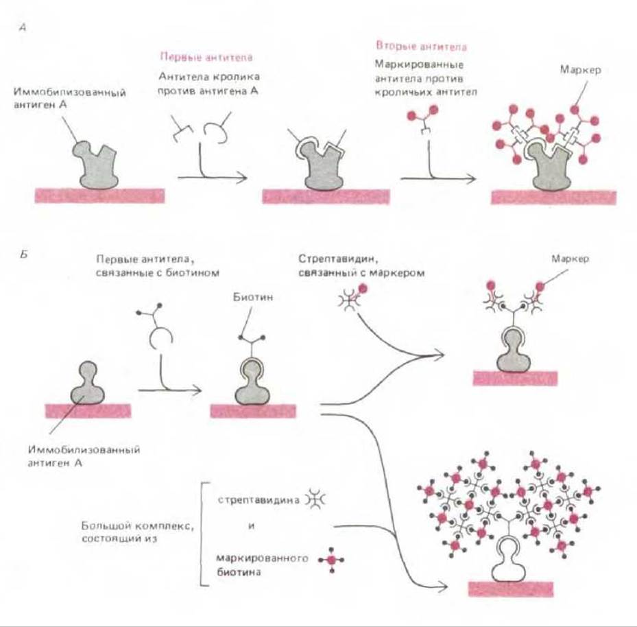
Fig. 4-58. Use of antibodies to detect specific molecules with high sensitivity. A. Illustration of the indirect immunocytochemistry method, whose high sensitivity is due to the fact that the primary antibody (the antibody molecule that directly binds to the recognized antigen molecule) is recognized by multiple molecules of a secondary antibody. These secondary antibodies are pre-coupled to marker molecules, allowing their detection. Marker molecules include the dyes fluorescein and rhodamine (for fluorescence microscopy), the enzyme horseradish peroxidase (for electron and light microscopy), the iron-containing protein ferritin, or colloidal gold microspheres (for electron microscopy), and the enzyme alkaline phosphatase (for biochemical detection). B. Modification of the method shown in A: instead of secondary antibodies, the high-affinity interaction between biotin and streptavidin is used. Since each streptavidin molecule can bind four biotin molecules, it cross-links multiple biotinylated marker molecules to form a massive three-dimensional network. Below, an especially sensitive "sandwich" method is shown, where such networks are used to intensely label each primary antibody molecule.
In the most sensitive signal amplification methods, an enzyme is used as the marker molecule. For example, alkaline phosphatase catalyzes a reaction that produces inorganic phosphate, so coupling the enzyme to a secondary antibody allows a sensitive chemical test for phosphate to detect the antibody-antigen complex. Because such an enzyme molecule, due to its catalytic properties, generates many thousands of product molecules, the enzyme-linked immunosorbent assay (ELISA) method makes it possible to detect minute amounts of antigen. This method has proven highly effective in clinical medicine for diagnosing various types of infections.
Typically, antibodies are recovered from antibody-rich serum obtained by repeated injection of an antigen into an animal (such as a rabbit or goat). This antiserum contains a heterogeneous mixture of antibodies, each type produced by specific antibody-secreting cells (B lymphocytes). Different antibodies recognize different PARTS OF THE antigen molecule, as well as impurities in the antigen preparation. Sometimes, the specificity of an antiserum for different antigens can be improved by removing unwanted antibody molecules that bind to other molecules. For example, an antiserum raised against protein X can be passed through an affinity Column containing antigens Y and Z, thereby removing all contaminating anti-Y and anti-Z antibodies. However, even then, the serum remains heterogeneous, which limits its usefulness.
4.5.4. Hybridoma cell lines serve as a source of Monoclonal Antibodies [34]
The problem of antiserum heterogeneity was overcome in 1976 with The Development of a new method that revolutionized the study of intracellular processes using antibodies. This method involves cloning B lymphocytes that secrete only one specific type of antibody, thereby providing large quantities of homogeneous antibodies. The lifespan of B lymphocytes in culture is typically very limited. Therefore, B lymphocytes secreting individual types of antibodies are obtained from immunized mice and fused with "immortal" tumor cells of B-lymphocyte origin. This results in a heterogeneous mixture of hybrid cells, from which hybrids capable of proliferating in culture and synthesizing a specific type of antibody are selected. These so-called hybridomas are cloned individually to obtain clones, each of which serves as a permanent source of a single type of monoclonal antibody (Fig. 4-59).

Fig. 4-59. Scheme for obtaining hybrid cells, or "hybridomas," that synthesize homogeneous monoclonal antibodies against a specific antigen (X). The selective medium used for cell growth contains an inhibitor (aminopterin) that blocks the normal pathways of nucleotide Biosynthesis. Therefore, to synthesize Nucleic Acids, cells must use a salvage pathway (shunt) of biosynthesis. However, it is precisely this salvage pathway that is defective in the mutant cells used for fusion with normal B lymphocytes. Since none of the parental cell lines used in the experiment can proliferate in this medium, only hybrid cells survive.
Monoclonal antibodies are produced by B lymphocytes of a single clone, i.e., cells descended from a single progenitor cell. Therefore, all antibody molecules of a given type possess identical antigen-binding specificity. A single such binding site can recognize, for example, a specific conformation of a particular group of 5–6 Amino Acids in the side chain of a protein molecule, or a similar number of sugar residues in a polysaccharide. Due to their strict specificity, monoclonal antibodies offer a significant advantage over conventional antiserum, which typically contains antibodies that recognize many different antigenic sites (epitopes) even on a relatively small macromolecule.
The main advantage of the hybridoma method lies in The ability to obtain monoclonal antibodies against unpurified molecules present as minor components in a complex mixture. This advantage is made possible by the feasibility of selecting individual hybridomas producing a specific type of antibody from a complex mixture of different hybrid cells that produce many different antibodies. In this way, it is theoretically possible to obtain monoclonal antibodies against any protein present in the cell. Each type of antibody can then be used as a specific probe both for localizing proteins using cytological methods and for Protein Purification. Once the proteins are obtained in pure form, we can study their Structure and function. To date, no more than 5% of the 1,000 or more different proteins estimated to be present in a typical mammalian cell have been isolated. The use of MONOCLONAL ANTIBODIES AND Gene cloning technology (see below) resolves many of the difficulties in identifying and characterizing new proteins and genes. The remaining challenge for researchers is to determine their Functions.
4.5.5. Antibodies and Other macromolecules can be injected into living cells [35]
Antibody molecules can be used to determine the function of the molecules to which they bind. For example, newborn rats injected with antibodies against a protein factor that stimulates neuronal growth fail to develop a specific type of nerve cell that requires this growth factor for survival. Similarly, antibodies that react with surface molecules on certain cell types can be used to destroy these cells; by specifically depleting a particular cell type from a mixed cell population, one can determine The Importance of that cell type in carrying out various biological functions.
Because The Plasma Membrane of cells is impermeable to large molecules, proteins located inside living cells cannot interact with antibodies added externally. If such proteins need to be targeted, antibodies and other molecules can be introduced into the cytoplasm of Eukaryotic cells by injecting them through the plasma membrane using a fine Glass pipette. The punctured plasma membrane has the ability to "reseal" shortly after injection. Using this method, it was established that when antibodies to Myosin are injected into a fertilized sea urchin egg, its division is arrested, although nuclear division proceeds normally. This indicates that myosin plays a key role in the contractile processes that drive cytoplasmic division during mitosis, but does not participate in the function of the mitotic spindle. Monoclonal antibodies possess high specificity, are easily obtained in concentrated form, and are therefore particularly well-suited for such studies.
Any molecule in a cell can be labeled by incorporating one or more radioactive atoms into it. Unstable radioactive atoms decay, emitting radiation that allows The Fate of the molecules under study to be traced. The use of radioisotopes in cell biology is largely centered on Two Types of experiments: the analysis of metabolic pathways using pulse-chase methods, and the localization of labeled molecules within the cell using autoradiography. Antibodies represent a highly convenient and sensitive tool for localizing specific biological macromolecules. Vertebrate organisms produce millions of different antibodies, each containing binding sites that recognize specific molecular groups. The hybridoma method makes it possible to produce monoclonal antibodies of identical specificity in virtually unlimited quantities. In principle, monoclonal antibodies can be generated against any macromolecule in the cell and then used to localize or purify these specific macromolecules, and in some cases, to analyze their intracellular properties.
Last update: 12/08/2026
Editorial and Educational Adaptation: This material has been compiled based on the primary/original source text. The project team performed an editorial review, corrected technical inaccuracies, structured sections, and adapted the content for an educational format.
What was processed:
- elimination of formatting defects (OCR errors, structural breaks, corrupted characters);
- editorial organization of content;
- standardization of terminology in accordance with academic sources;
- verification of factual statements against the original source text.
All mentions of the author, publication year, and origin of the primary text have been preserved in accordance with the source.