Review of Medical Physiology - William F. Ganong 2002
Endocrine System, Metabolism, and Reproduction
Endocrine Function of the Kidneys, Heart, and Pineal Gland
Cardiac Hormones and Other Natriuretic Factors
The existence of various natriuretic Hormones
has been recognized only recently. Two of them are secreted by The Heart. Muscle Cells in the atria, and to a lesser extent in the ventricles, contain secretory granules (Fig. 24-6), the number of which increases with higher sodium chloride intake and extracellular fluid volume. Atrial tissue extracts are capable of enhancing urinary sodium excretion.
The first natriuretic hormone isolated from the heart was atrial natriuretic peptide (ANP), a polypeptide with a characteristic 17-amino-acid ring formed by a disulfide bond between two Cysteine residues. The circulating form of this polypeptide contains 28 amino acid residues (Fig. 24-7). It is derived from a large precursor molecule containing 151 amino acid residues, including a 24-amino-acid signal peptide. ANP has subsequently been isolated from other Tissues, notably the Brain, where it exists in two forms smaller than vascular ANP.
A second natriuretic peptide was isolated from the porcine brain and named brain natriuretic peptide (BNP). In humans, it is also present in the brain, but is found in greater amounts in the heart, including its ventricles. The circulating form of this hormone contains 32 amino acid residues and possesses the same 17-amino-acid ring as ANP, although some of these Amino Acids differ from those in the analogous ANP ring (see Fig. 24-7). A third member of this family has been designated C-type natriuretic peptide (CNP) because it was the third to be isolated (hence denoted by the third letter of the alphabet). It contains 22 amino acid residues (see Fig. 24-7), although a larger 53-amino-acid form also exists. CNP is present in the brain, Pituitary Gland, Kidneys, and vascular endothelial cells. However, its amounts in cardiac tissue and the bloodstream are very low; it is thought to function primarily as a paracrine transmitter.
Class="center">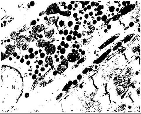
Fig. 24-6. ANP granules (g) interspersed among Cell/35.html">Mitochondria (m) in a rat atrial muscle cell; G, Golgi apparatus; N, Nucleus. Granules in human atrial cells have a similar appearance. x17,640 (courtesy of M. Cartin).
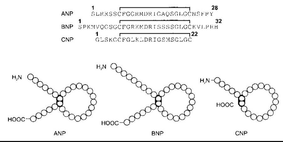
Fig. 24-7. Human ANP, BNP, and CNP. Top: One-letter amino acid codes showing conserved Amino acid sequences (shaded). Bottom: Molecular conformation. Note that the carboxy-terminal residue of CNP is cysteine; in this way, the carboxy terminus of this polypeptide chain is closed to form a 17-membered ring (modified from Imura H, Nakao K, Itoh H: The natriuretic peptide system in the brain: Implication in the central control of cardiovascular and neuroendocrine Functions. Front Neuroendocrinol 1992; 13:217).
Function
Circulating ANP and BNP act on the kidneys to increase Na+ excretion; injected CNP produces a similar effect. They presumably exert their action by dilating afferent arterioles and relaxing mesangial cells. Both of these effects enhance Glomerular Filtration (see Chapter 38) and also act on the renal tubules to inhibit Na+ reabsorption. Other effects include increased capillary permeability, leading to fluid extravasation and a drop in Blood pressure. In addition, they promote the relaxation of smooth muscle elements in arterioles and venules. CNP has a more pronounced vasodilatory effect on Veins than ANP and BNP. These Peptides also suppress renin secretion and counteract the pressor effects of catecholamines and angiotensin II.
In the brain, ANP is present in Neurons. A neural pathway containing ANP extends from the anteromedial Hypothalamus to Brainstem areas responsible for the neural Regulation of The Cardiovascular system.
In general, the central effects of ANP are opposite to those of angiotensin II, and ANP-containing nerve fibers most likely mediate a reduction in blood pressure and enhanced urinary sodium excretion. The Significance of CNP and BNP in the brain is presumably similar to that of ANP, although more detailed information on this matter is not yet available.
Natriuretic Peptide Receptors
Today, three distinct types of natriuretic peptide receptors (NPRs) have been identified and characterized (Fig. 24-8). Two of these receptors—NPR-A and NPR-B—span The Cell membrane and possess cytoplasmic domains that are chemically guanylyl cyclases. ANP shows the highest affinity for the NPR-A receptor, whereas CNP is most affine for NPR-B. The third receptor, NPR-C, binds all three natriuretic peptides but has a markedly truncated cytoplasmic domain. There is some evidence that it operates via G Proteins to activate phospholipase C and inhibit adenylyl cyclase. However, it remains a matter of debate whether this receptor initiates any Intracellular Signaling cascades or merely acts as a clearance receptor that simply binds circulating natriuretic peptides and subsequently releases them, helping to maintain stable blood concentrations of these hormones.
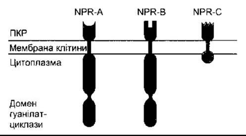
Fig. 24-8. Schematic representation of natriuretic peptide receptors. NPR-A and NPR-B molecules contain intracellular guanylyl cyclase domains, whereas the clearance receptor NPR-C possesses only a small cytoplasmic domain.
Secretion and METABOLISM
The plasma concentration of ANP in individuals consuming a moderate amount of sodium is normally about 5 fmol/mL. ANP secretion increases when extracellular fluid volume expands As a result of intravenous isotonic sodium chloride infusion, a high-sodium diet, or HEAD-out Water immersion (Fig. 24-9) (this Procedure counteracts the effects of gravity on Circulation by increasing central venous pressure and, consequently, atrial pressure). Notably, water immersion also decreases renin and aldosterone secretion. Conversely, changing posture from recumbent to standing causes a slight yet detectable drop in plasma ANP levels, which correlates with a decrease in central venous pressure. In vitro strips of atrial Muscle tissue release ANP upon artificial stretching. Therefore, it is likely that the atria in vivo respond directly to stretch, and The rate of ANP secretion is proportional to the degree of atrial distension caused by elevated central venous pressure.
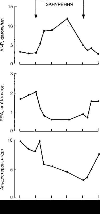
Fig. 24-9. Effect of neck-deep water immersion on plasma concentrations of ANP and aldosterone, and plasma renin activity (PRA) (modified and reproduced with permission from Epstein M et al: Increases in circulating atrial natriuretic factor during immersion-induced central hypervolaemia in normal humans. Hypertension 1986; 4 [Suppl 2]:593).
Circulating ANP has a short half-life. It is metabolized by neutral endopeptidase (NEP), which is inhibited by Tryptophan. Consequently, tryptophan administration increases blood levels of ANP.
Much less is known about the cardiac tissue secretion of BNP and its metabolism, but they are likely similar to those of ANP.
Na+-K+-ATPase Inhibitor Factor
Another natriuretic factor is present in the blood, which stimulates urinary sodium excretion by inhibiting the Na+-K+-ATPase enzyme and tends to raise blood pressure rather than lower it. Recent studies have demonstrated that this substance is ouabain, a digitalis-like steroid synthesized in the Adrenal Glands. However, its physiological significance remains to be elucidated.
Last update: 10/08/2026
Editorial and Educational Adaptation: This material has been compiled based on the primary/original source text. The project team performed an editorial review, corrected technical inaccuracies, structured sections, and adapted the content for an educational format.
What was processed:
- elimination of formatting defects (OCR errors, structural breaks, corrupted characters);
- editorial organization of content;
- standardization of terminology in accordance with academic sources;
- verification of factual statements against the original source text.
All mentions of the author, publication year, and origin of the primary text have been preserved in accordance with the source.