Review of Medical Physiology - William F. Ganong 2002
Endocrine System, Metabolism, and Reproduction
Sexual Differentiation and Development
Male Reproductive System - Spermatogenesis and Ejaculation
The walls of the convoluted seminiferous tubules are lined with primary Germ Cells (see below), Sertoli cells, and large, complex Glycogen-containing cells that extend from the basement membrane to the lumen (see Fig. 23-17). Tight junctions between adjacent Sertoli cells near the basement membrane form the blood-testis barrier, which prevents many large molecules from the interstitial fluid from entering the tubular lumen. However, Steroids cross this barrier readily. Evidence suggests that certain Proteins also pass via paracrine pathways from Sertoli cells to Leydig cells and vice versa. Furthermore, maturing germ cells must cross the barrier as they migrate toward the lumen. This passage occurs without disrupting the barrier's integrity through the temporary disassembly of tight junctions above the germ cells and the subsequent formation of new tight junctions below them. The fluid within the lumen of the convoluted seminiferous tubules differs from plasma; it contains low Levels of Protein and glucose, yet is rich in androgens, estrogens, K+, Inositol, glutamic acid, and aspartic acid. Maintaining this composition relies on the blood-testis barrier, which protects germ cells from harmful blood-borne agents and prevents antigenic products of germ Cell Division and maturation from entering the bloodstream and triggering an autoimmune response. This barrier also helps maintain an osmotic gradient that facilitates fluid movement into the tubular lumen.
Class="center">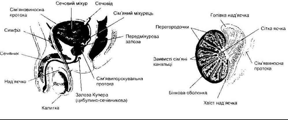
Fig. 23-15. Left: Male Reproductive System. Right: Duct System of the Testes.

Fig. 23-16. Section of a human testis.
Spermatogonia are primary germ cells located near the basement membrane of the convoluted seminiferous tubules that mature into primary spermatocytes (see Fig. 23-17). This process begins at Puberty. Primary spermatocytes undergo meiotic division, which reduces their chromosome number. During this two-stage process, they divide into secondary spermatocytes and subsequently into spermatids, which contain a haploid set of 23 Chromosomes. Spermatids mature into spermatozoa (sperm). Throughout the division and maturation of a single spermatogonium, its progeny remain interconnected via cytoplasmic bridges until the late spermatid phase. This presumably ensures the synchronized differentiation of each germ cell clone. The estimated number of spermatids produced from a single spermatogonium is 512. In humans, The formation of a mature spermatozoon from a primary germ cell via normal spermatogenesis takes an average of 74 days. Each spermatozoon is a complex, motile cell rich in DNA, with a HEAD composed primarily of chromosomal material (Fig. 23-18). The head is capped by an acrosome, a lysosome-like organelle containing Enzymes involved in sperm penetration of the oocyte and other Fertilization processes. In its proximal region, the motile tail of the spermatozoon is sheathed with numerous Mitochondria. The membranes of late spermatids and spermatozoa contain a specialized small form of angiotensin-converting enzyme known as germinal angiotensin-II-converting enzyme (see Chapter 24). The function of this enzyme in spermatozoa remains unknown, although male mice with disrupted angiotensin-converting enzyme Gene function have been shown to exhibit reduced fertility.

Fig. 23-17. Epithelium of a convoluted seminiferous tubule. Note that the maturing germ cells are interconnected by cytoplasmic bridges at the early spermatid stage and are densely enveloped by the Cytoplasm of Sertoli cells as they migrate from the basement membrane toward the lumen (reproduced with permission from Junqueira LC, Carneiro J, Kelley RO: Basic Histology, 9th ed. McGraw-Hill, 1998).

Fig. 23-18. Human spermatozoon in profile. The acrosome is an organelle covering the anterior half of the sperm head within The Plasma Membrane (reproduced with permission from Junqueira LC, Carneiro J, Kelley RO: Basic Histology, 9th ed. McGraw-Hill, 1998).
Spermatids mature into spermatozoa within deep cytoplasmic recesses of Sertoli cells (see Fig. 23-17). Mature spermatozoa are released from Sertoli cells into the tubular lumen. Sertoli cells secrete androgen-binding protein (ABP), inhibin, and MIH; they do not secrete androgens, but they contain aromatase (CYP19), the enzyme responsible for converting androgens into estrogens. Furthermore, they can produce estrogens. The function of ABP is presumably to maintain a steady supply of androgens to the tubular fluid; inhibin suppresses FSH secretion (see below); and MIH induces the regression of the Mullerian ducts in males during embryonic life (see above).
FSH and androgens support the gametogenic function of the testes. Following hypophysectomy, administration of LH induces a marked increase in the local concentration of androgens within the testes, which in turn promotes gametogenesis. The developmental stages from spermatogonia to spermatids are androgen-dependent. Maturation from spermatids to spermatozoa relies on the action of androgens on the Sertoli cells surrounding the developing sperm. FSH acts on Sertoli cells to support the final stages of spermatid maturation and also stimulates the secretion of ABP.
Emerging evidence indicates a high concentration of estrogen in the rete testis fluid (see Fig. 23-15) and significant levels of estrogen receptors alpha in the walls of the rete. In this region, fluid is reabsorbed and spermatozoa become concentrated. If this process fails, spermatozoa enter the Epididymis suspended in a large volume of fluid, leading to infertility.
Spermatozoa exiting the testis are not fully motile. They continue to mature and acquire motility during their passage through the epididymis. If the percentage of ova fertilized by spermatozoa from the tail of the human epididymis (see Fig. 23-15) is taken as 100, the corresponding value for spermatozoa from the body of the epididymis is 50, and for those from the head of the epididymis, it is 0. Fertilization is possible if a spermatozoon from the head of the testis is microinjected directly into an oocyte. Therefore, the primary impairment is the lack of sperm motility.
The fertilizing capacity of spermatozoa is enhanced upon subsequent entry into the female reproductive tract. This process, known as capacitation, is not yet fully understood and comprises two main components: an increase in sperm motility and preparation for the acrosome reaction. The Role of capacitation is presumably to facilitate and simplify the fertilization process, as In vitro fertilization is also feasible. Subsequent stages of fertilization are discussed below in the context of Pregnancy.
Temperature Effects
Spermatogenesis requires a temperature significantly lower than that of the core body. Normally, testicular temperature is maintained at around 32°C. Cooling is facilitated by air Circulation around the Scrotum and, presumably, countercurrent heat exchange between the spermatic Arteries and Veins. When testes are retained in the Abdominal cavity, or in experimental animals where they are positioned close to the body due to tight tissue attachments, degeneration of the tubular walls and infertility ensue. Hot baths (43–45°C for 30 min daily) and heavy physical exertion lead to a reduction in human sperm count, in some cases by as much as 90%. However, the reduction achieved in this manner is insufficient to make this Procedure a viable form of Male Contraception. Additionally, there is evidence of seasonal variations in men, with sperm counts increasing in winter when temperature conditions are more favorable for the scrotum.
Semen
Semen is the fluid expelled during orgasm. It contains spermatozoa and secretions from the Seminal Vesicles, prostate, Bulbourethral Glands, and presumably the urethral glands (Table 23-4). The average volume of a single ejaculate ranges from 2.5 to 3.5 mL after several days of abstinence. Semen volume and sperm count decrease rapidly with repeated ejaculations. Although only a single spermatozoon is required to fertilize an ovum, the normal concentration is approximately 100 million per milliliter of semen. Fifty percent of men with sperm counts of 20–40 million/mL and virtually all of those with counts below 20 million/mL are sterile. Semen also exhibits a high concentration of Prostaglandins, which originate primarily from the seminal vesicles. The function of these fatty acid derivatives in semen is not yet fully elucidated. Their Structure and diverse effects on other PARTS OF THE body are discussed in Chapter 17.
Table 23-4. Composition of human semen

Human spermatozoa travel through the female reproductive tract at a speed of about 3 mm/min, reaching the uterine tubes 30 to 60 minutes after intercourse. In some animal species, contractions of the FEMALE REPRODUCTIVE Organs aid in The transport of spermatozoa toward the uterine tubes, though it remains unclear whether such contractions occur in humans.
Erection
Erection occurs As a result of the dilation of the penile arterioles. During this process, the erectile tissue of the Penis engorges with blood, and the veins are compressed, blocking outflow and giving the organ turgor. Integration centers in the lumbar segments of the Spinal Cord are activated by impulses from afferents originating in the genitalia and by descending pathways that mediate erection in response to erotic psychological stimulation. Efferent parasympathetic fibers run in the pelvic splanchnic nerves. It is likely that they contain acetylcholine and VIP as cotransmitters (see Chapter 4). Some fibers terminate presynaptically on noradrenergic Neurons, where acetylcholine acts on muscarinic receptors, reducing the release of norepinephrine by vasoconstrictors. In addition, VIP induces vasodilation. However, the administration of VIP alone does not fully induce erection. Intraurethral administration of PGE1 (alprostadil) also relaxes the smooth Muscle of the penis and is used clinically to facilitate erection.
There are also nonadrenergic, noncholinergic fibers in the pelvic nerves. They contain a high concentration of NO synthase, the enzyme that catalyzes the formation of nitric oxide (NO; see Chapter 31). NO activates guanylyl cyclase, thereby increasing The production of cGMP, a potent vasodilator. Administration of NO synthase inhibitors to experimental subjects prevents the erection normally stimulated by the pelvic nerve. This clearly demonstrates the crucial role of NO in achieving an erection. Viagra (sildenafil) inhibits The breakdown of cGMP by phosphodiesterases and is therefore widely used today for the Treatment of erectile dysfunction. Multiple mammalian phosphodiesterases (PDEs) are divided into seven isoenzyme families. Sildenafil is most active against PDE5, a phosphodiesterase isoform found in the corpus cavernosum. However, it should be noted that sildenafil also appreciably inhibits PDE6. This phosphodiesterase isoform is found in the retina. Consequently, one of the side effects of sildenafil is transient blue-green color discrimination impairment (see Chapter 8).
Normally, erection subsides as a result of sympathetic vasoconstrictor impulses acting on the penile arterioles.
Ejaculation
Ejaculation is a two-stage spinal reflex comprising emission (the movement of sperm into the Urethra) and true ejaculation (the expulsion of semen from the urethra during orgasm). The afferent pathways consist primarily of fibers from sensory receptors in the glans penis that reach the spinal cord via the pudendal nerves. Emission is a sympathetic response integrated in the upper lumbar segments of the spinal cord, which affects the contraction of the smooth muscle of the vas deferens and seminal vesicles in response to hypogastric nerve stimulation. Semen is propelled from the urethra through the contraction of the bulbospongiosus Skeletal Muscle. The spinal reflex centers for this phase are located in the upper sacral and lower lumbar segments of the spinal cord, with motor pathways operating at the level of the first through third sacral roots and the internal pudendal nerves. Carbon monoxide is involved in The regulation of ejaculation, provided that the HO-2 enzyme catalyzing its production in The Nervous system (see Chapter 4) is present in sufficient quantities within the pathways associated with ejaculation. Ejaculatory capacity is diminished in the absence of the HO-2 gene.
Prostate-Specific Antigen
The Prostate Gland produces and secretes into the semen and bloodstream a 30-kDa Serine protease known as prostate-specific antigen (PSA). The PSA gene contains two androgen-responsive elements. It hydrolyzes seminogelin, an inhibitor of sperm motility in semen, and has several plasma substrates, although its physiological function in the circulation remains unclear. Plasma PSA levels become elevated in prostate Cancer, making it a widely used screening test for this condition. PSA is also elevated in benign prostatic hyperplasia and prostatitis.
Vasectomy
Bilateral ligation of the vas deferens (vasectomy) has proven to be a relatively safe contraceptive procedure. However, restoring duct patency in individuals seeking to regain fertility is known to be technically challenging. The success rate of such reversal Procedures, measured by subsequent pregnancies, is approximately 50%. Following vasectomy, half of all men develop anti-sperm Antibodies. In monkeys, the presence of these antibodies is associated with a high rate of persistent infertility after vasectomy reversal. No other Adverse effects of anti-sperm antibodies have been identified.
Last update: 10/08/2026
Editorial and Educational Adaptation: This material has been compiled based on the primary/original source text. The project team performed an editorial review, corrected technical inaccuracies, structured sections, and adapted the content for an educational format.
What was processed:
- elimination of formatting defects (OCR errors, structural breaks, corrupted characters);
- editorial organization of content;
- standardization of terminology in accordance with academic sources;
- verification of factual statements against the original source text.
All mentions of the author, publication year, and origin of the primary text have been preserved in accordance with the source.