Orthopedics - Oleksa A.P. 2006
Congenital Spinal Deformities
Spondylolysis and Spondylolisthesis
The term "spondylolysis" refers to a defect or non-union of the vertebral arch in the pars interarticularis region (Fig. 254).
There are various opinions regarding the Etiology of spondylolysis. It was traditionally considered a congenital anomaly that remains clinically silent until spondylolisthesis develops under METABOLISM/18.html">The Influence of physical overloads in this spinal region.
Congenital spondylolysis occurs As a result of the failure of fusion of the ossification centers that form one-half of the vertebral arch, or the entire arch in cases of bilateral non-union.
We examined a lumbar spine specimen from an adult human featuring bilateral non-union of the L5 arch. The bony fragments of the arch were interconnected vertically by a very thin layer of Cartilage-like tissue; after its removal, uneven yet congruent transverse bone-to-bone Contact surfaces were revealed.
According to Friberg, this localization is the most frequent, accounting for 67.7%, while the incidence of spondylolysis decreases at higher vertebral levels (L4 — 25.8%, L3 — 4.7%).
Nathan attributes The Development of spondylolysis to a primary horizontal orientation of the sacrum, followed by a forward tilt of the L5 vertebral body and arch. This shifts the vector of Muscle tension and overload along the articular processes, exerting a shearing force on the arch. When the sacrum is normally angled, the L5 body and arch lie in a more horizontal plane, and vertical forces act upon the vertebra without any directional deviation.
Class="center">
Fig. 254. Diagram of vertebral arch non-union (spondylolysis).
When the sacrum is positioned horizontally (at an angle of 130–150°), shearing forces act upon the pars interarticularis region of the L5 arch. Under the influence of muscular strain and physical overload—especially during periods of rapid child growth—these forces can prevent the Ossification of the cartilaginous portion of the arch.
The possibility of spondylolysis resulting from acute trauma or repeated microtraumas acting along the spinal axis—with force concentrated on the articular processes and arches—cannot be ruled out. A single acute trauma may cause an arch fracture leading to pseudoarthrosis, whereas microtraumas induce structural remodeling of the arch.
Reinberg S.A. (1964) views spondylolisthesis strictly as a bone remodeling process of the vertebral arch in the pars interarticularis region caused by excessive mechanical stress. The author regards spondylolysis as a constitutional variant rather than a developmental anomaly. He compares it to Baastrup's disease—a condition characterized by lesion and remodeling of the spinous processes of the lumbar, and sometimes thoracic or even cervical vertebrae, featuring Looser's zones, which are occasionally interpreted as pseudoarthroses of the processes.
Cases of familial spondylolysis have been documented (Friberg, Bailey) in two or even three family members, which can be explained by an inherited degree of sacral inclination.
Unilateral and atypical variants of spondylolysis also occur (spondylolysis retroisthmica, retrosomatica, interarticularis posterior).
Spondylolisthesis occurs in the presence of bilateral spondylolysis of the vertebral arch and may also result from impaired osteogenesis of the arch (spondylolisthesis congenita) or cumulative shearing forces from microtraumas. Additionally, there are cases caused by a combination of both factors: Developmental anomalies of the arch and excessive mechanical loading.
As spondylolisthesis progresses, pathological changes occur within the intervertebral disc, leading to its degeneration. In advanced cases, degenerative changes affect the adjacent vertebral endplates, subsequently developing into spondylosis. If the slippage of the L5 vertebral body occurs during the patient's growth phase, a wedge-shaped vertebral deformity develops, accompanied by an increase in its transverse dimensions.
Diagnosis of spondylolisthesis. As previously mentioned, spondylolysis may remain clinically asymptomatic; therefore, patients are typically examined only after spondylolisthesis develops and they begin to complain of lower back pain following physical exertion.
The Clinical symptoms of spondylolisthesis depend on the rate and degree of anterior Displacement of the overlying vertebra. The more gradual the vertebral slip, the milder the clinical signs. In the vast majority of patients, slippage occurs progressively over years, precluding severe or acute disorders. Radicular pain or sciatica typically appears after spinal overloads, during Pregnancy, and similar circumstances. Later on, pain is experienced during sitting, standing, and trunk flexion.
Physical examination may reveal an increased lumbar lordosis and localized tenderness upon Palpation. Spinal range of motion is generally unrestricted, though trunk flexion may show a protective guarding of the paraspinal Muscles. Only in cases of pronounced spondylolisthesis are flexion and lateral bending restricted.
Severe spondylolisthesis (Fig. 255) can be complicated by persistent neuralgia-type radicular pain, and even paresis or paraplegia. Although we observed a case where the L5 vertebral body had slipped completely relative to S1 across the entire surface of the vertebral body, the patient experienced no complications other than pain, a shortened trunk, and limited spinal mobility, and remained fully ambulatory.

Fig. 255. Severe Clinical presentation of spondylolisthesis.
X-ray imaging is paramount in diagnosing spondylolysis and spondylolisthesis. Standard anteroposterior (AP) radiographs of the spine may fail to detect spondylolysis because the defect in the vertebral arch does not coincide with the trajectory of the X-ray beam.
Oblique radiographs also offer limited diagnostic value; therefore, computed tomography (CT) or Magnetic Resonance imaging (MRI) are currently utilized to accurately diagnose spondylolysis.
Spondylolisthesis of any grade is readily visible on lateral plain radiographs of the spine, as well as during functional fluoroscopy in the Cytology/cytology/16.html">Early stages of L5 vertebral slippage.
Based on clinical and radiological findings, Meyerding classified spondylolisthesis into five grades: Grade I involves displacement of the vertebral body by 1/4 of the sacral base; Grade II by 1/2; Grade III by 3/4; Grade IV involves displacement across the entire surface of the S1 vertebral body; and Grade V (spondyloptosis) involves displacement of the L5 vertebral body below the level of the S1 body (Fig. 256).
Brelsford and others distinguish three degrees of spondylolisthesis: the first is when contact between the body of the L5 vertebra and The surface of the S1 body is preserved (spondylolisthesis); the second occurs when this contact is lost and the vertebra tilts over the anterior edge of the S1 body (spondylolisis); and the third degree involves the displacement of the L5 vertebral body into the pelvis with its contact against the anterior surface of the sacrum (spondylopthosis) (Fig. 257).
Treatment. Upon diagnosing spondylolisthesis, the question of conservative or surgical management must be addressed immediately to prevent the progression of vertebral slippage.
Conservative treatment is administered as a preparatory stage prior to surgery. Depending on the degree of vertebral displacement and its duration, the preparation time varies.
In cases of acute spondylolisthesis, an attempt is made to reduce the displaced vertebra immediately under general anesthesia. To achieve this, the patient is positioned on the table so that the pelvis and thighs can be displaced anteriorly, allowing the displaced vertebra to snap back into place (Fig. 258).
However, acute spondylolisthesis is extremely rare in clinical practice, and therefore the displaced vertebra must usually be reduced gradually. The patient is placed supine in bed, and the pelvis and lower limbs are progressively elevated using slings. The slings are secured to Balkan frames. One sling must be positioned exclusively over the sacral region and must not shift onto the fifth lumbar vertebra. With the hips and knees flexed at 90°, the lower legs are placed on higher-positioned slings. With relaxed muscles, the weight of the torso causes the lumbar spine to shift posteriorly, thereby achieving reduction of the displaced vertebral body.
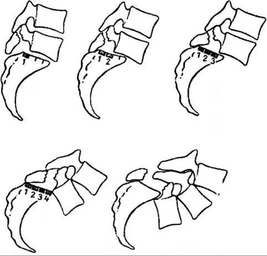
Fig. 256. Meyerding grading of spondylolisthesis.

Fig. 257. Magnetic resonance imaging of spondylolisthesis with displacement of the L5 vertebra into the pelvis.

Fig. 258. Single-stage reduction of a displaced vertebra in spondylolisthesis (under anesthesia).
Similarly, the displaced L5 vertebra can be gradually reduced by elevating the pelvis and applying lower leg traction using a Perlscha device or the apparatus shown in Fig. 259.
The reduced or partially reduced vertebra must be surgically stabilized to prevent recurrence of displacement.

Fig. 259. Device for gradual reduction of a vertebra in spondylolisthesis.
According to Weissflog (1968), all surgical interventions for spondylolisthesis can be divided into four groups.
1. Palliative operations used in the early stages of spondylolisthesis, which consisted of wedging a bone autograft into the sagittal cleft of the spinous processes (after Albee) or inserting an autograft between the corticated spinous processes and the arches of L3-S2 (after Henie).
2. More modified Procedures for posterior spinal fusion:
a) according to Gruca — splitting the spinous processes in the sagittal plane, retracting them laterally along with the adjacent muscles, and decorticating the L3-S1 arches bilaterally, into which tibial bone autografts are inserted and secured with sutures to the ligaments (Fig. 260);
b) according to Gruca — a similar operation, differing from the previous one in that cancellous bone chips harvested from the iliac wing are implanted;
c) according to Meyerding — placing autografts on both sides of the decorticated spinous processes and L3-S1 arches, with packing of the gaps in the lower region using cancellous bone;
d) transpedicular fixation of the L4-L5 vertebral bodies with potential correction of the displaced L5 body using a central screw should be classified as palliative.

Fig. 260. Anterior spinal fusion according to A. Gruca for spondylolisthesis.
3. Radical procedures for anterior spinal fusion.
Gruca performed the surgery with the patient in the prone position with the hips flexed. The author made an arcuate Skin incision extending from the mid-sacrum to the coccyx. He then separated and retracted the soft-tissue flap and excised the coccyx. Next, he dissected the proper Fascia of the pelvis anterior to the sacrum up to the body of the L5 vertebra. He incised the periosteum transversely along the inferior border of the S1 body and drilled a channel through it, passing through the disc into the body of the L5 vertebra and half of the L4 body. Into this channel, he impacted a bone graft harvested from the Fibula.
Several variants of posterior corporedesis exist, which are now rarely used.
Today, anterior corporedesis of the vertebrae via anterior transabdominal or lateral extraperitoneal approaches is generally performed for spondylolisthesis.
Transabdominal corporedesis of L5-S1 was first performed by Burns in 1953, and later by Kellog and Speed. They drilled a 12 mm diameter channel from the upper margin of the L5 body obliquely into the S1 body and impacted a slightly wider tibial bone graft into it.
We consider transabdominal anterior corporedesis—fixation of the L5 vertebral body to the S1 body using a bone graft (according to Burns)—to be the optimal surgical Procedure.
Surgical technique. The operation is performed under endotracheal anesthesia. The Abdominal cavity is opened through a lower midline incision; the greater omentum and Small Intestine are retracted upward and protected along with the Urinary Bladder using surgical Sponges. The posterior Peritoneum is incised to expose the common iliac Arteries and Veins. Ligatures on clamps are passed beneath them to allow for immediate Hemorrhage control in the event of vascular injury during surgery. After exposing the bodies of the fourth and fifth lumbar and the first sacral vertebrae, a channel is carefully drilled through them in an oblique direction, passing through the intervertebral disc (Fig. 261).
Then, an autogenous graft corresponding to the diameter and length of the channel is harvested from the tibial metaepiphysis and wedged into the channel. The ligatures are removed from the vessels, the peritoneum is closed, and the laparotomy wound is closed in layers.
Postoperatively, the patient remains in bed for 2 to 2.5 months in the same position as before surgery (on slings supporting the pelvis and calves). Afterward, a supportive brace is applied, and the patient is discharged for outpatient treatment.
Transabdominal corporedesis has fully met the expectations of orthopedists.
B. Tsyrlyuk (1978) improved the Burns procedure by excising the intervertebral disc, wedging a bone wedge graft into the intervertebral space, and subsequently placing a bone dowel.
M.O. Korzh, in cases of significant unreduced vertebral displacement, transabdominally exposes the L5-S1 vertebral bodies, incises the anterior longitudinal ligament of the spine, and resects a thin plate from the Inferior surface of the displaced vertebra, removing it along with the intervertebral disc. He harvests a similar plate from the S1 body and creates a recess in it. He inserts a bone graft into the defect, surrounding it with cancellous bone (Fig. 262). Next, he sutures the longitudinal ligament, then the peritoneum, and closes the laparotomy wound in layers.
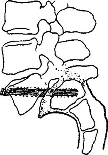
Fig. 261. Anterior transabdominal spondylodesis for spondylolisthesis.

Fig. 262. Transabdominal method of vertebral fixation in spondylolisthesis according to Korzh-Khvysyuk.
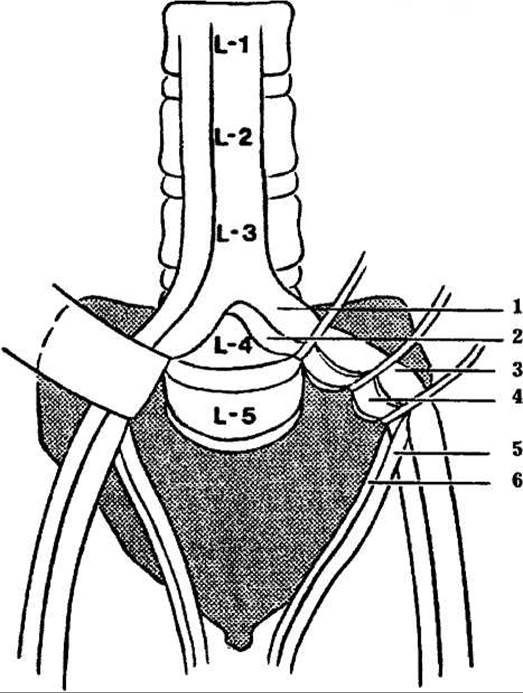
Fig. 263. Schematic drawing of vessel retraction and exposure of the L4-L5 vertebral bodies during the transabdominal surgical approach.
If the displacement of the L5 vertebra is so severe that performing the aforementioned surgery is difficult, the fixation method described by M.O. Korzh and M.I. Khvysyuk (1986) is employed. During the operation, an attempt is made to correct THE POSITION OF the vertebra using the lever principle, followed by fixation using the previously described method. If correcting the vertebral displacement fails, grafts are used to fix the fifth lumbar vertebra to the first sacral vertebra within a trough, and superficial to them, the body of the fourth lumbar vertebra is fixed to the second sacral vertebra.
Some spine surgeons (Chaklin, Osna, Tsyvyan) performed anterior corporedesis via an anterolateral extraperitoneal approach, which is technically more demanding and carries the risk of damaging the major iliac vessels (Fig. 263).
Surgical technique. The operation is performed under anesthesia with the patient in the supine position. An ilioinguinal tissue incision is made on the left side, starting from the upper third of Poupart's ligament, extending past the anterior superior iliac spine obliquely upward almost to the costal margin. The external and internal oblique muscles, as well as the transversus abdominis muscle, are split along the fiber direction. The muscles are bluntly separated, and the parietal peritoneum is retracted laterally toward the abdomen along with the adjacent bowel loops. It is essential to avoid injuring the peritoneum and the iliac vessels, which are deflected during the exposure of the L5-S1 vertebral bodies.
Thin plates are osteotomized from the anterior half of the vertebral bodies and removed along with the intervertebral disc. Afterward, an attempt is made to correct the degree of displacement of the L5 body. A bone graft is wedged into the triangular defect between the vertebrae. The torso is flexed forward, which tightens the position of the wedge. Hemostasis is checked, and the wound is closed in layers.
N.R. Voronovich, A.M. Petrenko, O.N. Dulub, and V.N. Nikolayev (1987) perform anterior spondylodesis in lumbar spondylolisthesis using a vascularized autograft harvested from the iliac wing, based on the nourishing iliolumbar artery. The procedure is performed via a costro-inguinal approach. For posterior spondylodesis, they utilize an autograft harvested from the dorsal region of the ilium, incorporating a portion of the quadratus lumborum muscle and the posterior branch of the lumbar artery.
Postoperatively, the patient remains in the same position as during preoperative preparation for two and a half months. Thereafter, the patient is allowed to lower their legs, and after three months, to walk wearing an unloading brace.
Today, supported by advanced technology and comprehensive Anesthetic Management, anterior corporedesis for spondylolisthesis—regardless of the degree of L5 vertebral body displacement—should be the method of choice. Long-term outcomes of such surgeries confirm their efficacy.
Palliative surgeries should not be used in the routine practice of orthopedists and spine surgeons.
If the patient refuses the proposed surgery, conservative treatment is administered. Such treatment involves traction, as applied during preoperative preparation, and The Use of an unloading brace. Patients are given torso muscle massages, physiotherapeutic and balneological procedures in the lumbosacral region, encouraged to swim, and forbidden from performing heavy physical labor, carrying loads, etc.
Unlike anterior spondylolisthesis, retrolisthesis (spondylolisthesis posterior or reversa) is possible, which involves the displacement of the L4 vertebral body relative to L5, as well as posterior pseudospondylolisthesis (Junghans' pseudospondylolisthesis), caused not by vertebral displacement, but by the elongation of the entire vertebra in the sagittal plane.
Last update: 10/08/2026
Editorial and Educational Adaptation: This material has been compiled based on the primary/original source text. The project team performed an editorial review, corrected technical inaccuracies, structured sections, and adapted the content for an educational format.
What was processed:
- elimination of formatting defects (OCR errors, structural breaks, corrupted characters);
- editorial organization of content;
- standardization of terminology in accordance with academic sources;
- verification of factual statements against the original source text.
All mentions of the author, publication year, and origin of the primary text have been preserved in accordance with the source.