Human Anatomy - Kotsan I. Ya. 2009
Olfactory organ
The Organ of Smell (organum olfactus) represents the peripheral portion of the olfactory analyzer. The olfactory analyzer is a complex morphophysiological system that provides the reception, conduction, analysis, and innervation of olfactory stimuli.
In human life, The Organ of smell is of lesser importance compared to the Organs of Vision and Hearing. However, its role is invaluable in environmental orientation; it monitors the quality of food and inhaled air, plays a significant role in digestive Reflexes, and so on. Structure/19.html">The Importance of the olfactory analyzer for humans and animals is much broader than it sometimes appears. Its role cannot be reduced merely to the simple perception of signals regarding odorous substances present in inhaled air. The presence of olfactory memory in humans is also of great importance, making it possible to recognize odors. Furthermore, the ability for remote environmental assessment allows for the evaluation of certain properties of objects that are inaccessible to the organs of hearing and vision.
The organ of smell is of ectodermal origin. In phylogenesis, it initially forms alongside the oral opening, and subsequently separates from the Oral Cavity and shifts to the initial sections of the Upper Respiratory Tract.
In most vertebrate animals, the organ of smell is represented by specialized receptor Cells located in the mucous membrane of the olfactory pits. In most fish, the organ of smell is formed by two olfactory pits. In the course of evolutionary development, these pits deepen, transforming in lungfish into a paired Nasal cavity separated from the oral cavity by a primary palate. In amphibians, the organ of smell is surrounded by a cartilaginous capsule. In terrestrial vertebrates, the nasal cavity performs two Functions: olfactory and respiratory. Accordingly, the mucous membrane of the nasal cavity is divided into two regions: the olfactory and the respiratory. Glands appear within the mucous membrane, followed by The formation of cartilaginous (in reptiles) and later bony nasal conchae.
Depending on The Development of the organ of smell, mammals are divided into three groups: anosmic, macrosmatic, and microsmatic. In anosmic animals (e.g., dolphins), the olfactory apparatus is atrophied, nasal conchae are absent, and the SENSE OF SMELL is entirely lacking. In macrosmatic animals, the olfactory apparatus is complex and well-developed, with olfactory centers predominating over other Brain regions (marsupials, insectivores, rodents, ungulates, predators). In microsmatic animals, the sense of smell is present but poorly developed, whereas visual and auditory centers are most prominent. Microsmatic animals include pinnipeds, primates, and humans.
In humans, the organ of smell is located in the superior region of the nasal cavity (Fig. 276). The olfactory region of the nasal mucosa (regio olfactoria tunicae mucossae nasi) includes the mucous membrane covering the superior nasal concha, the upper part of the nasal septum, the walls of the superior nasal meatus, and the upper PARTS OF THE middle nasal meatus. The olfactory region of the mucosa is covered by olfactory epithelium, among the cells of which three types are distinguished: olfactory receptor, supporting, and basal cells (Fig. 277). Olfactory receptor cells are scattered throughout the entire olfactory region, which is why the Olfactory nerve, unlike other sensory Cranial Nerves, lacks a ganglion (nerve knot). Supporting cells lie between the olfactory cells, separating them; they possess short cilia and exhibit secretory features. Basal cells lie deeper on the basement membrane, surrounding the axon bundles of the olfactory receptor cells. Even deeper, beneath the basement membrane, lie tubular-alveolar glands whose ducts open onto The surface of the olfactory mucosa. The olfactory glands (glandulae olfactoriae) produce a predominantly serous secretion that moistens the surface of the olfactory epithelium and dissolves the molecules of odorous substances.
Class="center">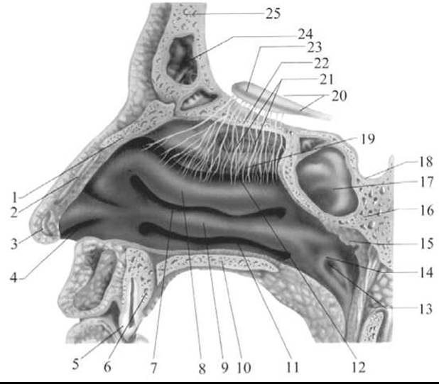
Fig. 276. LATERAL VIEW OF the nasal cavity wall
1 — Nasal bone; 2 — superior nasal Cartilage; 3 — inferior nasal cartilage; 4 — nasal vestibule; 5 — Teeth; 6 — Maxilla; 7 — middle nasal meatus; 8 — middle nasal concha; 9 — Inferior nasal concha; 10 — Palatine bone; 11 — inferior nasal meatus; 12 — superior nasal meatus; 13 — opening of the Eustachian tube; 14 — torus tubarius; 15 — pharyngeal tonsil and adenoids; 16 — Sphenoid bone; 17 — sphenoid sinus; 18 — sella turcica; 19 — superior nasal concha; 20 — olfactory tract; 21 — olfactory fibers; 22 — Ethmoid bone; 23 — olfactory bulb; 24 — frontal sinus; 25 — Frontal bone
In the olfactory region of the human nasal mucosa, there are approximately 40 million olfactory receptor cells, whereas in macrosmatic animals their number can reach 200 million or more. Olfactory receptor cells represent the first Neurons of the olfactory pathway. They possess short peripheral and long central processes. The peripheral process—the dendrite—terminates in a thickening called the dendritic vesicle (olfactory knob), on the apex of which 10–12 motile olfactory cilia are located. Each cilium contains 9 peripheral pairs and 2 central microtubules that interact with dissolved substances.
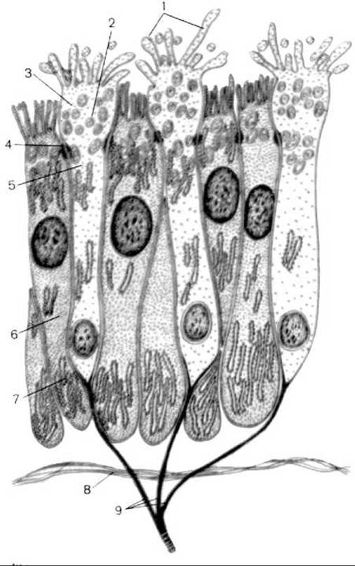
Fig. 277. Diagram of the ultramicroscopic STRUCTURE OF THE olfactory epithelium (according to V. G. Eliseev et al.)
1 — microvilli; 2 — vesicles; 3 — olfactory club; 4 — junctional complex (desmosome); 5 — body of the olfactory neurosensory Cell; 6 — supporting cell; 7 — Endoplasmic reticulum; 8 — basement membrane; 9 — axons of olfactory neurosensory cells forming olfactory nerve filaments
The central processes—axons—of the olfactory receptor cells pass between the supporting cells, gather into bundles, and form 15–20 olfactory filaments (nerves). The olfactory nerves penetrate the cranial cavity through the openings of the cribriform plate of the ethmoid bone and then enter the olfactory bulb, where they terminate in the olfactory glomeruli. Here, the second-order neurons (mitral cells) begin. The axons of the mitral cells travel as part of the olfactory tract and terminate in the Cells of the Gray matter of the olfactory tract, the olfactory trigone, the anterior perforated substance, and the septum pellucidum. The majority of fibers reach the cortex of the parahippocampal gyrus and the uncus, where the cortical end of the olfactory analyzer is located. The conduction pathway of the organ of smell is shown in Fig. 278.
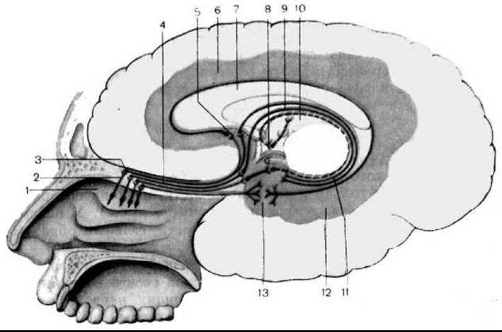
Fig. 278. Conduction pathway of the organ of smell (according to M. R. Sapin)
1 — superior nasal concha; 2 — olfactory nerves (cranial nerve I); 3 — olfactory bulb; 4 — olfactory tract; 5 — subcallosal area; 6 — cingulate gyrus; 7 — corpus callosum; 8 — mammillary body; 9 — fornix; 10 — posterior thalamus; 11 — dentate gyrus; 12 — parahippocampal gyrus; 13 — uncus
In clinical practice, a weakened sense of smell known as hyposmia, a complete loss of smell known as anosmia, and heightened Olfaction known as hyperosmia are observed. Diseases of the nasal cavity (rhinitis, polyps, etc.) are frequently accompanied by hypo- or anosmia. Allergic conditions can lead to the development of hyperosmia. Tumors in the region of the anterior surface of the frontal lobe result in unilateral anosmia or hyposmia due to mechanical pressure on the olfactory bulb and tract. Olfactory hallucinations are observed in cases of tumors located in the parahippocampal gyrus.
Last update: 08/08/2026
Editorial and Educational Adaptation: This material has been compiled based on the primary/original source text. The project team performed an editorial review, corrected technical inaccuracies, structured sections, and adapted the content for an educational format.
What was processed:
- elimination of formatting defects (OCR errors, structural breaks, corrupted characters);
- editorial organization of content;
- standardization of terminology in accordance with academic sources;
- verification of factual statements against the original source text.
All mentions of the author, publication year, and origin of the primary text have been preserved in accordance with the source.