Human Anatomy - Kotsan I. Ya. 2009
Doctrine of Internal Organs
Digestive System
Large Intestine
The Large Intestine (intestinum crassum) is a derivative Structure OF THE hindgut. It represents the final section of the digestive tract. In the large intestine, with the help of saprophytic microflora, the digestive process is completed, and fecal masses are formed and excreted. Furthermore, partial absorption of Water and mineral salts takes place here.
The large intestine is located in the abdominal and pelvic cavities. Its length ranges from 1.5 to 2 m. The diameter of the large intestine reaches 7 cm in its initial section and then gradually decreases to 4 cm.
The large intestine comprises three main sections: the cecum with the vermiform Appendix, the colon, and the rectum. The colon, in turn, is further divided into four parts: the ascending, transverse, descending, and sigmoid colons.
Class="center">
Fig. 153. Large intestine (segment of the transverse colon)
1 — omental Appendices; 2 — haustra of the colon; 3 — semilunar folds; 4 — omental tenia; 5 — mesocolic tenia; 6 — free tenia
The large intestine has several distinct features that differentiate it from the Small Intestine. Firstly, the large intestine has a significantly larger diameter than the small intestine. Secondly, the longitudinal layer of the muscular coat of the large intestine is not distributed evenly along its entire length, but is gathered into three bands (teniae) running at specific intervals from one another (Fig. 153). They originate at the Base of the vermiform appendix and extend to the rectum. Each of these bands is about 1 cm wide and has its own name. The mesocolic tenia (taenia mesocolica) runs along the attachment site of the transverse mesocolon (on the transverse and sigmoid colons) and the line of attachment of the intestine to the posterior abdominal wall (on the ascending and descending colons). The omental tenia (taenia omentalis) is located on the transverse colon near the attachment site of the greater omentum, and on the ascending and descending colons along their posterolateral surfaces. The free tenia (taenia libera) is situated on the anterior surface of the cecum and ascending colon; however, in the region of the transverse colon, it shifts to its inferior surface, and on the descending colon, it runs again along the anterior surface. Upon reaching the rectum, these bands widen, converge, and once again enclose the rectum in a continuous layer. Thirdly, between the colonic teniae, there are numerous sac-like protrusions known as colonic haustra (haustra coli), which are formed primarily because the longitudinal teniae are shorter than the length of the intestine between them. These pouches are separated from each other by deep transverse grooves, which correspond internally to the semilunar folds (plicae semilunares) of the mucous membrane. Fourthly, on the external surface of the large intestine along the free and omental teniae, there are finger-like outpocketings of the serous coat—the omental appendices (appendices epiploicae (omentales))—which contain adipose tissue. Their length reaches 4–5 cm. The omental appendices protect the intestinal wall from pinching between areas containing dense contents. Such appendices are never found on the small intestine.
The large and small intestines also differ in the color of their walls. In the large intestine, it has a grayish tint, whereas in the small intestine, it is pink.
Unlike the small intestine, the mucous membrane of the large intestine lacks villi; it is smooth and features semilunar folds, intestinal glands (crypts of Lieberkühn), and solitary lymphatic follicles. Peyer's patches are absent in the large intestine. The mucous membrane is lined with a single-layered columnar epithelium containing A large number of goblet Cells. The goblet cells and tubular intestinal Glands of the mucosa secrete mucus and, together with bacterial metabolic products, create a mildly acidic environment within the lumen of the large intestine.
The muscular coat of the large intestine consists of two layers: an outer longitudinal layer and an inner circular layer, but the longitudinal layer is not continuous. As mentioned above, it forms three longitudinal teniae.
The serous coat does not cover the large intestine uniformly throughout. The peritoneal relationships of the various sections of the large intestine will be discussed below in the description of each respective section.
The cecum (caecum) is the initial segment of the large intestine, located in the right part of the Abdominal cavity below the opening where the ileum empties into the large intestine (Fig. 154). The posterior surface of the cecum rests against the iliopsoas Muscle, while the anterior surface abuts the anterior abdominal wall. The cecum is covered by Peritoneum on all sides, meaning it is located intraperitoneally with respect to the peritoneum. The cecum has no mesentery. Its length ranges from 3 to 8 cm, and its width from 4 to 7 cm.
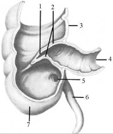
Fig. 154. Cecum with vermiform appendix (anterior wall removed)
1 — ileocecal orifice; 2 — ileocecal valve; 3 — ascending colon; 4 — ileum; 5 — orifice of the vermiform appendix; 6 — vermiform appendix; 7 — cecum
The transition from the ileum to the cecum—the ileocecal orifice (ostium ileocaecale)—appears as a horizontal slit bounded above and below by high folds of the mucous membrane that form the ileocecal valve (valva ileocaecalis). The base of this valve contains circular muscle fibers, but they do not form a true sphincter, and the orifice remains constantly open, allowing undigested food residues to pass from the small intestine into the large. The folds of the ileocecal valve close automatically when pressed upon by masses of food residue coming from the direction of the large intestine. The larger this mass, the tighter the folds close, preventing regurgitation back into the small intestine.
Approximately 3 cm below the ileocecal orifice, on the inner surface of the posteromedial wall of the cecum, lies the orifice of the vermiform appendix (ostium appendicis vermiformis).
The vermiform appendix (appendix vermiformis) is a tubular outpocketing of the cecum, the length of which varies in different individuals from 2 to 20 cm, and its diameter from 0.5 to 1.0 cm. THE POSITION OF the appendix is highly variable and depends on its length and the Location OF THE cecum. It is primarily situated in the right iliac fossa, but it may drape over the iliac vessels and descend into the pelvic cavity, or turn upward and lie posterior to the cecum—all of which complicates the Diagnosis of appendicitis. The vermiform appendix is covered by peritoneum on all sides (is located intraperitoneally) and possesses a mesentery. Blood Vessels and nerves run within the thickness of the mesentery to the appendix.
The mucous membrane of the vermiform appendix contains a large number of lymphatic follicles, allowing it to be classified as part of The Immune System of the digestive tract.
The ascending colon (colon ascendens) is the continuation of the cecum. In adults, its length reaches 14–20 cm. The ascending colon is located in the right region of the abdominal cavity. Ascending upward, it reaches the Liver, beneath which it turns sharply to the left, forming the right colic flexure (flexura coli dextra), and transitions into the transverse colon. Posteriorly, the ascending colon is adjacent to the quadratus lumborum and transversus abdominis Muscles; anteriorly, it abuts the anterior abdominal wall; medially, it comes into contact with loops of the ileum; and laterally, it borders the right wall of the abdominal cavity. The ascending colon is covered by peritoneum anteriorly and laterally, but lacks a peritoneal covering posteriorly (making it retroperitoneal / mesoperitoneal in anatomical Classification).
The transverse colon (colon transversum) is the longest part of the large intestine. Its length ranges from 30 to 83 cm (averaging 50 cm). Beginning at the right colic flexure, it runs toward the left hypochondriac region and there, forming the left colic flexure (flexura coli sinistra), transitions into the descending colon. The length of the transverse colon exceeds the distance between its points of origin and termination; consequently, its middle portion sags somewhat downward. The position of the transverse colon is highly variable and depends on the individual's body type, the length of the intestine, and age. In individuals with a brachymorphic body type, the transverse colon typically lies horizontally, whereas in those with a dolichomorphic body type, it sags significantly, sometimes dropping even below the umbilicus. The transverse colon is covered by peritoneum on all sides (is located intraperitoneally) and has a transverse mesocolon (mesocolon), which provides this part of the colon with considerable mobility. Superiorly, the transverse colon is adjacent to the liver, Stomach, and Spleen; inferiorly, to loops of the small intestine; and posteriorly, to the duodenum and Pancreas. When The Stomach is empty, the anterior surface of the transverse colon abuts the anterior abdominal wall, but when filled, it is displaced downward by the stomach and moves away from it.
The descending colon (colon descendens) is located in the left region of the abdominal cavity. Its length in adults is about 22 cm. The descending colon begins at the left colic flexure, descends along the posterior abdominal wall, and reaches the pelvic cavity, where, at the level of the posterior segment of the iliac crest, it transitions into the sigmoid colon. The anterior surface of the descending colon is in contact with the anterior abdominal wall.
Its posterior surface is adjacent to the quadratus lumborum muscle and the lower pole of the left Kidney. To the right of the descending colon lie loops of the jejunum, and to the left is the left abdominal wall. The peritoneum covers the descending colon anteriorly and laterally (mesoperitoneal position).
The sigmoid colon (colon sigmoideum) is situated in the left iliac fossa and partially within the pelvic cavity. It extends from the end of the descending colon, located at the level of the iliac crest, to the beginning of the rectum, located at the level of the third sacral vertebra. The length of the sigmoid colon averages 20 cm, but in certain cases, depending on individual anatomical variations, its dimensions can vary considerably (from 15 to 67 cm). The sigmoid colon is covered by peritoneum on all sides (is located intraperitoneally) and has a long mesentery, which accounts for its S-shape and considerable mobility.
The rectum (rectum) is the terminal segment of the large intestine and the digestive tract as a whole; it serves to store and subsequently excrete fecal masses from the body. The rectum is located in the pelvic cavity. In men, the Urinary Bladder, Prostate Gland, Seminal Vesicles, and ampullae of the Ductus Deferentes lie anterior to the rectum; in women, the Vagina and Uterus are located there. The posterior wall of the rectum, in both men and women, is adjacent to the sacrum. The length of the rectum in an adult is 14–18 cm, and its diameter in various parts ranges from 2.5 to 7.5 cm (Fig. 155).
The rectum is not actually straight; instead, it forms two anteroposterior curves: the sacral flexure and the perineal flexure. The sacral flexure (flexura sacralis) follows the concavity of the anterior surface of the sacrum. The perineal flexure (flexura perinealis) bulges forward and corresponds to the position of the coccyx.

Fig. 155. The rectum (anterior wall removed)
1 — lymphoid follicles (nodules); 2 — levator ani muscle; 3 — anal columns; 4 — hemorrhoidal zone; 5 — anal canal; 6 — anal sinuses; 7 — Skin; 8 — external anal sphincter; 9 — internal anal sphincter; 10 — rectal ampulla; 11, 15, 16 — transverse folds; 12 — mucous membrane; 13 — muscular coat; 14 — peritoneum
In addition to the two anteroposterior curves, the rectum features three or four transverse curves, though these are inconstant.
The rectum consists of two parts: the pelvic part and the anal part. The pelvic part lies above the pelvic floor (Diaphragm) within the lesser pelvis and is further subdivided into the narrower supra-ampullary region and the broad rectal ampulla (ampulla recti). The anal part represents the terminal segment of the rectum, known as the anal canal (canalis analis). It passes through the pelvic floor and terminates at the anus. The length of the pelvic part is 12–15 cm, and that of the anal canal is 2.5–3.7 cm.
Structure of the rectal wall. The mucous membrane of the rectum contains intestinal glands and solitary lymphatic follicles, forming transverse and longitudinal folds. There are 2 to 3 transverse folds of the rectum (plicae transversales recti) located within the rectal ampulla. They are well pronounced, resembling the semilunar folds of the sigmoid colon, but have a spiral course and are formed by the mucous membrane along with the circular layer of the muscular coat. Besides the transverse folds, the rectal ampulla contains inconstant longitudinal folds that flatten out when the rectum is distended.
In the anal canal, the mucous membrane forms 8 to 10 permanent longitudinal folds that thicken distally, known as anal columns (columnae anales). Shallow longitudinal depressions, the anal sinuses (sinus anales), lie between the anal columns. Inferiorly, the anal sinuses are bounded by small transverse folds of the mucous membrane known as anal Valves (valvulae anales), which form a ring-like elevation near the anus called the anorectal line (linea anorectalis). Within the submucosa and mucosa forming the anorectal line lies the rectal venous plexus (plexus venosus rectalis). This is the exact site where the intestinal epithelium transitions into the skin (the anocutaneous line, linea anocutanea).
The muscular coat of the rectum is composed of two layers of muscle cells: an outer longitudinal layer forming a continuous, uniform sheet, and an inner circular layer extending along the entire length of the intestine, which thickens significantly in the anal region to form the internal (involuntary) anal sphincter (m. sphincter ani internus). It is 2–3 cm high, and its lower border corresponds to the transition of the anal canal mucosa into the skin. Superficial to this muscle, directly beneath the skin surrounding the anus, lies the external (voluntary) anal sphincter (m. sphincter ani externus), which is composed of Striated Muscle tissue (part of the pelvic diaphragm muscles). Both sphincters close the anus and open during defecation.
The serous coat (peritoneum) covers the upper part of the rectum on all sides ( intraperitoneal position). In its middle third, the rectum is covered by the peritoneum on three sides (mesoperitoneal position), while the lower third is entirely devoid of peritoneal covering (lying extraperitoneally). Here, the outer coat is represented by the adventitia.
Blood supply to the large intestine. The cecum, ascending colon, and the greater part (proximal 2/3) of the transverse colon receive their blood supply from Branches of the superior mesenteric artery, whereas the distal part of the transverse colon, descending colon, and sigmoid colon are supplied by branches of the inferior mesenteric artery. Venous blood from these parts drains via similarly named Veins into the PORTAL VEIN SYSTEM.
The rectum is supplied by the unpaired superior rectal artery (a branch of the inferior mesenteric artery) and the paired middle and inferior rectal Arteries (branches of the internal iliac artery). Venous blood from the rectum drains via the unpaired superior rectal vein into the portal vein system (via the inferior mesenteric vein) and via the paired middle and inferior rectal veins into the INFERIOR VENA CAVA system (via the internal iliac veins).
Lymphatic vessels of the cecum and colon drain into the mesenteric Lymph Nodes. Lymphatic Vessels of the rectum run to the pararectal lymph nodes, located on the rectal wall, and to the superior rectal lymph nodes, situated along the course of the superior rectal artery. Lymph from the skin of the anus drains into the superficial inguinal lymph nodes.
Innervation of the large intestine. The cecum and colon are innervated by branches of the vagus nerves (the sigmoid colon receives fibers from the pelvic splanchnic nerves) and sympathetic nerves from the superior and inferior mesenteric plexuses. Innervation of the rectum is provided by the pelvic splanchnic nerves (parasympathetic) and sympathetic nerves from the inferior mesenteric plexus, as well as from the superior and inferior hypogastric plexuses.
Last update: 08/08/2026
Editorial and Educational Adaptation: This material has been compiled based on the primary/original source text. The project team performed an editorial review, corrected technical inaccuracies, structured sections, and adapted the content for an educational format.
What was processed:
- elimination of formatting defects (OCR errors, structural breaks, corrupted characters);
- editorial organization of content;
- standardization of terminology in accordance with academic sources;
- verification of factual statements against the original source text.
All mentions of the author, publication year, and origin of the primary text have been preserved in accordance with the source.