HUMAN MEDICAL BIOLOGY, ANATOMY, PHYSIOLOGY AND PATHOLOGY - Y.I. Fedoniuk 2010
ANATOMY, PHYSIOLOGY, PATHOLOGY
CHAPTER 3. ANATOMICAL AND PHYSIOLOGICAL ASPECTS OF BODY FUNCTION SELF-REGULATION
ENDOCRINE GLANDS AND THEIR PATHOLOGY
THE ENDOCRINE PART OF THE PANCREAS
2. DIABETES MELLITUS
Diabetes Mellitus is a chronic hyperglycemia syndrome that develops under METABOLISM/18.html">The Influence of genetic and exogenous factors.
The prevalence of diabetes mellitus reaches 1-3%. Today, more than 60 million people suffer from this disease, and the number of patients doubles every 10-15 years.
Not only the genetic, but also the pathophysiological heterogeneity of diabetes mellitus has been convincingly proven. According to the disease Classification proposed by the WHO Expert Committee (1981), There are two main pathogenetic forms of the disease: type I diabetes (Insulin-dependent) and type II diabetes (non-insulin-dependent).
Type I diabetes is caused by the presence of mutant diabetes genes on chromosome 6, which are associated with the HLA (human leukocyte antigen) system. This system determines the individual, genetically predetermined response of the Organism to various Antigens. According to pathogenetic features, type I diabetes is divided into two subtypes: Ia and Ib.
Subtype Ia is associated with a defect in antiviral Immunity, therefore a viral infection is the main pathogenetic factor. It is believed that adenovirus, smallpox virus, and some others have a tropism for the tissue of the pancreatic islets.
Class="center">Table 3.1. Characteristics of type I diabetes mellitus
Criteria |
Features |
|
Clinical manifestations |
Juvenile type, occurs predominantly in children and adolescents, insulin-dependent |
|
Etiological factors |
Association with HLA, impaired Immune Response to Viruses |
|
B-Cell destruction, insufficient regeneration |
||
Type Ia |
Type Ib |
|
Viruses |
Impaired organ-specific immunity |
|
Total prevalence of diabetes, % |
10 |
1 |
Gender |
Equal ratio |
Females predominate |
Association with autoimmune diseases |
None |
Frequent |
Time of first detection of islet tissue Antibodies |
After a past viral infection |
Several years before the first clinical manifestations of diabetes |
Subtype Ib is considered a manifestation of an autoimmune disease, which is confirmed by the frequent combination of diabetes with other autoimmune endocrine and non-endocrine diseases: autoimmune thyroiditis, toxic goiter, rheumatoid Arthritis. The pathogenesis of diabetes subtype Ib is associated with a genetically determined defect in the immunological surveillance system, i.e., the inadequacy of T-suppressor lymphocytes, which normally prevent The formation of T-lymphocyte clones whose activity is directed against the body's own Proteins. Circulating autoantibodies are detected in the Blood of patients even before the appearance of the first symptoms of the disease.
Thus, diabetes subtype Ia is caused by a disturbance in the body's immune response to certain exogenous antigens (viruses), while subtype Ib is an organ-specific autoimmune disease (Table 3.1).
Lesions of the islet apparatus of the Pancreas vary depending on the duration of diabetes mellitus. As the duration of the disease increases, patients with type I diabetes show a decrease in the number of b-Cells against the Background of unchanged a-cell content. This process is a consequence of lymphocyte infiltration of the islets and is called insulitis. Insulitis results from primary or secondary (against the background of viral infections) autoimmune damage to the pancreas. Diffuse fibrosis of the pancreatic islet apparatus (Fig. 3.73) is also characteristic of insulin-deficient diabetes, especially when diabetes is combined with other autoimmune diseases. In the Cytology/cytology/16.html">Early stages of the disease, foci of b-cell regeneration are revealed, which completely disappear as the duration of the disease increases.
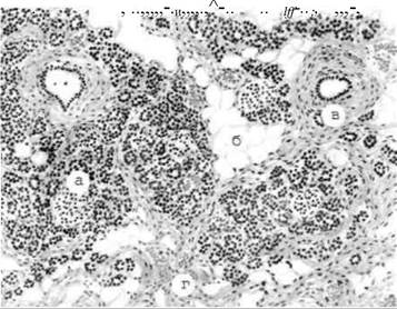
Fig. 3.73. Diabetes mellitus, pancreatic atrophy
Under conditions of absolute insulin deficiency, CARBOHYDRATES (glucose coming from food) are metabolized to a lesser extent in The Liver and insulin-dependent Tissues than in healthy individuals. Amino Acids (Alanine) are used for glucose synthesis in the liver. The source of amino acids is tissue proteins, which undergo accelerated breakdown. Thus, patients experience hyperglycemia (elevated blood glucose levels) and aminoacidemia (elevated blood amino acid content). Increased utilization of Amino Acids and tissue proteins is accompanied by a negative nitrogen balance and is one of the causes of weight loss in patients. When the glucose concentration in the blood exceeds the renal threshold (9.5-10 mmol/L), glucosuria occurs (excretion of glucose in the urine).
The Osmotic Pressure of urine increases, which causes polyuria (increased daily diuresis). Fluid loss through urine, which can reach 3-6 L/day, leads to dehydration of the body and polydipsia (excessive thirst). When intravascular blood volume decreases, blood pressure drops and the hematocrit value increases.
Under conditions of insulin deficiency, free Fatty acids, formed in adipose tissue As a result of enhanced lipolysis, serve as the main source of energy for Muscle tissue. The consequence of the latter is an excessive influx of glycerin and free fatty acids into the blood. The latter, being oxidized in the liver, serve as a source of Ketone Bodies (acetone, beta-hydroxybutyric and acetoacetic acids), which accumulate in the blood, causing ketoacidosis. In ketoacidosis, blood pH decreases and tissue Hypoxia occurs. Partially, free Fatty acids are used in the liver for the synthesis of triglycerides, which cause fatty liver infiltration and also enter the bloodstream. Excess triglycerides and free fatty acids in the blood form The basis of hyperlipidemia in diabetes mellitus.
Type II diabetes (non-insulin-dependent) is characterized by a high frequency of familial forms of the disease and a correlation with overweight. Since this type of diabetes is combined with a high blood insulin content, Lipogenesis processes predominate in such patients, contributing to obesity. On the other hand, obesity is a risk factor for this form of diabetes. Non-insulin-dependent diabetes mellitus is also pathogenetically heterogeneous.
Regardless of the type of diabetes mellitus, one of its most severe manifestations is diabetic macro- and microangiopathy, as well as neuropathy. Hyperglycemia plays the primary role in their pathogenesis. Thickening of the basement membrane due to the excessive deposition of proteins with an altered Structure is detected in microcirculation vessels. Autoantibodies (immune complexes) can form against these vascular wall proteins. An increase in blood clotting activity and microcirculation disorders also play a role in the pathogenesis of diabetic microangiopathy. All these processes culminate in sclerosis and hyalinosis.
Stereotypical microvessel changes are most frequently observed in the Kidneys (diabetic glomerulosclerosis), the retina of the eye (diabetic retinopathy), Skin, skeletal Muscles, pancreas, and Brain.
Diabetic macroangiopathy manifests as atherosclerosis of elastic and muscular-elastic type Arteries.
Pathological vascular changes lead to such frequent complications of diabetes mellitus as lower extremity gangrene, myocardial infarction, blindness, and renal failure.
Diabetic neuropathy is characterized by segmental demyelination of nerve fibers and degenerative changes in axons.
Diabetic nephropathy—manifested as nodular glomerulosclerosis and tubular nephrosis—is characteristic of diabetes mellitus. Other Kidney diseases are not specific to diabetes.
Last update: 08/08/2026
Editorial and Educational Adaptation: This material has been compiled based on the primary/original source text. The project team performed an editorial review, corrected technical inaccuracies, structured sections, and adapted the content for an educational format.
What was processed:
- elimination of formatting defects (OCR errors, structural breaks, corrupted characters);
- editorial organization of content;
- standardization of terminology in accordance with academic sources;
- verification of factual statements against the original source text.
All mentions of the author, publication year, and origin of the primary text have been preserved in accordance with the source.