Plant Physiology - Musienko M. M. 2001
Adaptation and Plant Stress Resistance Mechanisms
Radiation Stress
Stress in plant organisms can be caused by elevated levels of ultraviolet or ionizing radiation. Following the Chornobyl Nuclear Power Plant disaster (1986), this issue became exceptionally relevant for Ukraine. In recent years, a vast body of experimental data has been accumulated, explaining The Diversity of phenomena induced in plants by ionizing radiation (Grodzinsky, 1989). It turned out that among the various reactions of plant organisms to irradiation, one can distinguish processes that are common to all living things, as well as effects characteristic exclusively of plants. Plant-specific reactions arise As a result of the manifestation of molecular damage within the metabolic and physiological systems of Cells and Tissues.
The physical properties and characteristics of ionizing radiation determine the type of radiobiological response of an Organism — ranging from the acceleration of its GROWTH AND DEVELOPMENT (stimulation) to death. A distinction should be made between the radiation energy incident on a specific object and the energy actually absorbed, which causes the physical impact. It is important to know the units of ionizing radiation dose and radioactivity (Gudkov, 1991):
|
Name |
Obsolete unit |
SI unit |
RELATIONSHIP BETWEEN UNITS and |
|
Exposure dose |
Roentgen (R) |
Coulomb per kilogram (C/kg) |
1 R = 2,58×10⁻⁴ C/kg, 1 C/kg = 3876 R |
|
Absorbed dose |
Rad |
Gray (Gy) |
1 rad = 0,01 Gy, 1 Gy = 100 rad |
|
Equivalent dose |
Rem |
Sievert (Sv) |
1 rem = 0,01 Sv, 1 Sv = 100 rem |
|
Radioactivity |
Curie (Ci) |
Becquerel (Bq) |
1 Ci = 3,7×10¹⁰ Bq, 1 Bq = 2,7×10¹¹ Ci |
To characterize the distribution of ionizing radiation over time, METABOLISM/2.html">THE CONCEPT OF dose rate is used, which is understood as The amount of energy absorbed per unit of time. Depending on the dose rate, a distinction is made between acute irradiation (over the course of seconds, but at high doses—hundreds of grays per hour, or minutes) and fractionated/prolonged irradiation (over a long period, at fractions of a gray per hour).
The concepts of Radiosensitivity and Radioresistance
D.M. Grodzinsky and I.M. Gudkov (1973) proposed defining radiosensitivity as the ability of an organism to respond to minimal doses of radiation, registering low levels of exposure through non-lethal radiobiological effects. By the term radioresistance, they characterized the organism's ability to withstand high doses of exposure, expressed, for example, in terms of semi-lethal and lethal doses.
A measure of radioactivity is the lethal dose (LD) of irradiation, which causes Cell inactivation or organism death within specific timeframes following exposure. Typically, the LD50 index is used — the radiation dose that results in the survival of 50% of the individuals.
Literature contains data regarding the radiosensitivity of over 2,000 plants. It is believed that the highest radiosensitivity is found in lilies (LD50 — 10 Gy), and in conifers such as pine and spruce (LD50 — 10-20 Gy and 20-60 Gy, respectively). Grapevines (LD50 — 10-90 Gy) and fruit-berry species are quite sensitive. Among herbaceous plants, legumes are rather sensitive (broad beans — LD50 — 100 Gy).
Cereal crops are characterized by high radioresistance, while maximum radioresistance is observed in Brassicaceae (radish — LD50 — 1000-1500 Gy). The radioresistance of vegetative plants is always 10-15 times lower than that of seeds.
The unit of absorbed dose (Gray) is the dose at which 1 J of ionizing radiation energy is absorbed by 1 kg of irradiated material in 1 s. The rate of absorbed dose accumulation is called the absorbed dose rate, expressed in Gy × second-1:
Class="center">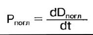
Due to the fact that the biological effect of irradiation depends not only on the absorbed dose but also on its quality, the concept of equivalent dose was introduced, according to which: the dose of a specific type of irradiation that produces the same biological effect as a dose of 1 R constitutes 1 roentgen equivalent man — 1 rem. This unit of equivalent dose was used until the Introduction of the modern SI unit, the sievert (Sv).
1 Sievert is equal to the dose of a specific type of radiation at which the biological effect is identical to that produced by 1 Gy of X-rays or γ-radiation. To calculate the radiation dose from external sources of γ-radiation and internal exposure from radionuclides incorporated in tissues (which emit Different types of radiation), the particle or quantum flux intensity must be determined from the radionuclide activity, and the exposure dose rate must be calculated. These calculations are based on radionuclide activity.
The mass of 1 Ci of a radionuclide is smaller the shorter its half-life. Knowing the radioactive decay constant K (s-1), one can calculate the mass of pure radionuclide of a given activity. For example, the mass of 1 µCi, in grams, is equal to 6,15 × 10⁻²⁰ A/λ, where A is the mole of the radionuclide. The mass of 1 µCi of 14C is only 2,17 × 10⁻⁷ g, and of 32P — 3,49 × 10⁻¹⁷ g.
Among radionuclides, there are those that emit β- and α-particles, accompanied by the generation of γ-quanta. For example, β-decay accompanied by γ-radiation is characteristic of 134Cs, 137Cs, 132I, and 131I. There are also radionuclides that emit only β-particles (35S, 45Ca, 90Sr, etc.).
The primary event in the interaction of ionizing radiation with a living organism is ionization, as a result of which electrons are stripped from atoms, creating an ionized state of atoms and molecules that triggers various chemical and biological reactions in tissues and Organs.
Radiation injury is a multi-stage process. Typically, two main classes of radiobiological effects are distinguished — somatic and genetic. Somatic effects include changes that occur in the organism during its ontogenesis, whereas genetic effects manifest in subsequent generations. D.M. Grodzinsky presents the following sequence of stages in The Development of radiation injury:
|
Levels of radiobiological processes |
Subcellular structures |
1. Irradiation: interaction of radiation with cellular substances 2. Formation of damage to biologically important molecules 3. Inactivation of subcellular structures |
|
Cell |
4. Impairment of cellular Functions 5. Disruption of Structural and functional Features of the cell |
|
|
Tissues and organs |
6. Disruption of "positional information" and donor-acceptor relationships |
|
|
Organism |
7. Disruption of morphogenesis, tissue metabolism, and integral regulation of ontogenesis. Formation of delayed somatic and genetic consequences |
|
|
Cenosis |
8. Disruption of phytocenoses |
Among plant tissues, Meristems are the most sensitive to radiation. A typical PLANT RESPONSE TO irradiation is an alteration in growth processes, with both inhibitory and stimulatory effects observed depending on the dose. A vast amount of experimental data has been accumulated indicating that low doses of radiation induce radiation stimulation.
It has been proven that under The Influence of stimulatory doses, the content of Plant HORMONES—growth activators—increases in plants, which triggers metabolic activation and ensures the acceleration of Plant Growth and Development. According to A.M. Kuzin, the enhancement of the phytohormonal system's activity is the result of nonspecific depression and the Activation of a specific group of genes under the influence of ionizing radiation.
The stimulatory effect of low doses (5 Gy) is utilized in crop production, for instance, for pre-sowing irradiation of corn, which provides a 10-12% increase in yield (Table 20).
Table 20. Stimulating and critical doses of gamma radiation for seeds of certain plant species (Gudkov, 1991)
|
Species |
Stimulating dose, Gy |
Critical dose, Gy |
Species |
Stimulating dose, Gy |
Critical dose, Gy |
|
Peas |
3 |
75-250 |
Flax |
7,5-10 |
400-1000 |
|
Corn |
5-10 |
100-200 |
Radish |
10 |
1000-2500 |
|
Wheat |
5-8 |
150-250 |
Cucumbers |
3 |
500 |
|
Tomatoes |
5-10 |
200 |
Oxygen Effect
Changes in the gas COMPOSITION OF THE atmosphere surrounding a plant during irradiation significantly affect its radioresistance. Thus, as early as the 1920s, E. Petri discovered that irradiation in a CO2 atmosphere without access to oxygen reduces radiosensitivity. It was later established that replacing oxygen with any other gas exerts a radioprotective effect. This phenomenon became known as the "oxygen effect", which manifests at all Levels of biological Organization—from molecular to tissue levels.
English radiobiologist L.H. Gray, after whom the unit of absorbed dose of ionizing radiation is named, first made an in-depth Study of the oxygen effect back in the 1930s.
There are two Structure/97.html">Definitions of the "oxygen effect": first, it is The phenomenon of enhanced radiation damage when the oxygen concentration in the environment increases compared to that observed under irradiation in anaerobic conditions (anoxia); second, it is the protective effect of reduced oxygen content (Hypoxia) during the irradiation of living organisms. The "oxygen effect" is a universal phenomenon in radiobiology. The Mechanism of the protective action of hypoxia is explained by the fact that during irradiation in the presence of O2, peroxide free radicals are formed, which amplify The Effect of radiation on vital molecules (DNA) and cellular structures while reducing the efficiency of intracellular defense systems. The discovery of this effect revolutionized concepts in the field of radiation damage modification. It demonstrated that the processes of development and realization of radiation injury can be managed and even mitigated.
The degree to which the "oxygen effect" manifests in seeds depends on their moisture content. Maximum radioresistance in seeds is achieved at a Water content slightly higher than that found in air-dry seeds. The Role of moisture in the radioresistance of vegetative plants remains poorly understood. It is believed that the development of a certain xeromorphism, water loss, and a decrease in metabolic rate positively correlate with radioresistance.
Radioprotective Effects
Experiments with plants have analyzed the effects of substances exhibiting radioprotective properties (aminoethylisothiouronium, dithiothreitol, Glutathione, thiourea, cystamine, Cysteine, cystine, etc.).
Radioprotectors are defined as substances whose administration into the organism via various pathways prior to irradiation reduces the severity of radiobiological effects. Thousands of compounds with radioprotective properties are known, but since the discovery of radioprotectors, sulfhydryl compounds have remained the most effective and well-studied class of such substances to this day. Their protective action is presumably based on the fact that sulfhydryl compounds, acting as strong reducing agents, scavenge free radicals and prevent their interaction with nucleic acid and protein macromolecules.
Radiobiologists Z. Bacq and A. Alexander put forward a hypothesis regarding the protective action of sulfhydryl compounds, known as the "biochemical Shock" or "metabolic protection" hypothesis. According to their view, sulfhydryl compounds at protective concentrations induce a shift in normal metabolism, which manifests as the inhibition of DNA, RNA, protein, and carbohydrate synthesis, as well as the suppression of energy processes. The inhibition of DNA Synthesis correspondingly reduces the rate of Cell Division and disrupts the growth and development of various tissues and organs. This temporary "shock" inhibition and the subsequent morphological changes serve as the primary cause of radioprotection.
The membrane hypothesis links cell death to radiation damage of membranes. Damage to the Plasmalemma, nuclear, and thylakoid membranes increases their permeability and disrupts the intracellular organization of metabolism. Other hypotheses regarding the causes of differential radioresistance in organisms and the mechanisms of radioprotector action are also known, including those concerning donor-acceptor substances in The Cell, the presence of antioxidants in cellular compartments, the maintenance of hypoxic states, and others.
Such a multitude of hypotheses concerning The Nature of radioresistance and its modification highlights the complexity and multiplicity of its mechanisms.
Last update: 07/08/2026
Editorial and Educational Adaptation: This material has been compiled based on the primary/original source text. The project team performed an editorial review, corrected technical inaccuracies, structured sections, and adapted the content for an educational format.
What was processed:
- elimination of formatting defects (OCR errors, structural breaks, corrupted characters);
- editorial organization of content;
- standardization of terminology in accordance with academic sources;
- verification of factual statements against the original source text.
All mentions of the author, publication year, and origin of the primary text have been preserved in accordance with the source.