MODERN BOTANY - P. RAVEN - 1990
SECTION IV. DIVERSITY
CHAPTER 16. BRYOPHYTES
Data from the previous chapter indicate that land plants (bryophytes and vascular plants) evolved from ancient groups of green Algae. Like the latter, plants share chlorophyll a as their primary photosynthetic pigment, along with chlorophyll b and carotenoids as accessory pigments. Across all three groups, the main reserve carbohydrate is starch, which is deposited within METABOLISM/14.html">Chloroplasts rather than the Cytoplasm, as seen in other photosynthetic eukaryotes. Cellulose serves as a crucial component of The Cell wall in both plants and certain green algae. Finally, during Cell Division, plants form a phragmoplast and a cell plate. Among other living organisms, this feature is found only in a single genus of brown algae and a few genera of green algae. Because all these traits are observed in both bryophytes (Fig. 16-1) and vascular plants, it is reasonable to suggest that both groups originated from a distant common ancestor that successfully colonized the land.
Class="center">Fig. 16-1. A dense growth of the moss *Fissidens* on limestone rocks near a waterfall. This photograph was taken in a nature reserve west of Yalta on the Crimean Peninsula in the USSR.

If this hypothesis is correct, the evolutionary lineages of bryophytes and vascular plants must have diverged very early. The oldest known fossils resembling bryophytes date to the Devonian period, approximately 400 million years ago (Fig. 16-2), whereas remains unambiguously classified as bryophytes are at least 370 million years old. Fossils resembling vascular plants have been discovered in Lower Silurian deposits approximately 430 million years old (see Fig. 1-5, p. 14). In light of these discoveries, it can be hypothesized that the hypothetical common ancestor of bryophytes and vascular plants—a relatively complex green alga (see Fig. 15-21)—colonized land more than 430 million years ago. Tetrads of spores and cuticle-like fragments from the Ordovician (dating back about 450 million years) also suggest an even earlier origin for plants.
Fig. 16-2. A. Cytology/practical/54.html">Longitudinal section of a gametophyte from chert beds near the village of Rhynie (Scotland). This plant lived during the Early Devonian, about 400 million years ago. The arrows indicate THE POSITION OF antheridia on the upper surface of the gametophyte. B. A section through an antheridium showing mature male Gametes. In size and structural complexity, such gametophytes closely resemble those of bryophytes. It remains unclear whether any Early Devonian sporophytes from these same deposits are related to these gametophytes.

Given that a well-defined Morphology/12.html">ALTERNATION OF GENERATIONS is observed in all bryophytes and vascular plants, this phenomenon was evidently characteristic of their common ancestor as well. Its gametophytes almost certainly formed multicellular gametangia similar to those found in all modern plants. All plants possess an embryo: zygotes begin dividing within the gametophyte and depend entirely upon it for Nutrition. Algae lack embryos, and it remains unknown whether the earliest plants possessed them. We also do not know whether the ancestral plants possessed conducting Tissues, and if so, which types. However, elongated Cells resembling tracheids, featuring annular or spiral thickenings, have been discovered in certain Lower Silurian fossils; some evidence points to the presence of Lignin-like substances in their cell walls. It is also possible that the earliest land plants formed endomycorrhizal symbioses, perhaps with a zygomycete fungus (see p. 206).
Organisms that successfully transitioned from Water to land developed structures that protect them from desiccation. One such adaptation is a sterile protective layer surrounding the cells that form sperm and egg cells within the male and female gametangia—namely, the antheridium and the archegonium. Similarly, a sterile layer formed around the spore-producing Cells of the sporangia.
The colonization of land by plant ancestors was accompanied by the retention of the zygote within the female gametangium, where it develops into an embryo. As a result, during critical early developmental stages, the young embryo (sporophyte) is protected by the female gametophyte. In contrast, in green algae, the Development of the Zygote is generally independent of the female gametophyte.
The aerial parts of most vascular plants are covered by a waxy protective layer, the cuticle, which helps prevent desiccation. Its presence is closely correlated with Stomata, specialized pores whose primary function is The regulation of gas exchange. The cuticle appears to be absent in most bryophytes, yet stomata are present on the sporophytes of hornworts and mosses. In some mosses, these stomata consist of a single doughnut-shaped guard cell, differing entirely from those of vascular plants. In hornworts, stomata likely remain open until late in development, whereas in mosses, they close only after the sporophyte has completely dried out. Thus, bryophytic stomata do not function in the same manner as those of modern vascular plants.
All plants are oogamous and exhibit an alternation of heteromorphic generations.
Characteristics of Bryophytes
Bryophytes—liverworts, hornworts, and mosses—are relatively small plants; many are less than 2 cm in length, and the majority are under 20 cm (Fig. 16-3). They often grow abundantly in relatively humid environments, where numerous species occur in large populations. Mosses sometimes dominate and displace other vegetation across vast polar regions and on rocky mountain slopes above the treeline. Many of them can even withstand prolonged periods of severe Antarctic cold (Fig. 16-4). Much like Lichens, bryophytes are highly sensitive to air pollution (especially sulfur dioxide) and are frequently absent or represented by only a few species in heavily polluted areas. A number of mosses occur in deserts, with some forming extensive patches on dry, open rocks that can heat up to very high temperatures. Many mosses remain viable in a dry state for years, rapidly resuming activity upon rehydration. Aquatic mosses and liverworts are also known, though they perish within roughly a day if allowed to dry out. A few bryophytes have even been found on wave-splashed marine rocks, although none are truly marine organisms. There are approximately 16,000 species of bryophytes—more than in any other plant group except the angiosperms.
Fig. 16-3. Hair-cap moss (*Polytrichum*). Sporophytes attached to female gametophytes are visible. Each sporophyte consists of a capsule, a seta, and a FOOT connecting it to the gametophyte. The leaves at the bottom of the photo belong to the gametophytes upon which the sporophytes developed following Fertilization, which occurred at least six months prior.

Fig. 16-4. A. Mount Melbourne in Antarctica (approx. 75° S). Here, at an altitude of 3,000 meters above sea level, daily summer temperatures fluctuate between —10 and —30 °C. In these unimaginably harsh conditions, on the snow-free patches visible in the photograph, New Zealand botanists discovered in 1984 populations of a moss belonging to the genus *Campylopus* (B), growing where volcanic activity warms the substrate to 30 °C. The growth of *Campylopus* in these locations demonstrates the remarkable dispersal capacity of mosses rather than merely their survival ability under harsh conditions, though the latter is also exceptionally high.

Two key features distinguish bryophytes from vascular plants. First is the absence of specialized conducting tissues—xylem and phloem. This means that all bryophytes, strictly speaking, lack true leaves, stems, and roots, since one of the defining characteristics of those structures is the presence of precisely such tissues. Nevertheless, the terms "leaf" and "stem" are commonly used in reference to the leaf-like and stem-like structures of liverwort and moss gametophytes. We will also follow this tradition.
The setae, or sporophores, of the sporophytes in most mosses contain a central strand of water-conducting cells called hydroids; similar cells are also present in the gametophytes of many of these species. Hydroids are elongated with tapering, oblique end walls that are easily permeable to water, making them efficient pathways for The transport of water and solutes. Similar to the tracheids and vessels of vascular plants, mature hydroids lack a protoplast and appear empty. However, they differ in their lack of specialized wall thickenings (Fig. 20-13). In some mosses, the strand of hydroids is surrounded by cells,
which transport nutrients, known as leptoids (Fig. 16-5), closely mirroring the spatial arrangement of phloem and xylem in certain vascular plants. Mature leptoids are elongated, with degenerated nuclei and a living protoplast. Some of them bear a striking resemblance to the phloem cells of primitive vascular plants. It is quite possible that the water- and nutrient-conducting cells of mosses and vascular plants evolved from identical cell types present in the common ancestor of both groups.
Fig. 16-5. Conducting strands in the sporophyte seta of the moss *Dawsonia superba*. A. Transverse section showing the general Structure OF THE seta (scanning electron micrograph). B. Transverse section featuring a central strand of hydroids, surrounded by a jacket of leptoids and cortical parenchyma. C. Longitudinal section of a portion of the central strand. From left to right: hydroids, leptoids, parenchyma.


In most bryophytes, the gametophyte is anchored to the substrate by elongated single cells or filaments of cells known as rhizoids. As a rule, these function solely to anchor the plant, since water and inorganic ions are generally absorbed directly and rapidly by the entire gametophyte. ROOT-like Organs are absent, although The structure of the subterranean stems in some mosses is quite complex.
The second major distinguishing feature of bryophytes is their pattern of alternation of generations: the gametophytes are always nutritionally independent, whereas the sporophytes remain permanently attached to the gametophytes and are dependent on them to varying degrees. In other words, the bryophyte gametophyte is the large, dominant generation, whereas in vascular plants this role is played by the sporophyte (see Figs. 10-11, B).
Bryophytes inhabit moist (at least seasonally) environments. For fertilization to occur, the biflagellate sperm must swim through water to reach the egg cell located within the archegonium. The archegonium is flask-shaped, sometimes stalked, with a long neck and an expanded basal portion—the venter—containing the egg (Fig. 16-6, A). At maturity, the so-called neck canal cells in the center of the neck break down, leaving a fluid-filled channel through which the sperm swims to the egg. The oblong or spherical antheridium (usually stalked) consists of a sterile jacket one cell thick that encloses numerous spermatogenous cells (Fig. 16-6, B). Each such cell gives rise to a single biflagellate sperm.
Fig. 16-6. Gametangia of Marchantia. A. Several archegonia at various Selection/3.html">Stages of development. B. Immature antheridium. In Marchantia, archegonia and antheridia are produced on separate gametophytes.

In liverworts and hornworts, the sperm often truly swim to the archegonium through a continuous film of water. Upon approaching, they are guided into the neck of the archegonium by chemical attractants released by it. In mosses, the antheridia are often clustered and surrounded by leaf-like structures that form a cup (Fig. 16-7). Here, the sperm are released into the water within the cup and are subsequently dispersed by splashing raindrops. Water droplets laden with sperm can be carried from plant to plant by insects. Because sperm can reach isolated female gametophytes situated relatively far away via these mechanisms, the latter frequently develop numerous sporophytes.
Fig. 16-7. Leafy male gametophytes of the moss Polytrichum piliferum, showing mature antheridia clustered into heads. Sperm are released into the water accumulating within these leaf-surrounded heads and are subsequently splashed out by raindrops, occasionally landing on another gametophyte bearing archegonia.

The fertilized egg, or zygote, remains within the venter of the archegonium, where it develops into an embryo. For a time, the cells of the venter divide in parallel with the growth of the young sporophyte developing inside. The enlarged archegonium forms a protective cap known as the calyptra. The mature sporophyte of many bryophytes consists of a capsule (sporangium) borne on a stalk (seta), which transitions into a "foot" embedded in the archegonium (Fig. 16-8).
Fig. 16-8. Nearly mature sporophyte of Marchantia. Elaters are clearly visible—spiral, filamentous structures inside the spore-filled capsule that aid in spore dispersal.

The cells of young and maturing sporophytes typically contain chloroplasts and are photosynthetically active; however, by the time Meiosis takes place in the capsule and spores are formed, chlorophyll usually disappears. In most mosses, as the seta elongates, the calyptra is carried upward along with the capsule; this protective cap is shed prior to spore dispersal, at which point the spores are released through the spontaneous splitting of the sporangium.
Bryophytes are traditionally divided into three classes: Hepaticae (liverworts, 6,000 species), Anthocerotae (hornworts, 100 species), and Musci (mosses, 9,500 species). These three groups differ markedly from one another, and their shared characteristics generally reflect those of simply organized plants. Most mosses possess conducting tissue and could well be considered reduced vascular plants with which they presumably shared a common ancestor, whereas liverworts and hornworts diverged earlier from this evolutionary Lineage. There is no evidence that liverworts evolved from ancestors with conducting tissue, nor that they are closely related to mosses. Hornworts differ sharply from both groups and remain distant from any known group of organisms. Given this, a growing number of botanists view the three bryophyte groups as distinct divisions, whose similarities are not evidence of close relationship, but rather the result of convergent evolution or the retention of primitive traits. In this book, however, we traditionally retain them within a single division.
Class Hepaticae
Liverworts are small plants, generally less conspicuous than mosses. Their name dates back to the 9th century, when the Liver-like outline of the gametophyte in some genera led to the belief that they would be useful in treating ailments of this organ (in accordance with the medieval doctrine of signatures, which held that a plant's appearance indicates its specific medicinal properties).
The gametophytes of some liverworts are dorsiventrally flattened thalli, or thalloid bodies (a term traditionally used to describe undifferentiated plant bodies, typically with distinct upper and lower surfaces) that grow via an apical meristem. In the majority of species within this class, however, the gametophytes are leafy. Growth is driven by the division of a single apical cell resembling an inverted pyramid with a base and three lateral faces that cut off daughter cells. Liverwort rhizoids are unicellular, unlike those of mosses, which are multicellular. Gametophytes develop directly from spores. The sporophytes of most liverworts are generally simpler than those of mosses, and their capsules exhibit more diverse mechanisms for spore dispersal.
Thalloid Liverworts
Thalloid (non-leafy) liverworts are highly diverse. They are found along damp, shaded riverbanks and in other suitable habitats, such as flowerpots in unheated greenhouses. The thallus is composed of numerous cell layers (about 30 at the midrib and roughly 10 in thinner regions) and is clearly differentiated into a thin, chlorophyll-rich upper (dorsal) region and a thicker, colorless lower (ventral) region (Fig. 16-9, A). The lower surface bears Two Types of rhizoids as well as rows of scales. The upper surface is divided into raised areas, each marking the dimensions of the underlying air chamber and featuring a large pore that leads into it (Fig. 16-9, B).
Fig. 16-9. A. Cross section of the thalloid liverwort gametophyte (Marchantia). Numerous chloroplast-bearing cells are visible in the upper layers, alongside several layers of colorless cells beneath them, and rhizoids anchoring the plant body to the substrate. Gas exchange within the air Chambers of the dorsal assimilation layer occurs via pores. The specialized cells surrounding the pore are typically arranged in 4 to 5 tiers of 4 cells each, forming a barrel-shaped structure. Under dry conditions, the cells of the lowermost tier, which typically protrude into the pore, close together, whereas they move apart as humidity increases. Pores thus perform a function analogous to that of stomata in vascular plants. B. Scanning electron micrograph of a pore on the dorsal surface of a Marchantia gametophyte.
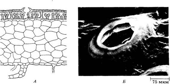
Fig. 16-10. Gametophytes of Marchantia. Antheridia (A) and archegonia (B) are elevated above the thallus on stalks.
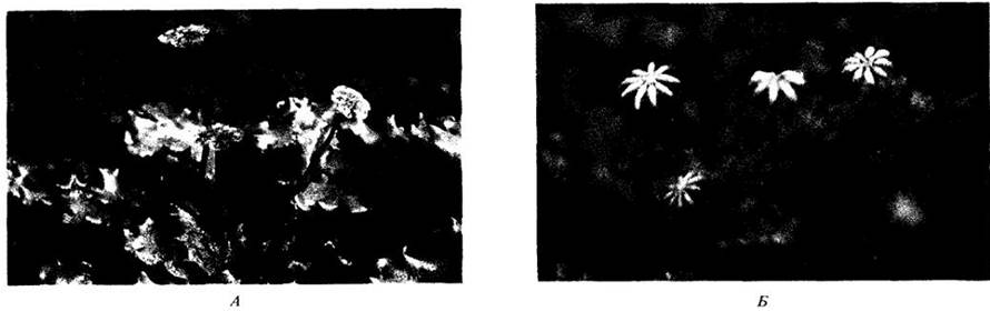
Fig. 16-11. Mature spores and elaters from the capsule of Marchantia.
One of the best-known liverworts is Marchantia, a widespread terrestrial genus growing on damp soil and rocks (Fig. 16-10). The dichotomously branched gametophytes mostly reach lengths of one to several centimeters, and the gametangia are concentrated on specialized upright structures known as gametophores. Marchantia gametophytes are unisexual; male and female individuals are easily distinguished by the differing shapes of their receptacles. In antheridial plants, these receptacles are disc-shaped and called antheridiophores, whereas in archegonial plants they are palmately lobed and called archegoniophores (Fig. 16-10). The life cycle of Marchantia is illustrated in Fig. 16-13. In this genus, the sporophyte generation consists of a foot, a short seta, and a capsule, or sporangium (see Fig. 16-8). In addition to spores, the mature sporangium contains elongated cells called elaters, which feature spiral hygroscopic (moisture-absorbing) wall thickenings (Figs. 16-8 and 16-11; see also the Appendix "Spore Dispersal in Liverworts"). The walls of these cells are sensitive to the slightest changes in humidity, twisting and untwisting in response. This action AIDS in spore dispersal after the capsule splits open into a number of petal-like segments.
The primary mode of asexual reproduction in liverworts is thallus fragmentation. Another widespread method of such reproduction in this class, as well as in mosses, is The formation of gemmae (brood buds)—multicellular bodies capable of giving rise to new gametophytes. In Marchantia, gemmae are produced within specialized cup-shaped structures called gemma cups on the dorsal surface of the gametophyte (Fig. 16-12). These brood buds are primarily dispersed by splashing rain.
Fig. 16-12. A. Gametophytes of Marchantia featuring cup-shaped gemma cups containing gemmae, which are splashed out by rain and can develop into new gametophytes. Each of these will be genetically identical to the parent plant from which the gemmae originated via mitosis. B. Longitudinal section of a gemma cup

Leafy Liverworts
This diverse group comprises more than 4,000 of the 6,000 known species in the class (Fig. 16-14). It is particularly abundant in tropical and subtropical regions, in areas with high rainfall or humidity (Fig. 16-15), though it is also quite common in temperate zones. These plants typically branch extensively and form small mats or tufts.
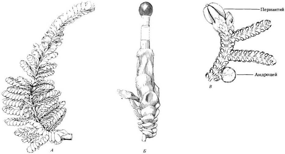
Much like those of mosses, liverwort leaves generally consist of a single layer of undifferentiated cells and, in many genera, are arranged in two rows, accompanied by a third row of reduced leaves along the lower surface of the gametophyte. The leaves are frequently bilobed, with each lobe growing via two apical cells. In Frullania, a common epiphytic liverwort, the leaves consist of a large, entire dorsal lobe and a smaller, helmet-shaped ventral lobe (Fig. 16-14, C).
In leafy liverworts, the antheridia are typically housed within a pouch-like outgrowth, the androecium, located on the lower part of a modified leaf. The developing sporophyte, like the archegonium from which it arises, is enclosed within a distinctive tubular sheath known as the perianth (Fig. 16-14, C).
Class Anthocerotae
This class includes only about 100 species grouped into six genera. Among them, Anthoceros is the most well-known, occurring worldwide, typically in moist, shaded habitats. The gametophytes of Anthoceros and thalloid liverworts appear superficially similar (Fig. 16-16, A), yet numerous features argue against a close relationship between these plants. For instance, in Anthoceros, each cell typically contains a single large chloroplast, much like many algae, rather than the numerous small, disc-shaped chloroplasts found in all other plants. Each chloroplast houses a pyrenoid, further enhancing its algal resemblance. The gametophyte structure is distinctly dorsiventral, often rosette-like, with a diameter typically of 1–2 cm. The large internal cavities of the Anthoceros gametophyte are filled with mucilage rather than air, as seen in thalloid liverworts. Nitrogen-fixing cyanobacteria of the genus Nostoc commonly inhabit these mucilage-filled cavities, supplying nitrogen to their plant hosts.
In some Anthoceros species the gametophytes are unisexual, while in others they are bisexual. The antheridia and archegonia are embedded within the tissue of the gametophyte's dorsal surface, with the antheridia grouped inside specialized chambers. Multiple sporophytes may develop on a single plant.
The upright, elongated sporophyte of Anthoceros consists of a foot and a long cylindrical sporangium (Fig. 16-16, B and 16-17, A). At a very early stage of development, a meristem—a zone of actively dividing cells that remains active as long as environmental conditions favor growth—becomes established between the foot and the sporangium. As a result, the sporophyte continues to elongate for a prolonged period. It is green, possessing several layers of photosynthetic cells, and is covered by a cuticle bearing stomata (Fig. 16-16, C). Spore maturation and subsequent dehiscence of the sporangium begin at the apex and progress toward the base (Fig. 16-16, D, E). Interspersed among the spores are sterile, elongated, often multicellular structures resembling the elaters of liverworts (Fig. 16-17, B). The sporangium splits longitudinally into ribbon-like halves.
Class Musci
In many plant groups, certain species are popularly and incorrectly referred to as mosses. For instance, "reindeer moss" and "Iceland moss" are actually lichens, "beard moss" is a vascular plant, and "irish moss" is an alga. True mosses, however, belong to the class Bryopsida, which comprises three subclasses: Bryidae (true or true mosses), Sphagnidae (peat mosses), and Andreaeidae (lantern mosses).
True Mosses
The gametophytes of all mosses are represented by two distinct phases: the protonema (from the Greek words protos, meaning first, and nema, meaning thread), which develops directly from a germinating spore, and the leafy gametophyte. In true mosses, the protonema consists of a single layer of cells, branches out, and resembles a filamentous green alga (Figs. 16-18). Leafy gametophytes develop from tiny bud-like structures on the Branches of the protonemata. In a few moss genera, the protonema is long-lived and performs the primary photosynthetic function, whereas the leafy shoots remain very small. Protonemata, characteristic of all mosses, are also found in some liverworts.
In true mosses, the leafy gametophyte is typically erect rather than dorsiventrally flattened like that of leafy liverworts; nevertheless, it also grows from an apical cell resembling an inverted three-sided pyramid. Although three rows of leaves are initially formed, subsequent twisting of the axis shifts these rows, creating the appearance of a spiral leaf arrangement. In some genera (such as the aquatic moss Fontinalis), the original three-ranked leaf arrangement is still discernible even in the mature gametophyte.
Moss gametophytes feature a complex structure and range in length from 0.5 mm to 50 cm or more. All of them possess multicellular rhizoids, and their leaves are generally only a single cell thick, except for the midrib (which is absent in some genera). As mentioned above, the stems of many mosses contain a central strand of water-conducting hydroids, and some also feature organic-nutrient-conducting leptoids.
Moss gametophytes exhibit two main growth habits. In some mosses (which often form dense cushions), the gametophytes are erect and sparsely branched, typically bearing terminal sporophytes. In others, the gametophytes are heavily branched ("feathery") and creeping, with lateral sporophytes (Figs. 16-19). This second growth type is found in many mosses that hang in masses from tree branches in humid regions.
Upon maturation, most leafy gametophytes produce gametangia either at the apex of the main axis or on a lateral branch. In some genera, the gametophytes are dioicous (Figs. 16-20), whereas in others, archegonia and antheridia arise on the same individual.
Fig. 16-13. Life Cycle of Marchantia, a widespread thalloid liverwort. Like other liverworts, Marchantia is characterized by an alternation of generations with a gametophyte (n) dominant over the sporophyte (2n). The two most critical events in the life cycle are meiosis and syngamy. The gametophytic generation begins with meiosis, which produces haploid spores. Some of these spores germinate into male gametophytes, others into female ones. The sporophytic generation begins with syngamy, producing a diploid zygote. Motile sperm require an aqueous medium to reach the egg cell. Within the calyptra, or venter, of the archegonium, the zygote develops into an embryo, or young sporophyte. As the embryo grows, the calyptra also increases in size. Ultimately, the maturing sporophyte ruptures the calyptra, elevating the sporangium into the external environment. Throughout its life, the sporophyte remains attached to the gametophyte by an expanded foot. Details of the structure of this liverwort are shown in Figs. 16-6 and 16-8 through 16-12.
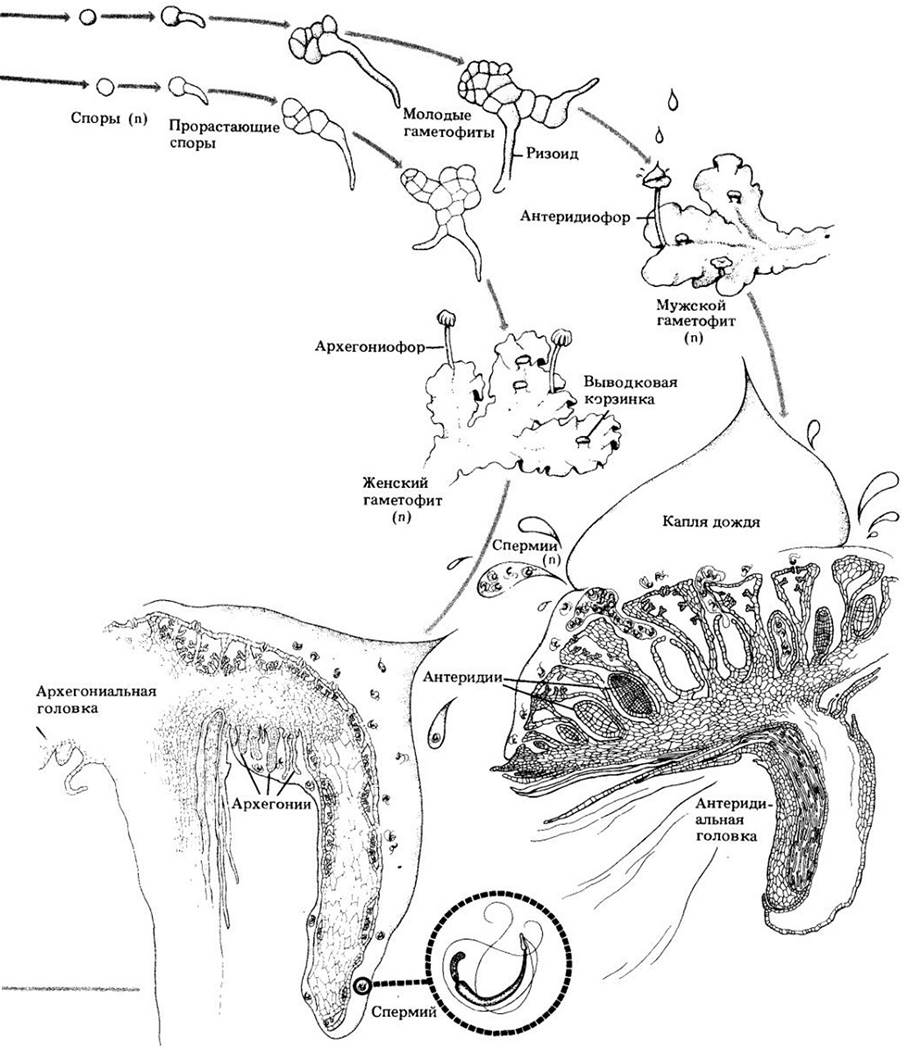
Fig. 16-14. Leafy liverworts. A. Clasmatocolea puccionana, showing the characteristic leaf arrangement. B. Tip of a Clasmatocolea humilis branch, showing the capsule and the long seta of the sporophyte. C. Part of a Frullania branch with its characteristic phyllotaxy.
In temperate species, sporophytes typically require 6 to 18 months to reach maturity; they arise on the gametophytes (see Fig. 16-3). The capsules are elevated on a seta that sometimes reaches 15–20 cm in length, though it is entirely absent in some species. A short foot at its base is embedded in the gametophytic tissue. The seta usually elongates early in The Development of the sporophyte, which is capable of Photosynthesis at this stage and is therefore generally less dependent on the gametophyte for nutrition than liverwort sporophytes are (in liverworts, the sporophyte typically remains surrounded by gametophytic tissue until maturity). Stomata, which are absent in liverwort sporophytes, are normally present in moss sporophytes. Furthermore, the sporophytes of mosses possess a more complex internal Organization. The setae of most species feature a central strand of hydroids, and many also characterize organic-nutrient-conducting leptoids (see Fig. 16-5).
Fig. 16-15. Leafy liverwort growing on the leaf of an evergreen tree in a rainforest of the Amazon basin near Manaus, Brazil.
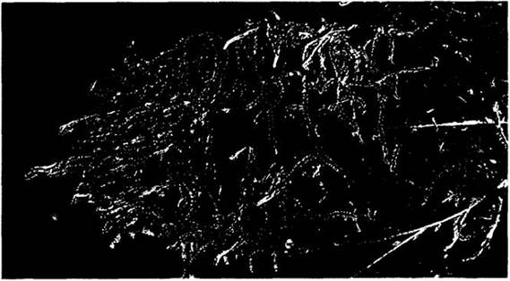
Fig. 16-16. Anthoceros. A. Gametophyte with attached sporophytes. B. Mature sporangium splitting to release spores. C. Stoma, which are abundant on the green, photosynthetic sporophytes of hornworts. D. Developing spores. E. Mature spores

Fig. 16-17. Anthoceros. A. Cross section of a sporophyte showing its foot embedded in the gametophyte tissue. B. Longitudinal section of a sporangium showing spore tetrads interspersed with elater-like structures. The central Column in the lower part of the sporangium consists of tissue that may function as conducting tissue

Fig. 16-18. Moss protonema with a bud-like structure. The protonema represents The First stage of the gametophyte generation in mosses and some liverworts, often resembling filamentous green algae
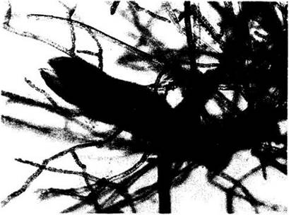
Fig. 16-19. Two main forms of gametophyte growth in mosses. A. "Cushion-forming" (e.g., Polytrichum juniperinum); gametophytes are erect and sparsely branched, with sporophytes (capsules on long, slender stalks) rising above them. B. "Feather-like," with creeping, carpet-forming gametophytes (e.g., Thuidium delicatulum)
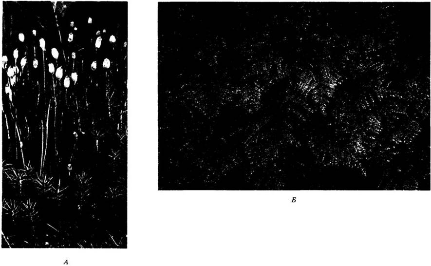
Fig. 16-20. Gametangia of the moss Mnium. A. Longitudinal section of an archegonial HEAD, showing purplish archegonia surrounded by sterile structures called paraphyses. B. Longitudinal section of an antheridial head, showing antheridia surrounded by paraphyses

As a moss sporophyte matures, it gradually loses its photosynthetic capacity, turning first yellow, then orange, and finally brown. Eventually, the operculum (lid) of the capsule falls off, revealing a ring of Teeth known as the peristome, which surrounds the opening (Fig. 16-21). The peristome teeth are formed by the splitting of a cell layer near the apex of the capsule along lines of weakness. This mechanism, which regulates spore dispersal, is characteristic of the subclass Bryidae and is absent in the other two subclasses. Each capsule releases up to 50 million haploid spores, each capable of giving rise to a new gametophyte. The typical life cycle of true mosses is illustrated in Fig. 16-22.
Fig. 16-21. Peristome teeth in true mosses. A. In Brachythecium, the peristome consists of two rings of teeth that open to release spores in response to changes in atmospheric humidity. Under moist conditions, the teeth of both rings interlock, whereas when the capsule dries, the outer teeth bend outward, allowing wind dispersal of the spores. B. Scanning electron micrograph of the peristome teeth of the genus Orthotrichum in dry weather, showing the inward-curved inner teeth and outward-bent outer teeth

Fig. 16-22. In the life cycle of a true moss, spores are released from the capsule upon the shedding of the operculum.
A haploid spore germinates into a branched, filamentous protonema, from which the leafy gametophyte develops. Sperm cells released from a mature antheridium may reach the vicinity of the archegonium, where they are guided into the neck canal by chemical attraction. Inside the archegonium, one of the sperm fuses with the egg cell to form a zygote, which divides mitotically to form the sporophyte. Simultaneously, the venter of the archegonium divides to form the calyptra. The sporophyte consists of a capsule—typically elevated rapidly on a seta (stalk, which is also part of the sporophyte)—and a foot through which it absorbs nutrients from the gametophyte. Meiosis occurs inside the capsule, producing haploid spores. The figure illustrates the life cycle of a species of the genus Polytrichum.


Asexual reproduction in these mosses typically occurs through fragmentation. In essence, any part of the gametophyte, including the sterile PARTS OF THE reproductive organs, is capable of regeneration, and many species produce gemmae that can give rise to new gametophytes.
Sphagnum Mosses
Approximately 350 species of mosses belonging to the single genus Sphagnum form a distinct group that diverged very early from the main evolutionary line of this division (Fig. 16-23). The stems of their leafy gametophytes bear whorls of branches, often five per node, which are more densely clustered at the apex of the plant to form a head-like structure. The gametophytes form large, light-green or sometimes reddish cushions in boggy habitats. They arise from gemmae along the margin of a thalloid protonema rather than a filamentous one. Sphagnum leaves lack a midrib, and mature plants lack rhizoids. In their typical wetland habitats, these plants almost always maintain high turgor pressure, keeping them erect and usually densely packed. The leaves of this genus consist of broad, dead cells surrounded by narrow, green or occasionally red living cells (Fig. 16-23, B). The former feature pores and wall thickenings and fill with water easily, giving Sphagnum a water-holding capacity roughly 20 times its dry weight (by comparison, absorbent cotton can absorb only 4 to 6 times its own dry weight in water).
Fig. 16-23. Peat moss Sphagnum. A. Gametophyte with numerous attached sporophytes. Some of the capsules (e.g., the two on the left) have already dispersed their spores. B. Leaf structure. Large dead cells are surrounded by smaller living cells containing chloroplasts. C. Dehiscence of the capsule. As it dries, the capsule fills with air. The stomata, through which air enters, close upon drying. With further contraction of the capsule, increasing internal pressure forcefully pops off the operculum and discharges a cloud of spores into the air

Owing to their exceptional absorptive properties, these mosses have been used in Europe since the 1880s as dressing material for wounds and sores, though since World War I they have been almost entirely replaced by cotton, likely due to the neater appearance of cotton products. Gardeners mix peat moss into soil to increase its water retention and acidity.
The sporophytes of Sphagnum (Fig. 16-23, A) are also highly distinctive: ranging in color from red to brownish-black, the capsules are nearly spherical and are elevated on a pseudopodium—a stalk derived from the gametophyte that reaches up to 3 mm in length, whereas the sporophyte's own stalk is very short. Spore dispersal occurs through a highly efficient mechanism (Fig. 16-23, C). At the apex of the capsule, a disc-shaped operculum is separated from the rest of the structure by a circular groove. As the capsule matures, its internal tissues shrivel, and air is drawn inside, possibly through non-functional stomata. As the wall dries, the air remains trapped within. Further contraction of the ripening capsule increases internal pressure until the operculum finally pops off with an audible snap, typically occurring in warm, sunny weather. The escaping gas forcefully expels a cloud of spores from the capsule. The most diagnostic Features of the subclass Sphagnidae are the absence of a peristome and the unique Morphology of the gametophyte.
Sphagnum Ecology
Members of this genus form extensive peat bogs, which are a defining feature of the cold and temperate Regions of the globe. These mosses contribute to increasing the acidity of their habitat by releasing hydrogen ions; consequently, the pH in the center of a bog is often below 4, which is highly unusual for a natural environment. Peat is formed through the accumulation and compaction of the sphagnum mosses themselves, alongside sedges, grasses, and other plants that grow alongside them. In Ireland and certain other northern regions, dry peat is widely used as fuel. By the most conservative estimates, peatlands cover at least 1% of the Earth's land surface, which is equivalent to half the area of the United States. Because global peat reserves are so vast, interest in peat as a potentially significant source of industrial energy grows every year. However, this necessitates a careful Assessment of the potential impact of peat extraction on the regions where it takes place, as well as the consequences of releasing large amounts of carbon dioxide into the atmosphere from burning such fuel.
Andreaea Mosses
The genus *Andreaea* comprises approximately 100 species of small, blackish-green or dark reddish-brown rupicolous mosses that form tufts (Fig. 16-24) and are no less distinctive in their own right than *Sphagnum*. Representatives of this group occur in mountainous or arctic regions, frequently on granites. Although their gametophyte closely resembles that of true mosses, it develops from a thick, multilobed protonema rather than a filamentous structure. The sporophyte lacks a true seta and is elevated above the leaves on a stalk derived from gametophytic tissue—that is, a pseudopodium, similar to that of sphagnum mosses. Along the small capsule of *Andreaea*, four vertical rows of less durable cells run, along which the capsule dehisces while remaining intact above and below the lines of splitting. The resulting four Valves are highly sensitive to ambient humidity, spreading widely when the air is dry (allowing spores to be dispersed far by the wind) and closing when it is damp. This mechanism of spore dispersal through the longitudinal splitting of the capsule differs from that observed in all other mosses (Fig. 16-24).
Fig. 16-24. A — *Andreaea rothii* on granite rocks in Devon, England. B — Sporangium of the genus *Andreaea* contracting upon drying and splitting into four valves, enabling spore dispersal in dry weather.

Andreaeobryum — a second genus of Andreaean mosses, discovered in Alaska in 1976 — differs from *Andreaea* by the presence of a true seta in the sporophyte and the splitting of the capsule all the way to the apex.

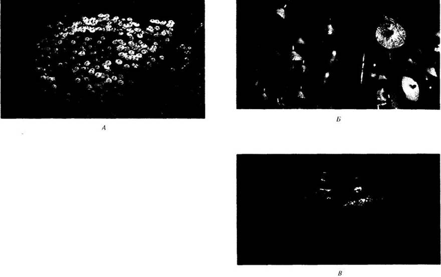
A — *Splachnum luteum*. Plants of this species, featuring cream-colored apophyses, resemble small flowers. B — The coloration of the plants and the chemical compounds they secrete attract insects. C — *Splachnum rubrum*, photographed on moose dung in the province of Alberta (Canada).
Last update: 07/08/2026
Editorial and Educational Adaptation: This material has been compiled based on the primary/original source text. The project team performed an editorial review, corrected technical inaccuracies, structured sections, and adapted the content for an educational format.
What was processed:
- elimination of formatting defects (OCR errors, structural breaks, corrupted characters);
- editorial organization of content;
- standardization of terminology in accordance with academic sources;
- verification of factual statements against the original source text.
All mentions of the author, publication year, and origin of the primary text have been preserved in accordance with the source.